Introduction
Osteogenic malignant tumours are neoplasms that arise from osteoblasts, which are mainly characterised by the formation of osteoid by the tumour cells themselves.1,2 The reported annual incidence of bone sarcomas in North America and Europe in both sexes is estimated at 0.75 cases per 100,000 of the population.3
Osteosarcomas have a bimodal age pattern, primarily affecting children and adolescents during periods of rapid growth, but also occurring at a lower rate in adults over the age of 60.2,4,5 It can occur in any bone in the body, although it is more common in long bones of the limbs such as the humerus, femur or tibia.6,7
However, according to the World Health Organization (WHO) Classification of Tumours (WCT), malignant osteogenic tumours constitute a heterogeneous group characterised by histopathological and epidemiological variability.7 Osteosarcoma is the most common of the primary malignant bone tumours. The annual incidence rate for those aged 0–24 years is approximately 4.4 cases per million people, and approximately 4.2 cases per million people aged ≥60 years.8 Parosteal osteosarcoma accounts for about 4% of all osteosarcomas and is the most common type, followed by periosteal forms; low-grade central osteosarcomas account for 1–2% of cases, whilst high-grade surface osteosarcoma is the rarest, accounting for <1% of diagnosed cases.3,7
The accurate identification of osteosarcoma tumour types, as in other cancers, has important implications, as it defines pathological features, guides treatment decisions, and informs prognostic assessments. Recent multi-omics studies further support the existence of distinct molecular subtypes with different clinical prognoses and therapeutic vulnerabilities.9 For this reason, the international standard for tumour classification and cancer diagnosis, provided by the International Agency for Research on Cancer (IARC) through a series of publications known as the WHO Blue Books, is periodically updated through successive editions; the fifth edition of the classification of tumours of soft tissue and bone was published in 2020.3
The WCT provides an international standard for tumour classification and accurate cancer diagnosis in what are commonly referred to as the WHO Blue Books. In the process of compiling and editing of each new edition, the Blue Books editorial team, comprising mainly experts in the field of cancer pathology, faces a number of challenges, such as the large number and wide variability of published studies. To facilitate and assist with this task, and to ensure that the WCT editors have the best available evidence for the next edition, IARC promoted the WCT EVI MAP Project (World Health Organization (WHO) Classification of Tumours: A Living Evidence Gap Map by Tumour Type.10 The WCT EVI MAP project aims to produce Evidence Gap Maps (EGM) for different tumours to visualise the available evidence for various tumour characteristics, as well as to identify low-level evidence and research gaps.11 Beyond describing the evidence base as mapped, it is essential to analyse how the quantity and quality of evidence vary across different tumour types to understand in which cases classification decisions are well-founded and where they are based on limited or low-quality evidence.
Osteosarcoma is one of the tumours that can benefit most from these efforts as it presents two key challenges: low incidence rates and considerable morphological overlap among defined histological types, both of which amplify the impact of limited evidence. This is particularly relevant as the definition of classification boundaries may directly influence diagnosis, grading and subsequent treatment decisions.12 The aim of this study was to map the available evidence across osteosarcoma tumour types to inform the WHO Classification of Tumours.
Methods
Evidence Gap Map methodology was applied, adapting methods described by 3ie (International Initiative for Impact Evaluation) and the Campbell Collaboration to suit the requirements of the project.13,14,15
The study protocol was registered in the OSF:
https://osf.io/2ax94/overview
Evidence gap map framework
Within the context of the WCT EVI MAP project, a common framework was developed for evidence mapping, to provide a structured representation of the available evidence on tumour characterisation across different tumour types. The framework was developed by a multidisciplinary group of experts, including evidence synthesis specialists, epidemiologists, pathologists and molecular biologists.
The framework is operationalised as a matrix with two main dimensions, where the columns correspond to tumour types and the rows to tumour characteristics.
Tumour entities were defined in accordance with the WCT, adopting its hierarchical taxonomy. Specifically, three levels were incorporated: anatomical location, tumour category and tumour type. The tumour type constitutes the primary unit and is represented at the column level, whilst the higher-order groupings (location and category) provide an organisational structure to facilitate navigation and interpretation of the map.
The selection of tumour characteristics was based on theoretical criteria and aligned with the main objective of the EGM, namely, to synthesise the evidence on pathogenesis, histopathology (including cytology), diagnostic imaging, diagnostic molecular pathology, prognosis and prediction. Operational definitions for each domain were established prior to study selection to ensure consistent classification.
Each cell in the matrix represents the intersection between a tumour type and a tumour characteristic. The evidence for each cell is displayed using bubble plots, where bubble size is proportional to the number of studies included, and colour corresponds to the level of evidence.
Levels of evidence were assigned according to a further adapted Hierarchy of Evidence for Tumour Pathology (HETP), originally developed during the first phase of this project,16 ranging from level one (highest e.g. Systematic Reviews) to level five (lowest e.g. case Reports). Records were categorised as “unclassifiable” if no evidence level could be assigned. This included study designs classified as “level not applicable” in the HETP, as well as records for which software constraints prevented independent matching of multiple study designs to different characteristics. The adapted HETP table is available in a public repository (see Data Availability section).
Search strategy
Comprehensive searches in PubMed and Embase were designed and conducted by a scientific information specialist (A.I.), using tailored search strategies that combined database-specific subject headings and keyword terms aligned with the tumour types and characteristics defined in the EGM framework. The search was restricted to studies published from 2018 to 2024 inclusive, due to the large volume of evidence available and time constraints of the project. The full search strategies are publicly available in an open repository (see Data Availability section).
Eligibility criteria
Studies focused on malignant osteogenic tumours were included, specifically those reporting on osteosarcoma, parosteal osteosarcoma, periosteal osteosarcoma, high-grade surface osteosarcoma, and low-grade central osteosarcoma. Eligible studies were required to report at least one of the tumour characteristic domains defined in the EGM framework, including pathogenesis, histopathology (including cytology), diagnostic imaging, diagnostic molecular pathology, prognosis or prediction.
No restrictions were applied regarding age, sex, or geographical location.
Studies were excluded for the following reasons:
• Language and accessibility: studies not providing title, abstract, or not available in English.
• Publication type and study design: non-peer-reviewed literature, including opinion pieces, comments, unpublished data, retractions or corrigenda, grey literature, narrative reviews, conference abstracts, proceedings and study protocols and pure qualitative studies. Also, pre-clinical research except those reporting on pathogenesis or relevant predictive outcomes.
• Intervention and outcome relevance: studies exclusively assessing tumour effectiveness or focusing on machine-learning/software application not linked to biomarkers, studies not reporting defined tumour characteristics, as well as studies focusing on social determinants.
• Data requirements: studies using only public omics repositories without independent validation, studies not reporting disaggregated results for the included tumour types, purely in silico studies and those using only non-human cell lines.
• Tumour type: studies not using WCT terminology (5th edition) such as “low-grade” and “high-grade osteosarcomas” without specification of surface origin, due to terminological ambiguity as these terms may refer to different entities; or focusing on recurrence or metastasis in a different anatomical site.
A detailed visual representation of the eligibility criteria hierarchy was developed during the project and is available in the open repository (see Data Availability section).
Study selection
The search results were deduplicated and imported into EPPI-Reviewer® version 6.
The screening process was piloted during the early stages of the project’s mapping process to ensure consistency between reviewers and across all project teams.
Three reviewers (O.C., E.G.-O., and M.M.-M.) independently screened 10% of the references in duplicate applying the eligibility criteria. Records were classified as included, excluded or for discussion. Discrepancies and studies classified as ‘for discussion’ were resolved through discussion with a third reviewer, a senior reviewer or a pathologist from the WCT EVI MAP Project coordination team.
The remaining references were then screened by a single reviewer, with the assistance of the “priority screening” tool in EPPI-Reviewer®. This machine learning tool ranks records according to the likelihood of inclusion based on prior screening decisions made by the reviewers, thereby prioritising the most relevant studies earlier in the process. Screening proceeded in this ranked order. It was discontinued after reaching 50 consecutive exclusions, at which point the remaining records were excluded due to the low probability of meeting inclusion criteria according to the model’s ranking.
This approach is supported by evidence from machine learning-assisted screening, where stopping rules are applied to balance efficiency and the risk of missing relevant studies in reviews involving a large volume of evidence to be assessed.17
Data extraction
Included studies were coded using a pilot coding tool developed at an early stage of the project. Coding and data extraction were carried out concurrently in EPPI-Reviewer®.
The coding tool allowed reviewers to assign to each record the osteogenic tumour type, the main tumour characteristic for which the study provided evidence, and the study design. Study design was assigned by reviewers using a decision tree diagram specifically developed for this purpose.18,19
In cases where multiple tumour types were reported in a single study, or outcomes were provided for multiple characteristics, a prioritisation based on tumour and characteristics relevance was performed by pathology experts; this prioritisation is shown in
Table 1. When multiple study designs were identified within a record, the study design with the highest level of evidence according to the HETP for the coded characteristic was selected. The studies were coded based on the information contained in the abstract.
Table 1. Priority rules for tumour type and tumour characteristic coding.
| Coding category |
Priority Order |
|---|
| Tumour Type | 1. Osteosarcoma |
| 2. Parosteal osteosarcoma |
| 3. Periosteal osteosarcoma |
| 4. High-grade surface osteosarcoma |
| 5. Low-grade central osteosarcoma |
| Tumour characteristic | 1. Pathogenesis |
| 2. Histopathology (including cytopathology) |
| 3. Diagnostic molecular pathology |
| 4. Prediction |
| 5. Prognosis |
| 6. Diagnostic imaging |
Two reviewers independently coded 10% of the studies, after which any uncertainties or discrepancies were resolved with a third reviewer or a senior reviewer. The remaining records were then coded by a single reviewer.
Upon re-examining the records while coding, some were excluded under the same eligibility criteria as the screening.
Finally, once coding of all reports had been completed, a quality check report was generated to identify potential errors, such as uncoded records or instances of repeated coding.
Data synthesis and presentation
The report and metadata were downloaded from EPPI-Reviewer® as a JSON file.
Evidence levels were then assigned to each study in accordance with the aforementioned HETP, using a Python script, generating a JSON file. This final dataset was uploaded into EPPI-Mapper® to generate the EGM.
Results
The study selection process is summarised in the PRISMA 2020 flow diagram (
Figure 1), which illustrates the number of records identified, screened, excluded, and coded. The initial search in Embase and PubMed identified a total of 14,938 studies. After screening and selection, 2,571 studies were included in the EGM. The flow diagram also reflects the use of priority screening during study selection.
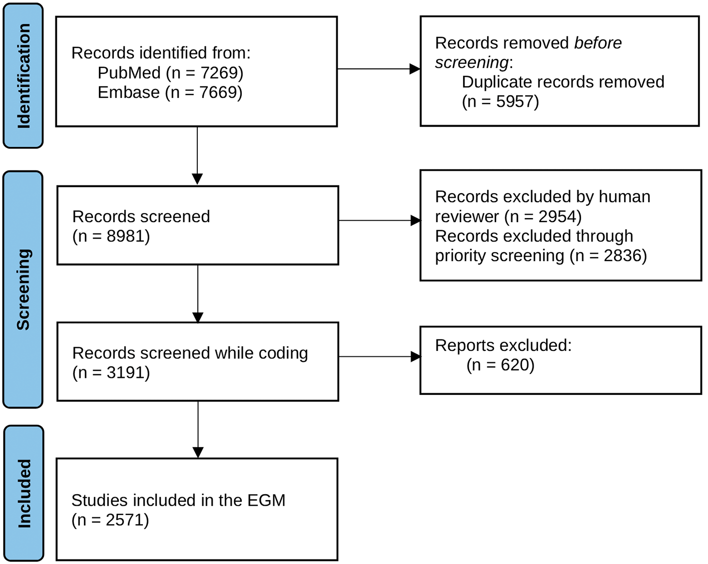
Figure 1. Adapted PRISMA 2020 flow chart for the osteosarcoma tumour types Evidence Gap Map.
The resulting EGM is shown in
Figure 2 and the interactive EGM can be consulted via the following link Evidence Gap Map of Osteogenic tumours
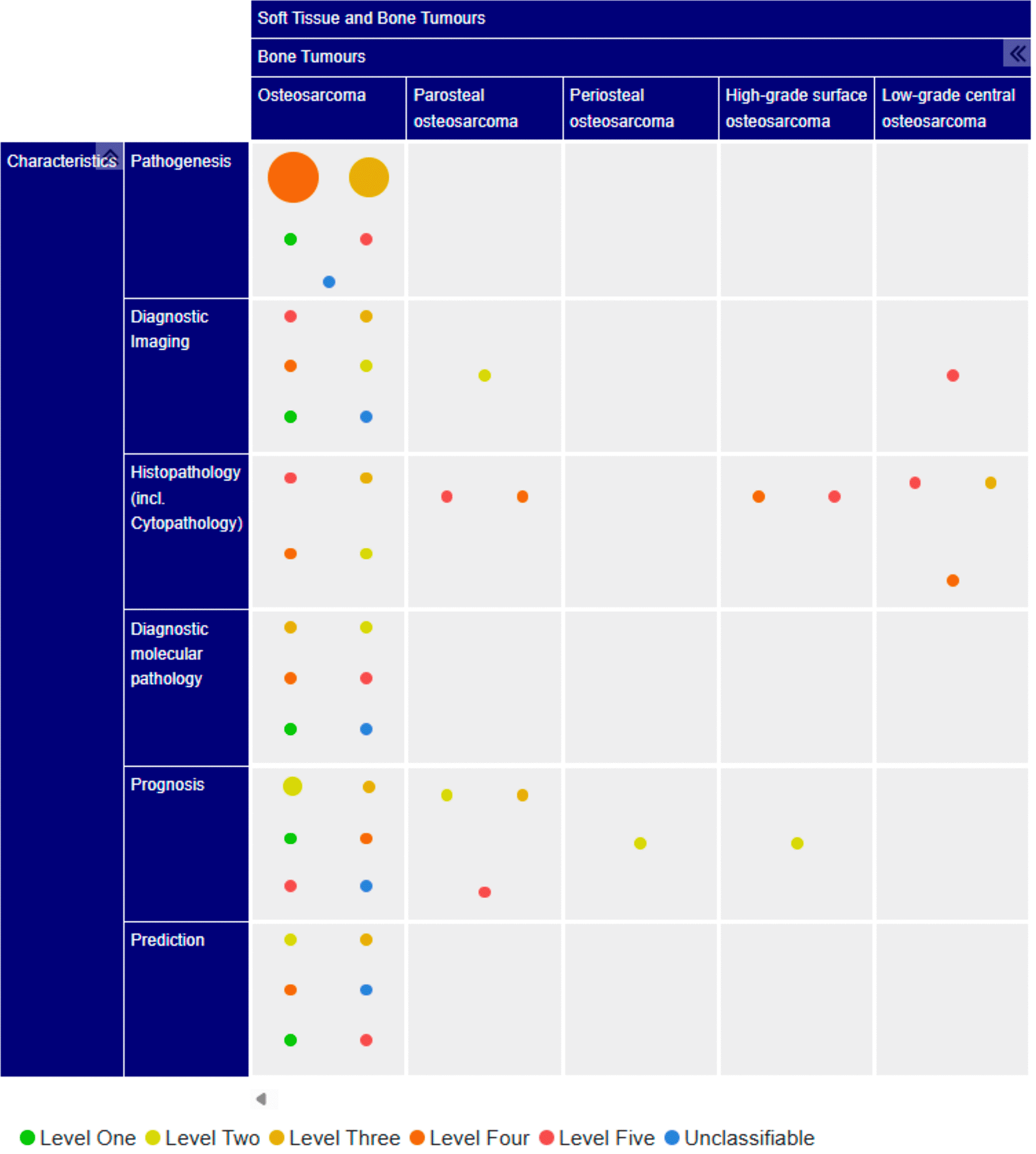
Figure 2. Malignant osteogenic tumour types evidence gap map.
Evidence distribution differed substantially by tumour type as shown in
Figure 3, with osteosarcoma being the most extensively represented entity (n = 2529), largely dominated by level four studies (n = 1440; 56.9%), followed by level three (n = 640; 25.3%). Higher-level evidence (levels one and two) accounted for 13.8% of studies (n = 348), with level one evidence representing 1.6% (n = 41).
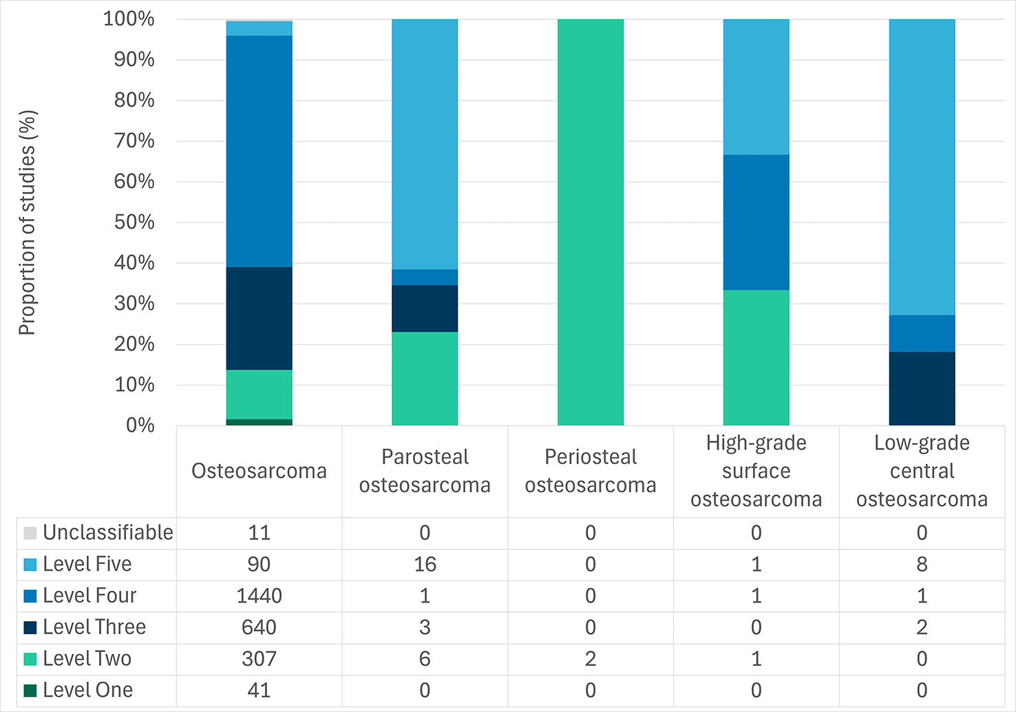
Figure 3. Studies found by osteogenic tumour type and level of evidence, with number of papers in each category listed below.
Evidence for rarer subtypes was limited and predominantly low level. Parosteal osteosarcoma was supported by 26 studies, predominantly at level five (n = 16), while periosteal osteosarcoma was supported by only two level two studies within the subset of papers coded. High-grade surface osteosarcoma was represented by three studies, evenly distributed across levels two, four, and five. Low-grade central osteosarcoma was mainly supported by level five studies (n = 8), with no high-level evidence identified (
Figure 3;
Table 2).
Table 2. Distribution of the evidence included in the evidence gap map for each type of malignant osteogenic tumour, by tumour characteristic and level of evidence.
| Osteosarcoma |
|---|
| Level of Evidence | All levels |
|---|
| Level One | Level Two | Level Three | Level Four | Level Five | Unclassifiable | Total |
|---|
| Tumour characteristics
| n | n | n | n | n | n | n |
| Pathogenesis
| 2 | 0 | 477 | 1370 | 2 | 1 | 1852 |
| Diagnostic imaging
| 2 | 5 | 13 | 7 | 15 | 1 | 43 |
| Histopathology (including cytopathology)
| 0 | 2 | 21 | 18 | 56 | 0 | 97 |
| Molecular Pathology
| 5 | 31 | 53 | 31 | 8 | 3 | 131 |
| Prognosis
| 31 | 248 | 68 | 9 | 8 | 2 | 366 |
| Prediction
| 1 | 21 | 8 | 5 | 1 | 4 | 40 |
| Total
| 41 | 307 | 640 | 1440 | 90 | 11 | 2529 |
| Parosteal osteosarcoma |
|---|
| Level of Evidence | All levels |
|---|
| Level One | Level Two | Level Three | Level Four | Level Five | Unclassifiable | Total |
|---|
| Tumour characteristics
| n | n | n | n | n | n | n |
| Pathogenesis
| 0 | 0 | 0 | 0 | 0 | 0 | 0 |
| Diagnostic imaging
| 0 | 2 | 0 | 0 | 0 | 0 | 2 |
| Histopathology (including cytopathology)
| 0 | 0 | 0 | 1 | 13 | 0 | 14 |
| Molecular Pathology
| 0 | 0 | 0 | 0 | 0 | 0 | 0 |
| Prognosis
| 0 | 4 | 3 | 0 | 3 | 0 | 10 |
| Prediction
| 0 | 0 | 0 | 0 | 0 | 0 | 0 |
| Total
| 0 | 6 | 3 | 1 | 16 | 0 | 26 |
| Periosteal osteosarcoma |
|---|
| Level of Evidence | All levels |
|---|
| Level One | Level Two | Level Three | Level Four | Level Five | Unclassifiable | Total |
|---|
| Tumour characteristics
| n | n | n | n | n | n | n |
| Pathogenesis
| 0 | 0 | 0 | 0 | 0 | 0 | 0 |
| Diagnostic imaging
| 0 | 0 | 0 | 0 | 0 | 0 | 0 |
| Histopathology (including cytopathology)
| 0 | 0 | 0 | 0 | 0 | 0 | 0 |
| Molecular Pathology
| 0 | 0 | 0 | 0 | 0 | 0 | 0 |
| Prognosis
| 0 | 2 | 0 | 0 | 0 | 0 | 2 |
| Prediction
| 0 | 0 | 0 | 0 | 0 | 0 | 0 |
| Total
| 0 | 2 | 0 | 0 | 0 | 0 | 2 |
| High-grade surface osteosarcoma |
|---|
| Level of Evidence | All levels |
|---|
| Level One | Level Two | Level Three | Level Four | Level Five | Unclassifiable | Total |
|---|
| Tumour Characteristics
| n | n | n | n | n | n | n |
| Pathogenesis
| 0 | 0 | 0 | 0 | 0 | 0 | 0 |
| Diagnostic imaging
| 0 | 0 | 0 | 0 | 0 | 0 | 0 |
| Histopathology (including cytopathology)
| 0 | 0 | 0 | 1 | 1 | 0 | 2 |
| Molecular Pathology
| 0 | 0 | 0 | 0 | 0 | 0 | 0 |
| Prognosis
| 0 | 1 | 0 | 0 | 0 | 0 | 1 |
| Prediction
| 0 | 0 | 0 | 0 | 0 | 0 | 0 |
| Total
| 0 | 1 | 0 | 1 | 1 | 0 | 3 |
| Low-grade central osteosarcoma |
|---|
| Level of Evidence | All levels |
|---|
| Level One | Level Two | Level Three | Level Four | Level Five | Unclassifiable | Total |
|---|
| Tumour Characteristics
| n | n | n | n | n | n | n |
| Pathogenesis
| 0 | 0 | 0 | 0 | 0 | 0 | 0 |
| Diagnostic imaging
| 0 | 0 | 0 | 0 | 1 | 0 | 1 |
| Histopathology (including cytopathology)
| 0 | 0 | 2 | 1 | 7 | 0 | 10 |
| Molecular Pathology
| 0 | 0 | 0 | 0 | 0 | 0 | 0 |
| Prognosis
| 0 | 0 | 0 | 0 | 0 | 0 | 0 |
| Prediction
| 0 | 0 | 0 | 0 | 0 | 0 | 0 |
| Total
| 0 | 0 | 2 | 1 | 8 | 0 | 11 |
The available evidence for different tumour characteristics is shown in
Figure 4.
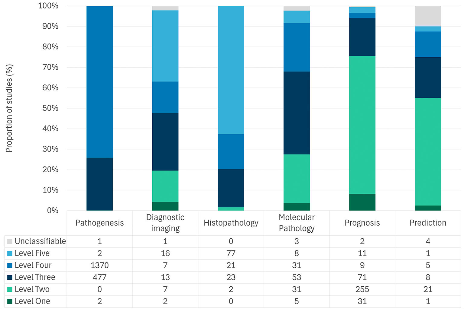
Figure 4. Distribution of studies by evidence level across tumour pathology-related characteristics.
The largest body of evidence relates to pathogenesis at level three (n = 477; 25.8%) and level four (n = 1370; 73.9%). This is followed by prognosis, where evidence is predominantly at level two (n = 255; 67.3%) and level one (n = 31; 8.2%) (
Figure 4;
Table 2).
Absolute gaps in published evidence were retrieved for all types of osteosarcomas other than osteosarcoma. None of these other types of osteosarcomas have level one evidence (
Figure 3;
Table 2).
No studies were found for any tumour type at level one or two for pathogenesis or at level one for histopathology (
Figure 3;
Table 2).
The breakdown by tumour type, tumour characteristics and level of evidence can be seen in
Table 2. For parosteal osteosarcoma, there was no evidence for molecular pathology or prediction. For periosteal osteosarcoma, only two studies on prognosis were included, and none were found for the remaining characteristics. For high-grade surface osteosarcoma, no studies were found that examined diagnostic imaging, molecular pathology or prediction. Finally, in low-grade osteosarcomas, all characteristics have absolute gaps in the evidence except for diagnostic imaging and histopathology.
Discussion
This study highlights a marked imbalance in both the quantity and the level of evidence across osteosarcoma subtypes, with the majority of studies and higher-level evidence largely confined to osteosarcoma, and a predominantly low-level evidence base supporting rarer variants during the period of study.
The predominance of studies on osteosarcoma reflects both its higher incidence and its historical role as the reference entity for biological and clinical research in osteogenic sarcomas. In the present map, osteosarcoma accounted for more than 90% of included studies. Population-based data and clinical guidelines show that osteosarcoma represents the majority of diagnosed cases, whereas surface and low-grade variants constitute a very small proportion of diagnoses, limiting the feasibility of large-scale or comparative study designs for these entities.20
Notably, prognosis was the only area in which the highest-level evidence (levels one and two) was relatively well represented. Across all tumour types, no level one evidence was identified for histopathology, nor was any level two evidence found for pathogenesis. This pattern reflects a broader methodological reality in tumour pathology, where some study designs are not always feasible, particularly for rare tumour types.11 Methodological work specific to tumour pathology has recognised that well-designed observational and descriptive studies provide appropriate and meaningful evidence for questions of tumour classification, histopathology and pathogenesis, even when higher-level comparative designs are not applicable.16
While these patterns largely affect established trends in osteosarcoma research, they should be interpreted also in the context of the methodological coding approach used in this EGM. Each record was assigned one single tumour type and primary characteristic based on expert prioritisation. Consequently, studies reporting multiple tumour types or tumour characteristics may be under-represented in secondary domains, particularly for less common osteosarcoma types.
One of the key findings of this EGM is the presence of absolute evidence gaps across multiple domains for parosteal, periosteal, high-grade surface, and low-grade central osteosarcomas. For these subtypes, several tumour characteristics, particularly molecular pathology and prediction, were entirely unrepresented, and no level one evidence was identified. These findings are consistent with the limited availability of robust evidence in osteosarcoma research.
Although numerous biomarkers have been investigated, their clinical utility and use in diagnosis is still considerably less developed than in other malignancies.21,22 In addition, the rarity of osteosarcoma and its subtypes result in small study populations, which restricts the feasibility of large, high-quality studies.21,23 Together, these factors likely contribute to the absence of high-level evidence and the observed gaps in domains such as molecular pathology and prediction. Recent methodological guidance on evidence gap mapping emphasises that identifying such gaps is not an endpoint but a mechanism to prioritise targeted evidence generation and secondary synthesis.11,15
When interpreting these results, certain methodological adjustments made during the coding phase should also be pointed out, as they help explain the reduction in the number of studies included, as shown in the PRISMA flow diagram following screening. These modifications included the exclusion of studies primarily focused on metastasis, the restriction of studies on prognosis and prediction that did not report on overall survival or equivalent outcomes, and the exclusion of studies evaluating diagnostic or prognostic factors following neoadjuvant therapy as a means of assessing treatment efficacy. Together, these decisions were made to improve conceptual consistency and comparability between studies and should be taken into account when interpreting study loss between the screening and coding stages.
By visualising the distribution of evidence across tumour types and characteristics, this EGM highlights those areas where WHO tumour classification could be supported by relatively robust evidence and areas where the supporting literature remains limited or based on lower-level evidence at least in recent years. Given that WCT tumour classification plays a central role in diagnosis, grading and treatment decisions, these findings highlight areas where certain tumour entities could benefit from further systematically accumulated empirical evidence, complementing the strong foundation of historical morphologic characterization and expert consensus.
A major strength of this study is the application of a standardised EGM framework aligned with WCT tumour taxonomy, combined with a pathology-specific hierarchy of evidence. The use of machine learning-assisted priority screening allowed efficient handling of a large evidence base while maintaining methodological rigour, consistent with emerging best practices in evidence mapping.15,17
Several limitations must also be acknowledged, data extraction based on title and abstract may have resulted in under-characterisation of study designs or tumour characteristics. Finally, as with all EGMs, this study maps the presence and level of evidence but does not assess its internal validity or synthesize effect estimates.11,15 Additionally, restriction to studies published from 2018 onwards may have excluded earlier foundational work, particularly for rare tumour entities.
By systematically mapping the evidence underpinning osteosarcoma tumour types, this study highlights that evidence is unevenly distributed across different tumour entities or characteristics. According to the articles identified, the most studied tumour type was osteosarcoma, while parosteal, periosteal, high-grade surface, and low-grade central osteosarcomas were supported by very limited literature, with few higher-level studies and no level one evidence identified across most characteristics. Future research should prioritise coordinated, multicentre efforts focusing on uncommon osteosarcoma types and under-represented tumour characteristics, with the aim of strengthening the foundations of tumour classification and reducing reliance on consensus in instances where evidence remains limited.
Ethical approval and informed consent
This study involved neither human participants nor animals; accordingly, ethical approval and informed consent were not required.
Disclaimer
The content of this article represents the personal views of the authors and does not represent the views of the authors’ employers and associated institutions. Where authors are identified as personnel of the International Agency for Research on Cancer / World Health Organization, the authors alone are responsible for the views expressed in this article, and they do not necessarily represent the decisions, policy, or views of the International Agency for Research on Cancer/World Health Organization.
Funded by the European Union. Views and opinions expressed are however those of the author(s) only and do not necessarily reflect those of the European Union.
Data Availability
The extended data for this publication is deposited and publicly available in OSF (Extended data_Evidence Distribution Across Osteosarcoma Tumour Types: An Evidence Gap Map to inform the WHO Classification of Tumours), accessible at: https://doi.org/10.17605/OSF.IO/6WY7E.24 License: CC0 1.0 Universal. This project contains the following extended data:
- Extended data 1. Adapted Hierarchy of Evidence for Tumour Pathology (HETP)
- Extended data 2. Search strategies for Embase and PubMed
- Extended data 3. Detailed eligibility criteria flow diagram
All data underlying the results are available as part of the article and its extended data. No additional datasets were generated or analysed.
Acknowledgments
The authors would like to thank Cécile Monnier, Project Assistant for the WCT EVI MAP Project, for her invaluable administrative and organisational support throughout the conduct of this study.
The authors also acknowledge the members of the WCT EVI MAP Group for their contributions to the development of the evidence gap mapping framework, methodological discussions, and expert input throughout the project.
WCT EVI MAP Group members: Sophie Gruber, Laura Knoblauch, Stefan Holdenrieder, Karolina Worf, Joanna Didkowska, Kateryna Maslova, Lukasz Taraszkiewicz, Paulina Kober, Mariusz Kulińczak, Maria Sromek, Christine Giesen, Ramon Cierco Jimenez, Nur Diyana Md Nasir, Zi Long Chow, Mervyn Ong, Denise Hsin Yi Thng and Clarissa Jing Wen Wong.
References
- 1.
Picci P:
Osteosarcoma (Osteogenic sarcoma).
Orphanet J. Rare Dis.
2007 Jan 23; 2: 6. PubMed Abstract
| Publisher Full Text
| Free Full Text
- 2.
Beird HC, Bielack SS, Flanagan AM, et al.:
Osteosarcoma.
Nat. Rev. Dis. Primers.
2022 Dec 8; 8(1): 77. Publisher Full Text
- 3.
Lazar AJ, Flanagan AM, Blay JY, et al.:
Bone Tumours.
Soft tissue and bone tumours.
Lyon (France):
WHO Classification of Tumours Editorial Board;
5th ed2020.
Reference Source
- 4.
Valery PC, Laversanne M, Bray F:
Bone cancer incidence by morphological subtype: a global assessment.
Cancer Causes Control.
2015 Aug; 26(8): 1127–1139. PubMed Abstract
| Publisher Full Text
- 5.
Mirabello L, Troisi RJ, Savage SA:
International osteosarcoma incidence patterns in children and adolescents, middle ages and elderly persons.
Int. J. Cancer.
2009 Jul 1; 125(1): 229–234. PubMed Abstract
| Publisher Full Text
| Free Full Text
- 6.
Bielack SS, Kempf-Bielack B, Delling G, et al.:
Prognostic Factors in High-Grade Osteosarcoma of the Extremities or Trunk: An Analysis of 1,702 Patients Treated on Neoadjuvant Cooperative Osteosarcoma Study Group Protocols.
J. Clin. Oncol.
2002 Feb; 20(3): 776–790. Publisher Full Text
- 7.
Misaghi A, Goldin A, Awad M, et al.:
Osteosarcoma: a comprehensive review.
SICOT J.
2018; 4: 12. PubMed Abstract
| Publisher Full Text
| Free Full Text
- 8.
WHO Classification of Tumours Editorial Board, editor.
Osteosarcoma.
Lyon (France):
WHO classification of tumours series;
5th ed.2020. Vol. 3. .
Reference Source
- 9.
Jiang Y, Wang J, Sun M, et al.:
Multi-omics analysis identifies osteosarcoma subtypes with distinct prognosis indicating stratified treatment.
Nat. Commun.
2022; 13: 7207. PubMed Abstract
| Publisher Full Text
| Free Full Text
- 10.
IARC:
Mapping the Evidence for the World Health Organization (WHO) Classification of Tumours: a Living Evidence Gap Map by Tumour Type (WCT EVI MAP).2024 [cited 2024 Jul 23].
Reference Source
- 11.
Campbell F, Tricco AC, Munn Z, et al.:
Mapping reviews, scoping reviews, and evidence and gap maps (EGMs): the same but different— the “Big Picture” review family.
Syst. Rev.
2023 Mar 15; 12(1): 45. PubMed Abstract
| Publisher Full Text
| Free Full Text
- 12.
Choi JH, Ro JY:
The 2020 WHO Classification of Tumors of Bone: An Updated Review.
Adv. Anat. Pathol.
2021; 28: 119–138. PubMed Abstract
| Publisher Full Text
- 13.
Saran A, White H:
Evidence and gap maps: a comparison of different approaches.
Campbell Syst. Rev.
2018; 14(1): 1–38. PubMed Abstract
| Publisher Full Text
| Free Full Text
- 14.
White H, Albers B, Gaarder M, et al.:
Guidance for producing a Campbell evidence and gap map.
Campbell Syst. Rev.
2020 Dec; 16(4): e1125. PubMed Abstract
| Publisher Full Text
| Free Full Text
- 15.
Khalil H, Welch V, Grainger M, et al.:
Methodology for mapping reviews, evidence maps, and gap maps.
Res. Synth. Methods.
2025; 16(5): 786–796. PubMed Abstract
| Publisher Full Text
| Free Full Text
- 16.
Colling R, Indave I, del Aguila J , et al.:
A New Hierarchy of Research Evidence for Tumor Pathology: A Delphi Study to Define Levels of Evidence in Tumor Pathology.
Mod. Pathol.
2024 Jan 1; 37(1): 100357. Publisher Full Text
- 17.
Repke T, Tinsdeall F, Danilenko D, et al.:
Don’t Stop Me Now, `Cause I’m Having a Good Time Screening: Evaluation of Stopping Methods for Safe Use of Priority Screening in Systematic Reviews.
Cochrane Evid Synth Methods.
2026 Jan 21; 4(1): e70068. PubMed Abstract
| Publisher Full Text
| Free Full Text
- 18.
Craciun OM, García-Ovejero E, Montes Mota M, et al.:
Decision tree and glossary for classifying study designs: interactive version (WCT EVI MAP Project).2024 Dec [cited 2025 May 21].
Reference Source
- 19.
Craciun OM, García-Ovejero E, Campbell F, et al.:
Development of a decision tree diagram for classifying study designs in tumour pathology research: a multidisciplinary approach.
J. Pathol. Clin. Res.
2026; 12(1): e70056. PubMed Abstract
| Publisher Full Text
| Free Full Text
- 20.
Biermann JS, Hirbe A, Ahlawat S, et al.:
Bone Cancer, Version 2.2025, NCCN Clinical Practice Guidelines In Oncology.
J. Natl. Compr. Cancer Netw.
2025 Apr; 23(4): e250017. PubMed Abstract
| Publisher Full Text
- 21.
Pekarek L, De la Torre-Escuredo B, Fraile-Martinez O, et al.:
Towards the Search for Potential Biomarkers in Osteosarcoma: State-of-the-Art and Translational Expectations.
Int. J. Mol. Sci.
2022 Nov 29; 23(23): 14939. PubMed Abstract
| Publisher Full Text
| Free Full Text
- 22.
Zamborsky R, Kokavec M, Harsanyi S, et al.:
Identification of Prognostic and Predictive Osteosarcoma Biomarkers.
Med. Sci.
2019 Feb 11; 7(2): 28. PubMed Abstract
| Publisher Full Text
| Free Full Text
- 23.
Wani AK, Prakash A, Sena S, et al.:
Unraveling molecular signatures in rare bone tumors and navigating the cancer pathway landscapes for targeted therapeutics.
Crit. Rev. Oncol. Hematol.
2024 Apr 1; 196: 104291. Publisher Full Text
- 24.
Craciun OM, Mota MM, García-Ovejero E, et al.:
Extended data_Evidence Distribution Across Osteosarcoma Tumour Types: An Evidence Gap Map to inform the WHO Classification of Tumours.
OSF.
2026.
Reference Source







