1. Introduction
A wide range of causes may lead to reproductive loss in ruminants, including abortion, premature birth, stillbirth, birth of weak offspring (hereafter referred to as “abortion”) and occasionally early embryonic death. Non-infectious causes such as nutritional deficiencies (e.g. selenium, iodine), trauma, genetic abnormalities, and ingestion of toxic plants usually occur sporadically and rarely cause abortion epidemics. In contrast, infectious causes may result in abortion “storms” affecting multiple animals in a herd or flock and may pose a significant zoonotic risk.
From an economic perspective, abortion represents a direct loss of offspring, reduced milk yield, and, occasionally, premature culling of dams, as well as indirect costs associated with veterinary interventions, diagnostics, and disrupted production cycles. The financial impact of abortions in ruminants worldwide is therefore considered significant (Ali et al., 2019; Palma et al., 2025).
From a zoonotic perspective, abortion, especially in small ruminants poses an immediate and tangible risk to human health. Brucellosis is non-endemic in most European countries as a result of strong surveillance and control programs. However, hotspots persist in certain areas, particularly in southern European and Balkan countries, where the risk of human infection remains higher (Laine et al., 2023), especially among shepherds, veterinarians, and abattoir workers. In northern Europe, Coxiella burnetii was the causative agent of a large human outbreak of a zoonotic disease linked to a ruminant abortifacient agent (the largest recorded Q fever epidemic), with profound long-term consequences for human health and substantial economic costs (van Asseldonk et al., 2013; Bronner et al., 2020). Pregnant women exposed to lambing sheep and goats infected with Chlamydia abortus face severe risks, including life-threatening systemic illness and abortion (Meijer et al., 2004). Globally, toxoplasmosis continues to represent a potential foodborne zoonosis, sometimes associated with the consumption of undercooked sheep or goat meat, although infectious stages of this parasite can also be shed at parturition (Holec-Gąsior and Sołowińska, 2022). Listeria monocytogenes is the causative agent of listeriosis in animals and humans, and it is regarded as one of the most serious foodborne infections in humans (WOAH, 2022). Rift Valley fever is an important zoonotic cause of abortion in several African countries and in the Middle East. Other causes of abortion, especially some bacteria, including Listeria ivanovii, can cause opportunistic human infection, particularly in immunocompromised individuals (Guillet et al., 2010). Whereas other pathogens, such as some viruses (e.g. pestiviruses, bluetongue virus (BTV), Schmallenberg virus (SBV), Akabane virus) and parasites (Neospora caninum), are host specific and non-zoonotic.
In this report, data collected during abortion surveillance in ruminants were compiled and analysed by the international coordination of research on infectious diseases (ICRAD)-project Q-NET-ASSESS consortium partners (www.q-net-assess.com). Previous work from the consortium reviewed and compared abortion diagnostic protocols across partner countries and proposed methodological guidance, with a specific focus on C. burnetii (Van den Brom et al., 2025). The present report does not revisit diagnostic best practices in detail, but instead compiles the findings generated by national and regional abortion diagnostic and reporting frameworks between 2022 and 2024, together with the organisational context needed to interpret them. The aims of this study were: 1) to describe the logistics and background of abortion diagnosis in participating countries, 2) to report the main abortifacient agents detected or attributed in ruminant abortion submissions between 2022 and 2024 in the participating countries, and finally, 3) to compile operational experiences across countries, identify differences and limitations of existing strategies, and formulate recommendations.
2. Methods
For this study, the consortium partners, from Belgium, France, Germany, Spain, the Netherlands, and the United Kingdom (UK), first compiled operational experiences related to abortion submission logistics (e.g. who submits the samples for investigation and laboratory structures), laboratory methods, system organisation (e.g. reporting procedures, subsidies for diagnostic testing), and national coverage, as well as other important factors related to abortion diagnosis in each country. Then, partners collated relevant data on causes of ruminant abortion diagnoses between 2022 and 2024 from unpublished sources and available reports in the countries represented in the Q-NET-ASSESS consortium. Based on these findings, differences and limitations of existing strategies were identified and recommendations were formulated to improve abortion diagnosis practices.
For this investigation, we referred to the protocols used by the different project partners and/or countries to diagnose abortion in domestic ruminants, which were previously reviewed in another study (Van den Brom et al., 2025). That earlier work developed guidelines for the detection of abortifacient agents in domestic ruminants, with a special focus on the diagnosis of C. burnetii as the causative agent of abortion. Those guidelines included a description of the essential and complementary samples required for a definitive diagnosis, the analytical techniques to be used, and the interpretation and validity of each type of sample and technique. They highlighted that the most comprehensive diagnostic approach to identify an infectious agent as the cause of abortion in ruminants should include histopathology, including immunohistochemistry (IHC), on the foetus and placental membranes including cotyledons (placentae), complemented by bacteriology, serology, and real-time PCR analyses of different sample types.
Collected data reflect investigations that were conducted within each national/regional framework. Where available, causes are attributed based on pathology and compatible findings; elsewhere, results reflect the detection of agents in an abortion context. This report should therefore be interpreted primarily as a comparison of diagnostic and reporting outputs generated by the different national/regional frameworks, rather than a direct comparison of the incidence or prevalence of abortifacients agents in ruminants across countries. Regulated disease programmes (e.g. for brucellosis) are mentioned only to describe the regulatory and diagnostic context; their surveillance outputs are not reported unless they were part of the abortion submission work-up.
3. Results
3.1 Legislation framework and findings from ruminant abortion submission from 2022–2024
In European Union Member States and the UK, the detection of suspected signs of notifiable diseases and diseases listed under the framework of the EU Animal Health Law (EU Regulation 2016/429) or UK statutory legislation, such as brucellosis, must be immediately notified to the competent animal health authorities. The six participating countries are officially recognised as free from brucellosis and maintain this status according to EU-or UK legislation through mandatory surveillance.
The information collected on the logistics and backgrounds of abortion diagnosis and reporting in participating countries is summarised in
Table 1, and the findings on the detected abortifacients in ruminant abortion submissions between 2022–2024 are presented in detail in
Tables 2a-f.
Table 1. Overview of the procedures governing the diagnostic investigation and reporting of ruminant abortions in the six European consortium countries.
| Country | Coverage | Subsidisation | Diagnostic Labs | Laboratory investigation | Data collection | Nationally representative |
|---|
| Belgium
| National official programme for ruminant abortions monitoring | Partial public subsidisation (certain tests; farmer payment for others) | Regional laboratories (ARSIA and DGZ) & Sciensano (mainly small ruminants) | No systematic implementation of histopathology; it can be performed at regional laboratories | National dataset used in this report, but no centralised reporting and publicly available data collection exist | Yes
|
| France
| No systematic national monitoring or surveillance system; OSCAR is voluntary, partial, and department-based
| Variable/partial; may be partly subsidised locally (e.g. livestock health associations GDS), otherwise farmer-paid
| Decentralised laboratory diagnostics | OSCAR protocols: harmonised but non-mandatory
No systematic histopathology | Partial reporting (OSCAR only); centralised data collection and curation (ESA platform)
https://www.plateforme-esa.fr/fr/observatoire-et-suivi-des-causes-davortements-chez-les-ruminants-oscar
| No
|
| Germany
| No systematic national monitoring or surveillance system; federal state based monitoring exists | Partial or full public subsidisation | Decentralised laboratory diagnostics | Diagnostic testing is defined by the federal states (no systematic histopathology) | Partial/regional | No
|
| Spain
| Surveillance system of uneven implementation | Partial public subsidisation | Decentralised laboratory diagnostics (private and public labs) | No harmonised protocols at national level (histopathology conducted at certain regional government laboratories) | No national centralisation | No
|
| The Netherlands
| National surveillance | Partial public subsidisation by government and farmers organisations | Royal GD centralises diagnostics | Harmonised protocols (including histopathology) | Centralised data collection at Royal GD (https://issuu.com/gezondheidsdienstvoordieren/stacks/428347f3f55746b88f1cc4a1ad591007
)
| Yes
|
| United Kingdom
| National scanning surveillance network (Great Britain) | Partial public subsidisation | Scanning surveillance via government-associated laboratories (network of Veterinary Investigation Centres and national post-mortem providers). Separate to this there are private veterinary laboratories | Harmonised protocols in government-associated laboratories for scanning surveillance (including histopathology) | Centralised data collection: quarterly and annual reports (VIDA)
APHA animal surveillance reports, publications and dashboards - GOV.UK (https://www.gov.uk/guidance/view-apha-surveillance-reports-publications-and-data#quarterly-and-annual-species-report) | Yes, for the Great Britain scanning surveillance network |
Table 2a. Abortifacients detected in ruminant abortion submissions in Belgium (Sciensano & ARSIA/DGZ) between 2022 and 2024.
| Sheep | Goat |
Cattle
|
|---|
| 2022A1 | 2023B1 | 2024C1 | 2022A2 | 2023B2 | 2024C2 | 2022A3 | 2023B3 | 2024C3 |
|---|
| Bacteria
| Coxiella burnetii
| 311 | 271 | 511 | 5 | 9 | 23 | 180 | 130 | 156 |
| Anaplasma sp. | NT | NT | NT | NT | NT | NT | 54 | 59 | 53 |
| Brucella sp. | 0 | 0 | 0 | 0 | 0 | 0 | 0 | 0 | 0 |
| Campylobacter sp. | 5 | 1 | 3 | 0 | 0 | 0 | 2 | 1 | 2 |
| Campylobacter fetus subsp. venerealis
| NT | NT | NT | NT | NT | NT | NT | NT | NT |
| Chlamydia abortus
| 61 | 41 | 61 | 0 | 1 | 0 | NT | NT | NT |
| Leptospira sp. | 0 | 0 | 1 | 0 | 0 | 0 | 3 | 6 | 10 |
| Mycoplasma sp. | NT | NT | NT | NT | NT | NT | NT | NT | NT |
| Ureaplasma sp. | NT | NT | NT | NT | NT | NT | 34 | 25 | 44 |
| Other bacteria: | Salmonella sp.
| 1 | 2 | 1 | 0 | 0 | 0 | 67 | 110 | 56 |
| Listeria sp. | 1 | 1 | 1 | 3 | 6 | 1 | 81 | 73 | 70 |
| Yersinia sp.
| 2 | 0 | 1 | 0 | 0 | 0 | 3 | 4 | 4 |
| Trueperella pyogenes
| 0 | 1 | 1 | 0 | 0 | 0 | 450 | 345 | 390 |
| Other bacteria (i.e. E. coli) | 23 | 9 | 8 | 8 | 1 | 0 | 1129 | 713 | 717 |
| Parasites
| Neospora caninum
| 6 | 3 | 6 | 0 | 0 | 1 | 370 | 384 | 433 |
| Toxoplasma gondii
| 18 | 11 | 12 | 2 | 4 | 6 | 0 | 0 | 0 |
| Tritrichomonas foetus
| NT | NT | NT | NT | NT | NT | NT | NT | NT |
| Viruses
| Bluetongue virus | 0 | 0 | 1 | 0 | 0 | 0 | 0 | 0 | 354 |
| Bovine herpesvirus | NT | NT | NT | NT | NT | NT | NT | NT | NT |
| Pestivirus (Border disease virus-BD; Bovine viral diarrhoea virus-BVD) | NT | NT | NT | NT | NT | NT | 16 | 1 | 23 |
| Schmallenberg virus | 15 | 1 | 4 | 0 | 0 | 0 | 46 | 2 | 32 |
| Viral abortion (virus not identified) | NT | NT | NT | NT | NT | NT | NT | NT | NT |
| Fungi
| Mycoses (particularly Aspergillus sp.) | 63 | 193 | 213 | | | | 33 | 49 | 48 |
| Other
| Indication of inflammation (placentitis/meningitis/..) | 763 | 763 | 733 | | | | 822 | 932 | 796 |
| Asphyxia | NT | NT | NT | NT | NT | NT | NT | NT | NT |
| Mummification | | | 113 | | | | | | |
| Congenital malformation | | | | | | | 123 | 132 | 939 |
| Trauma | | | | | | | 31 | 27 | 0 |
| No diagnosis
| 400 | 350 | 390 | 120 | 142 | 136 | 1683 | 2448 | 3211 |
| Total
| 508
| 410
| 486
| 138
| 163
| 167
| 5127
| 5441
| 7338
|
Table 2b. Abortifacients in ruminant abortion submissions in France between 2022 and 2024 based on OSCAR annual reports: strong (or possible) presumptive attribution according to OSCAR interpretation grids.
| Pathogen | Sheep | Goat | Cattle |
|---|
| 2022 | 2023 | 2024 | 2022 | 2023 | 2024 | 2022 | 2023 | 2024 |
|---|
| Bacteria
| Coxiella burnetii
| 35 (13) | 27 (11) | 29 (17) | 16 (11) | 18 (5) | 26 (8) | 23 (64) | 55 (95) | 65 (123) |
| Anaplasma sp. | | | | | | | 70 (23) | 66 (41) | 84 (98) |
| Brucella sp. | | | | | | | | | |
| Campylobacter sp. | | | | | | | 0 (3) | 0 (2) | 1 (2) |
| Campylobacter fetus subsp. venerealis
| | | | | | | | | |
| Chlamydia abortus
| 38 (9) | 43 (3) | 51 (8) | 15 (2) | 15 (3) | 12 (2) | 4 (30) | 2 (62) | 12 (73) |
| Leptospira sp. | | | | | | | | | |
| Mycoplasma sp. | | | | | | | | | |
| Ureaplasma sp. | | | | | | | | | |
| Other bacteria: | Salmonella sp.
| 15 (1) | 17 (2) | 33 (2) | 1 (0) | 5 (0) | 7 (1) | 11 (19) | 12 (23) | 12 (23) |
| | Listeria sp. | 2 (2) | 5 (1) | 6 (0) | 2 (0) | 3 (0) | 0 (1) | 3 (18) | 8 (15) | 11 (45) |
| | Yersinia sp.
| | | | | | | | | |
| | Trueperella pyogenes
| | | | | | | | | |
| | Other bacteria | | | | | | | | | |
| Parasites
| Neospora caninum
| | | | | | | 70 (76) | 74 (110) | 95 (137) |
| Toxoplasma gondii
| 26 (27) | 64 (32) | 32 (73) | 12 (16) | 13 (12) | 6 (11) | | | |
| Tritrichomonas foetus
| | | | | | | | | |
| Viruses
| Bluetongue virus | | | | | | | | | |
| Bovine herpesvirus | | | | | | | | | |
| Pestivirus (Border disease virus-BD; Bovine viral diarrhoea virus-BVD) | 0 (4) | 3 (2) | 1 (3) | 0 (0) | 0 (0) | 0 (0) | 5 (31) | 10 (26) | 1 (44) |
| Schmallenberg virus | | | | | | | | | |
| Fungi
| Mycoses (particularly Aspergillus sp.) | 4 (2) | 5 (1) | 7 (0) | 2 (1) | 2 (0) | 2 (0) | 7 (35) | 15 (27) | 16 (24) |
| Other
| Indication of inflammation (placentitis/meningitis/..) | | | | | | | | | |
| Asphyxia | | | | | | | | | |
| Mummification | | | | | | | | | |
| Congenital malformation | | | | | | | | | |
| Trauma | | | | | | | | | |
| No diagnosis
| 128 | 94 | 159 | 60 | 74 | 84 | 478 | 685 | 778 |
| Total
| 271
*
| 273
**
| 359
***
| 122
^
| 137
^^
| 149
^^^
| 849
#
| 1176
##
| 1402
###
|
Table 2c. Detection of selected zoonotic abortifacient pathogens in abortion-associated diagnostic submissions from German ruminants (2022–2024).
| Pathogen | Sheep | Goat | Cattle |
|---|
| 2022 | 2023 | 2024 | 2022 | 2023 | 2024 | 2022 | 2023 | 2024 |
|---|
| Bacteria
| Coxiella burnetii
| | | | | | | | | 10 |
| Anaplasma sp. | | | | | | | | | |
| Brucella sp. | | | | | | | | | |
| Campylobacter sp. | | | 3 | | 2 | | 5 | | |
| Campylobacter fetus subsp. venerealis
| | | | | | | 1 | | |
| Chlamydia abortus
| | | | | | | | | |
| Leptospira sp. | | | | | | | | | |
| Mycoplasma sp. | | | | | | | | | |
| Ureaplasma sp. | | | | | | | | | |
| Other bacteria: | Salmonella sp.
| 1 | | 1 | | | | 11 | 11 | 5 |
| | Listeria sp. | | | | | | | | | |
| | Yersinia sp.
| | | | | | | | | |
| | Trueperella pyogenes
| | | | | | | | | |
| | Other bacteria | | | | | | | | | |
| Parasites
| Neospora caninum
| | | | | | | | | |
| Toxoplasma gondii
| 1 | 5 | | | | 3 | 1 | | 1 |
| Tritrichomonas foetus
| | | | | | | | | |
| Viruses
| Bluetongue virus | | | | | | | | | |
| Bovine herpesvirus | | | | | | | | | |
| Pestivirus (Border disease virus-BD; Bovine viral diarrhoea virus-BVD) | | | | | | | | | |
| Schmallenberg virus | | | | | | | | | |
| Viral abortion (virus not identified) | | | | | | | | | |
| Fungi
| Mycoses (particularly Aspergillus sp.) | | | | | | | | | |
| Other
| Indication of inflammation (placentitis/meningitis/..) | | | | | | | | | |
| Asphyxia | | | | | | | | | |
| Mummification | | | | | | | | | |
| Congenital malformation | | | | | | | | | |
| Trauma | | | | | | | | | |
| No diagnosis
| | | | | | | | | |
| Total
| 2
| 5
| 4
| | 2
| 3
| 18
| 11
| 16
|
Table 2d. Abortifacients in ruminant abortion submissions investigated at NEIKER’s Diagnostic Service in Spain between 2022 and 2024: Attributed causes (pathology-based diagnoses: confirmed/presumptive)
| Pathogen | Sheep | Goat | Cattle |
|---|
| 2022 | 2023 | 2024 | 2022 | 2023 | 2024 | 2022 | 2023 | 2024 |
|---|
| Bacteria
| Coxiella burnetii
| 1 | | | (1) | (1) | 2
# (1
†) | | | |
| Anaplasma sp. | | | | | | | | | |
| Brucella sp. | | | | | | | | | |
| Campylobacter sp. | | | | | | | | | |
| Campylobacter fetus subsp. venerealis
| | | | | | | | | |
| Chlamydia abortus
| 3
* | | 1 | | | 2
# | | | |
| Leptospira sp. | | | | | | | (1) | (4
^) | (1
^) |
| Mycoplasma sp. | | | | | | | | | |
| Ureaplasma sp. | | | | | | | | | |
| Other bacteria: | Salmonella sp.
| 2
‡ | | | | | 1 | | | 2 |
| | Listeria sp.
| | | 1 | | | | | 1 | |
| | Yersinia sp.
| | | | | | | | | |
| | Trueperella pyogenes
| | | | | | | (1) | 2 (1) | (1) |
| | Bacillus sp.
| | | | | | | (2) | (1) | |
| | E. coli
| | | | | | | (3) | (1) | |
| | Staphylococcus aureus
| | | | | | | | (1) | (1) |
| | Other bacteria | 1 | | | | | | 1 | (1) | |
| Parasites
| Neospora caninum
| | | | | | | 1 (1) | 1 (3) | |
| Toxoplasma gondii
| 5 | (2) | 1 | 1 | | (2
†) | | | |
| Tritrichomonas foetus
| | | | | | | | | |
| Viruses
| Bluetongue virus | | | | | | | | | |
| Bovine herpesvirus | | | | | | | | | |
| Pestivirus (Border disease virus-BD; Bovine viral diarrhoea virus-BVD) | 1
* | | (1) | | 1 | | | 2 | |
| Schmallenberg virus | | | | | | | | | |
| Viral abortion (virus not identified) | | | | (1) | | | | | |
| Fungi
| Mycoses (particularly Aspergillus sp.) | | | | | | | | | |
| Other
| Indication of inflammation (placentitis/meningitis/..) | | | | | | | | | |
| Asphyxia | | | | | | | | | |
| Mummification | | | | | | | | | |
| Congenital malformation | | | | | | | | | |
| Trauma | | | 1 | | | | | | |
| Infectious abortion of undetermined etiology | | | 1 | | | | | | |
| No diagnosis
| 7 | 7 | 5 | | 3 | 2 | 8 | 21 | 21 |
| Total
§
| 19
| 9
| 11
| 3
| 5
| 8
| 18
| 39
| 26
|
Table 2e. Abortifacients in ruminant abortion submissions in the Netherlands (Royal GD) between 2022 and 2024: Attributed causes (pathology-based diagnoses).
| Pathogen | Sheep | Goat | Cattle |
|---|
| 2022 | 2023 | 2024 | 2022 | 2023 | 2024 | 2022 | 2023 | 2024 |
|---|
| Bacteria
| Coxiella burnetii
| | | | | | | 1 | 2 | 6 |
| Anaplasma sp. | | | | | | | | | |
| Brucella sp. | | | | | | | | | |
| Campylobacter sp. | 8 | 7 | 20 | | | | | | |
| Campylobacter fetus subsp. venerealis
| | | | | | | | | |
| Chlamydia abortus
| 11 | 7 | 8 | 2 | 5 | 2 | 1 | | |
| Leptospira sp. | | | | | | | | | |
| Mycoplasma sp. | | | | | | | | | |
| Ureaplasma sp. | | | | | | | | | |
| Other bacteria: | Salmonella sp.
| | | 2 | | | 5 | 8 | 14 | 12 |
| | Listeria sp. | 15 | 9 | 8 | 9 | 2 | 5 | 7 | 4 | 2 |
| | Yersinia sp.
| 2 | 5 | | | | | 1 | | 1 |
| | Trueperella pyogenes
| 2 | | | | 3 | | 34 | 27 | 23 |
| | Other bacteria | 7 | 8 | 3 | 3 | 2 | 3 | 41 | 26 | 7 |
| Parasites
| Neospora caninum
| | | | | | | 14 | 12 | 3 |
| Toxoplasma gondii
| 13 | 15 | 18 | | 11 | 1 | | | |
| Tritrichomonas foetus
| | | | | | | | | |
| Viruses
| Bluetongue virus | | | 1 | | | 5 | | | 32 |
| Bovine herpesvirus | | | | | | | | 2 | 2 |
| Pestivirus (Border disease virus-BD; Bovine viral diarrhoea virus-BVD) | | | | | | | 1 | 1 | |
| Schmallenberg virus | 9 | 1 | 9 | 2 | 2 | 1 | | | 1 |
| Fungi
| Mycoses (particularly Aspergillus sp.) | | | | | | | 2 | 1 | 3 |
| Other
| Indication of inflammation (placentitis/meningitis/..) | 6 | 13 | 13 | | 2 | 4 | 12 | 25 | 20 |
| Asphyxia | | 2 | 6 | | 1 | 2 | | | |
| Mummification | 2 | 3 | 1 | 1 | 2 | | 2 | 1 | 2 |
| Congenital malformation | 3 | 8 | | 2 | 2 | | 1 | 2 | 3 |
| Trauma | | | | | 2 | | | | |
| No diagnosis
| 56 | 33 | 30 | 39 | 32 | 51 | 114 | 147 | 93 |
| Total
| 134 | 111 | 119 | 58 | 64 | 79 | 240 | 264 | 210 |
Table 2f. Abortifacients in ruminant abortion submissions in Great Britain (England, Scotland and Wales), UK, through the APHA/SRUC scanning surveillance network between 2022 and 2024: Attributed causes (pathology-based diagnoses).
| Pathogen | Sheep | Goat | Cattle |
|---|
| 2022 | 2023 | 2024 | 2022 | 2023 | 2024 | 2022 | 2023 | 2024 |
|---|
| Bacteria
| Coxiella burnetii
| 1 | | | 2 | 1 | | 1 | 7 | 3 |
| Anaplasma sp. | | | | | | | | | |
| Brucella sp. | | | | | | | | | |
| Campylobacter sp. | 112 | 169 | 93 | | | | 8 | 7 | 7 |
| Campylobacter fetus subsp. venerealis
| | | | | | | | | |
| Chlamydia abortus
| 143 | 147 | 173 | 2 | 3 | | | 1 | |
| Leptospira sp. | | | | | | | 2 | 2 | |
| Mycoplasma sp. | | | | | | | | | |
| Ureaplasma sp. | | | | | | | | | |
| Other bacteria: | Salmonella sp.
| 19 | 20 | 28 | | | | 73 | 82 | 48 |
| | Listeria sp. | 29 | 16 | 21 | 1 | 2 | 2 | 18 | 19 | 18 |
| | Yersinia sp.
| 5 | 5 | 3 | | | | 1 | 2 | 2 |
| | Trueperella pyogenes
| 4 | 4 | 11 | | | | 35 | 40 | 41 |
| | Other bacteria | 19 | 18 | 23 | | | | 64 | 72 | 56 |
| Parasites
| Neospora caninum
| | | | | | | 42 | 33 | 41 |
| Toxoplasma gondii
| 136 | 168 | 115 | 1 | | 2 | | | |
| Tritrichomonas foetus
| | | | | | | | | |
| Viruses
| Bluetongue virus | | | | | | | | | |
| Bovine herpesvirus | | | | | | | | 1 | 1 |
| Pestivirus (Border disease virus-BD; Bovine viral diarrhoea virus-BVD) | 12 | 18 | 9 | | | | 13 | 11 | 9 |
| Schmallenberg virus | 27 | 3 | 262 | | | 2 | 5 | | 106 |
| Fungi
| Mycoses (particularly Aspergillus sp.) | 1 | 1 | | | | | 20 | 23 | 23 |
| Other
| Indication of inflammation (placentitis/meningitis/..) | | 3 | 3 | | | | 3 | 8 | 7 |
| Asphyxia | 13 | 12 | 17 | | 1 | | 35 | 18 | 30 |
| Mummification | | | | | | | | | |
| Congenital malformation | 8 | 6 | 20 | | 1 | | 23 | 30 | 38 |
| Trauma | | | | | | | 9 | 6 | 9 |
| No diagnosis
| 558 | 481 | 713 | 22 | 14 | 20 | 1144 | 1099 | 1245 |
| Total
† | 1087
| 1071
| 1491
| 28
| 22
| 26
| 1496
| 1461
| 1684
|
3.2 Diagnosis of ruminant abortions in Belgium
In Belgium, an official program for monitoring abortions in farm animals (primarily cattle, sheep, and goats) is in place. It is partly financed by the Federal Agency for the Safety of the Food Chain (FASFC), which covers the costs for certain diagnostic analyses. Additional analyses may be optional and are either fully paid by the farmer or partly covered through projects or offers available to members of farmer associations. Its primary objectives are to provide epidemiological surveillance, identify signals, support farmers, and maintain brucellosis-free status. The program is largely organized and supported by the regional laboratories (ARSIA for the Walloon region and DGZ for the Flemish region), which follow a broadly comparable approach, combining necropsy, bacteriology, mycology, serology and targeted molecular analyses. Sciensano supports the program by primarily conducting first-line diagnostic testing for C. burnetii in small ruminant abortions under the official Q fever program, with occasional additional testing for C. abortus and Brucella spp., and only limited involvement in cattle abortion diagnostics. In the event of an abortion, the attending veterinarian is contacted by the farmer to collect a blood sample from the dam and, when available, the foetus (es) and placental tissue. If neither the foetus nor placental tissue can be retrieved, a vaginal swab or an individual milk sample (only for the Walloon region) is collected from the dam. The provision of clinical history data is strongly recommended to support diagnostic interpretation of the laboratory results. The workflow from carcass collection to laboratory analysis and the pathogens investigated have been described previously (Van den Brom et al., 2025). Although officially established, the reporting of abortion cases remains voluntary, except for regulated diseases such as brucellosis and Q fever.
The data presented for Belgium reflect cumulative results from abortion cases submitted between 2022 and 2024 at the national level through a combined pathway involving the regional laboratories, ARSIA and DGZ, and Sciensano (
Table 2a). In total 19,778 abortion events (1,404 sheep, 468 goat and 17,906 cattle) were analysed over the period, with a possible causal agent being identified in 55.1% of the events (10,898/19,778). An additional 824 tested abortion events in small ruminants could not be stratified by species (i.e. sheep or goat) and were excluded from the total. In sheep, C. burnetii was the predominant abortifacient detected, followed by other bacteria (categorised as ‘other’ in
Table 2a) and T. gondii. Schmallenberg virus was the principal abortifacient virus in sheep and was mainly detected in 2022. In goats, C. burnetii, Listeria spp. and T. gondii were the main pathogens associated with abortion. Trueperella pyogenes and other bacteria (categorized as ‘other’ in
Table 2a) were the most frequently detected abortifacient bacteria in abortion submissions from cattle, while N. caninum was the leading parasitic agent. Bluetongue virus was the main viral cause of abortion in cattle in Belgium but was only detected in 2024.
3.3 Diagnosis of ruminant abortions in France
In France, there is no systematic national monitoring or surveillance system for abortion in ruminants, except for brucellosis (Bronner et al., 2015). The differential diagnosis of abortions in ruminants is voluntary and relies on initiatives of the farmer and their veterinarian. Samples are collected by veterinarians and analysed by local departmental laboratories (LVDs). Laboratories are encouraged to participate in national inter-laboratory proficiency programmes (EILA). There is no national reference laboratory for routine abortion diagnosis (except for brucellosis). Farmers must pay the cost of diagnosis themselves, but funds are sometimes available to partially subsidise this cost at the scale of a region or a department, through livestock health associations (called GDS for Groupement de Défense Sanitaire in French). Nationwide data are not collected, reported or available centrally at national level.
Since 2017, the Observatory of Causes of Abortions in Ruminants (OSCAR) provides local stakeholders (i.e., veterinarians, diagnostic laboratories and GDS) with standardised protocols for sampling, laboratory analyses, and results interpretation. OSCAR is a harmonised but non-mandatory national protocol intended for use only in cases of abortive series within a short period of time or repeated abortions in the same herd/flock. The network is coordinated by the French National Animal Health Surveillance (ESA) Platform (Dupuy et al. 2024), which centralises data collection and curation and publishes annual reports. The aim is to reduce the proportion of abortions for which a diagnosis remains inconclusive and thereby improve the management of ruminant abortions. The protocol is implemented variably on a voluntary basis at a local level, and may cover cattle and/or small ruminants, depending on the Departement. Out of 96 Departements in mainland France, 25 participated in 2022, 29 in 2023 and 29 in 2024. The protocol does not include systematic post-mortem examination but instead focuses on PCR analyses on vaginal swabs (preferably), placental cotyledons, foetal organs (spleen, liver), or foetal stomach fluid, along with serological testing of female serum. The pathogens investigated are described elsewhere (Van den Brom et al., 2025).
Given that comprehensive national-level data on the causes of abortion in ruminants are unavailable, the information presented here (
Table 2b) only includes the results of the OSCAR network, based on publicly available annual report summaries (https://plateforme-esa.fr/fr/observatoire-et-suivi-des-causes-davortements-chez-les-ruminants-oscar
). Because this network is based on a voluntary system with variable implementation, the data presented should not be extrapolated to the national level.
Across 2022–2024, a total of 4,738 abortion submissions/series were investigated within the OSCAR network (903 sheep, 408 goats, 3,427 cattle). Using the OSCAR interpretation grids, at least one infectious agent could be assigned (strong or possible causality) in 46.4% of overall submissions (2,198/4,738). This proportion was 57.8% in sheep (522/903), 46.6% in goats (190/408) and 43.4% in cattle (1,486/3,427) (
Table 2b; OSCAR annual reports 2022–2024). In small ruminants, T. gondii, C. abortus and C. burnetii were the most frequently assigned infectious causes. In cattle, N. caninum was most frequently assigned among the first-intention panel; C. burnetii and Anaplasma spp. were also frequently assigned when tested.
3.4 Diagnosis of ruminant abortions in Germany
In Germany, there is no nationwide systematic monitoring or reporting system for causes of abortions in ruminants. Abortion in ruminants may trigger diagnostic investigations, particularly when brucellosis or another notifiable disease is suspected. Livestock owners are required to consult a veterinarian when relevant health problems occur. The attending veterinarian assesses whether a regulated animal disease is suspected and, where appropriate, initiates sampling and diagnostic investigations in accordance with the German Animal Health Act (Tiergesundheitsgesetz, TierGesG) and associated legislation (Bundesanzeiger, 2014). In addition to testing within the framework of brucellosis surveillance, further diagnostic investigations are determined at the level of the Bundesländer (federal states) and may be partly or fully covered by the Animal Disease Fund (Tierseuchenkasse, TSK). Diagnostic testing may include gross pathology, bacteriological, virological, and parasitological testing as well as histopathology. Typically, samples include foetal and placental tissues and serum samples.
Confirmed diagnoses of notifiable animal diseases must be reported to the competent veterinary authority and are recorded in the Animal Disease Information System (Tierseuchennachrichtendienst, TSN). The data collected include animal species, herd size, geographic location, confirmed or suspected animal disease and test methods used. However, information on total number of samples and clinical signs is not recorded, limiting the usefulness of TSN for assessing abortion causes in domestic ruminants.
Additional information on the selected zoonotic abortifacient pathogens was obtained from the Federal Office of Consumer Protection and Food Safety (Bundesamt für Verbraucherschutz und Lebensmittelsicherheit, BVL). These data originate from diagnostic findings reported by the federal states and were screened for relevance to abortion investigations. The data should be interpreted with caution, as they originate from passive diagnostic submissions associated with suspected abortion events rather than from a structured surveillance program or a representative sampling design. Consequently, they are not representative of the German ruminant population and do not allow estimation of pathogen prevalence.
Table 2c summarizes the diagnostic findings extracted from the BVL dataset. Because the dataset was not specifically designed to investigate causes of abortion and covers only a limited number of pathogens included in the reporting scheme, the findings should be interpreted as diagnostic detections rather than confirmed causes of abortion.
Data obtained from the BVL were screened for their relevance to abortion diagnostics based on the reason for submission and the type of sample submitted. Between 2022 and 2024, a total of 3,511 samples were submitted in association with abortion in ruminants, whereof 61 (16 small ruminants, 45 cattle) were positive for one of the pathogens selected here. The available outputs are mainly based on PCR, serology and bacterial isolations rather than on attributed causes at post-mortem; therefore, a robust “diagnosis achieved (%)” cannot be derived from these data. Salmonella was the most frequently reported agent in cattle, alongside C. burnetii. In small ruminants, Campylobacter spp. and Toxoplasma were isolated sporadically.
3.5 Diagnosis of ruminant abortions in Spain
Systematic nationwide surveillance of abortion in ruminants associated with diseases other than brucellosis or Q fever is unevenly implemented in Spain. In the case of abortions in ruminants, veterinarians (on behalf of their farmer clients) may submit ruminant abortion material (including aborted fetuses, stillbirths and placentae) to different laboratories (private and public) throughout the country for diagnosis. Except for brucellosis and Q fever, there are no general harmonized protocols for the diagnosis of abortion in ruminants in Spain and, albeit officially established, the reporting of abortion cases other than Brucella spp. or C. burnetii remains voluntary. Nationwide data on the incidence and causes of abortion in ruminants are not collected, reported or available centrally at national level. Generally, farmers must pay the cost of diagnosis themselves, except when an abortion is notified to official veterinary services and samples are submitted to laboratories designated by the competent authorities for the diagnosis, at least, of brucellosis or Q fever. Public funds are available to partially subsidise this cost through national healthcare programmes managed at the regional level. For example, the Basque Government partially subsidises the cost for Basque farmers that submit their samples for diagnosis to NEIKER (Basque Institute for Agricultural Research and Development).
Given that comprehensive data on the causes of abortion in ruminants are not available at the country level, the information presented here (
Table 2d) only includes the results of the investigation of the causes of abortion in sheep, goats, and cattle for the cases referred to the Diagnostic Service of NEIKER and is not representative for the Spanish ruminant population. In all cases, placenta and/or foetal samples were requested for the investigation of the cause of the abortion, with serum and/or vaginal swab samples considered complementary to support the diagnosis. The placenta and foetus were subjected to an initial macroscopic examination, followed by necropsy to remove the foetal organs and take samples for further analysis. A combination of histopathology, modified Ziehl-Nielsen (MZN) staining, bacterial culture, serology and/or real-time PCR techniques aimed at detecting the main abortifacient agents were used for diagnosis as described elsewhere (Van den Brom et al., 2025).
Half of the cases submitted between 2022 and 2024 were from the Basque Country (50.4%) while the remaining originated from other regions of Spain. Only submissions that included placenta and/or foetal samples were considered suitable for the diagnosis of the cause of abortion and are presented in
Table 2d. Of a total of 138 abortion submissions investigated (39 sheep, 16 goats, 83 cattle), a diagnosis was achieved in 46.4% (64/138) of them, identifying the cause of the abortion in 33 cases and the presumptive cause of the abortion in 31 cases (
Table 2d). Toxoplasma gondii was the main detected abortifacient in abortion submissions in sheep, C. burnetii in goats, and N. caninum in cattle. Abortions associated with bacteria were common in cattle, the most prevalent being Leptospira spp. (mostly serovar Pomona), T. pyogenes, and E. coli. The only viruses detected were of the pestivirus genus.
3.6 Diagnosis of ruminant abortions in the Netherlands
In addition to the mandatory monitoring for brucellosis in cattle, sheep and goats, an increase in the number of abortions serves as the notification criterion for the investigation of Q fever in sheep and goats in the Netherlands. When such an increase is observed, the farmer and/or veterinarian notify the competent authorities, and official veterinarians visit the farm and collect samples, blood and vaginal swabs, from sheep and goats that aborted recently. These samples are tested for presence of C. burnetii and antibodies against Brucella spp. Furthermore, as part of the Dutch monitoring and surveillance system, farmers can submit abortion materials for post-mortem examination to Royal GD (Santman-Berends et al., 2016; Dijkstra et al., 2022). The post-mortem examination is financially supported by the funders (government and sector parties) of this surveillance system, and farmers pay only a part of the costs themselves. Submissions for post-mortem examination originate from the whole country and are performed by specialised pathologists accredited by the European Board of Veterinary Specialists (EBVS). The backgrounds of post-mortem examination and laboratory analyses and the pathogens investigated for abortion submissions are previously described (Van den Brom et al., 2025). Original data of outcomes of pathology on small ruminants in the Netherlands can be found at the website of Royal GD: https://issuu.com/gezondheidsdienstvoordieren/stacks/428347f3f55746b88f1cc4a1ad591007.
Of a total of 1,279 abortion submissions investigated between 2022 and 2024 (364 sheep, 201 goats, 714 cattle) in the Netherlands, a diagnosis was achieved in 53.5% (684/1,279) of them (
Table 2e). In sheep, T. gondii, C. abortus, Campylobacter spp., and Listeria spp. were the main detected abortifacients in abortion submissions, Listeria spp., C. abortus and T. gondii in goats, and T. pyogenes and Salmonella spp. in cattle. SBV was detected in sheep, goats and cattle, and in 2024, BTV was the main viral cause of abortion in cattle in the Netherlands.
3.7 Diagnosis of ruminant abortions in Great Britain of the United Kingdom
In Great Britain, veterinarians may submit ruminant abortion material to private veterinary laboratories or to government-associated laboratories. The latter carry out disease surveillance and collect data through scanning surveillance activities.
Data provided for this report is representative of the scanning surveillance network data for Great Britain whereby veterinarians (on behalf of their farmer clients) can arrange the submission of ruminant abortion material (including aborted foetuses, stillbirths and placentae) to the Animal and Plant Health Agency (APHA) Veterinary Investigation Centres (VICs) and APHA partner post-mortem providers in England and Wales, and to Scotland’s Rural College (SRUC) Veterinary Services in Scotland. Samples from ruminant abortions and stillbirths are submitted following APHA guidance available at
Livestock & Wildlife Disease Diagnosis at APHA Guidance on sample and test selection
. Data gathered by the network of VICs and partner post-mortem providers in England, Wales and Scotland are used for the production of the quarterly and annual report summaries available at APHA animal surveillance reports, publications and dashboards - GOV.UK. Quantitative diagnostic data for all of Great Britain are provided by the Veterinary Investigation Diagnosis Analysis (VIDA) surveillance system, with one entry recorded per incident. The investigation of ruminant abortions (including post-mortem examination and testing) is subsidised by government, and farmers pay only a part of the costs themselves. Investigations at VICs, SRUC and partner post-mortem providers are performed by specialist veterinarians, which at APHA and SRUC are called Veterinary Investigation Officers (VIOs). Additional tests are performed at specialised APHA and/or SRUC associated laboratories (professional staff include test consultants and histopathologists). The backgrounds of pathological techniques for abortion submissions are described by Van den Brom et al., 2025. The pathology results on abortion submissions of sheep, goat and cattle between 2022 and 2024 in the UK are presented in
Table 2f. Further information on scanning surveillance activities is available online:
Animal disease scanning surveillance at APHA - GOV.UK
and APHA animal surveillance reports, publications and dashboards - GOV.UK).
Of a total of 8,366 abortion data from 2022 to 2024 (3,649 sheep, 76 goats, 4,641 cattle) in the UK, a diagnosis was achieved in 36.7% (3,070/8,366) of them (
Table 2f). Chlamydia abortus was the main detected abortifacient in abortion submissions in sheep, C. abortus and Listeria spp. in goats, and Salmonella spp. in cattle. Abortions associated with bacteria were common in cattle, and besides Salmonella spp., T. pyogenes was also frequently detected. The main detected viral abortifacient in sheep, goats, and cattle was SBV, which was reported with particularly high frequency in 2024, when it became the main detected abortifacient. Toxoplasma gondii and Campylobacter spp. in sheep and N. caninum in cattle were also among the abortifacients most commonly reported.
3.8 Proposed surveillance harmonisation model
In order to improve comparability of abortion surveillance systems, a progressive harmonisation model is outlined in
Figure 1. This model illustrates a transition from heterogeneous national/regional frameworks towards a realistic shared core for harmonisation that supports more consistent interpretation of outputs and comparability within and across countries without requiring identical surveillance systems. Different steps are defined with varying levels of ease of harmonisation. Main outcomes from the proposed model would include clearer interpretation of national outputs, comparable reporting categories, and improved within and cross-country comparison. A shared harmonisation core could provide more transparent evidence to support decision-making and a stronger foundation for improved surveillance.
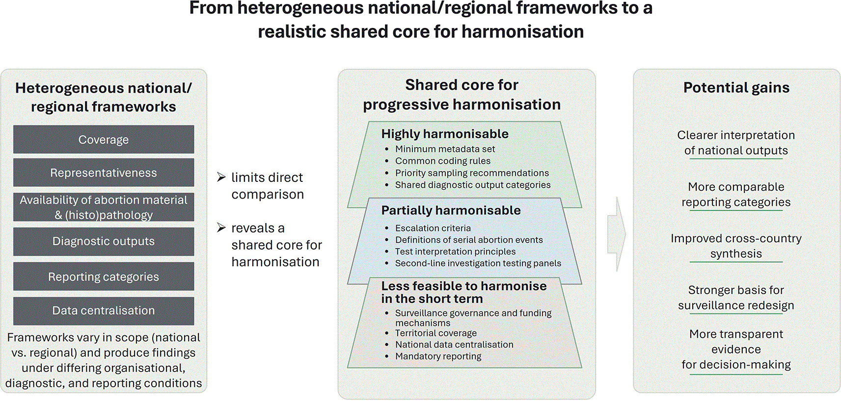
Figure 1. Proposed scheme of a progressive model for the harmonisation of ruminant abortion surveillance systems: stepwise transition from heterogeneous national/regional frameworks toward a feasible shared core approach, highlighting levels of harmonisation and associated potential gains.
4. Discussion
This report provides an overview of the procedures governing the sampling, diagnostic investigation, and reporting of ruminant abortions across six European countries, and summarises the predominant abortifacient agents identified within national/regional diagnostic frameworks between 2022 and 2024. It does not aim to report outcomes from official disease surveillance programmes (e.g. brucellosis), except where such testing was part of the abortion submission work-up. Previous work from the Q-NET-ASSESS consortium reviewed and compared abortion diagnostic protocols and proposed methodological guidance (Van den Brom et al., 2025). The present report describes the outputs generated by national/regional frameworks and the organisational and interpretive context in which they are produced, with explicit warnings about the limited comparability between countries.
The procedures compiled in this report enabled us to delineate both areas of convergence and points of divergence among national approaches across Europe. Although all countries partially subsidise abortion diagnostics, substantial differences were observed in the organisation of diagnostic investigations, including laboratory structures and analytical methods. Differences were also evident in the scope of surveillance and reporting systems, which range from nationwide to region-specific coverage. Hence, systematic nationwide monitoring or surveillance of ruminant abortions is implemented in Belgium, the Netherlands, and the United Kingdom, but not in France, Germany, or Spain. Diagnostic capacity is also organised differently across countries. In the Netherlands, a single laboratory performs almost all ruminant abortion analyses, whereas in Belgium regional laboratories and the federal national reference laboratory undertake the diagnostic workload. In contrast, multiple public and private laboratories operate across the territory in France, Germany, Spain, and the UK. Harmonised diagnostic protocols (including pathology) are applied in the Netherlands and the UK. In France, the OSCAR protocol provides harmonised methodologies (excluding pathology) and centralised data collection, but its implementation is voluntary and currently covers roughly one-third of the Departements. In Germany and Spain, diagnostic testing is not harmonised at the national level but can be at regional level (Bundesländer and Autonomous Community, respectively). In Belgium, surveillance is organised within an official framework, partly supported by public funds, but data collection is not consistently centralised nationwide and often relies on voluntary reporting. Additionally, definitions on repeated cases, series or increased numbers of abortion in different countries are currently not further specified. This compendium of practices highlights the heterogeneity of current frameworks, which limits the comparability of data across countries. The progressive harmonisation model presented in
Figure 1, which aims to move from heterogeneous national/regional frameworks toward a realistic shared core for harmonisation, would enhance the interpretation of national outputs and improve comparability without requiring identical surveillance systems.
In an attempt to explore the relative importance of the main abortifacient agents in the partner countries, data for the period 2022 to 2024 were compiled in accordance with the specifications of each national reporting framework. Under these frameworks, some countries document attributed causes when these can be established, whereas in other countries reporting is limited to agent detection, and it is not always possible to determine whether the agent detected was the cause of abortion from the available information and reports. Detailed methodological considerations on the interpretation of sample types and diagnostic approaches have been discussed previously (Van den Brom et al., 2025). In brief, a definitive diagnosis of the attributed cause of abortion requires not only the detection of the infectious agent but also the presence of compatible (histo) pathological changes. Isolation or PCR detection of the agent may support causality, or suggest, a presumptive causal role. However, it may also merely indicate exposure or incidental co-detection without clear attribution. Under these circumstances, the diagnostic value is strongly conditioned by the biological sample matrix used for detection; in general, recovery from foetal or placental tissues provides substantially greater interpretive weight than detection in matrices such as vaginal swabs or milk (Van den Brom et al., 2025). In this context, the data presented in
Tables 2a-f are not directly comparable across countries. Nonetheless, they provide a useful snapshot of the abortifacient agents most frequently identified.
Diagnosing ruminant abortion is challenging and often inconclusive, as in over approximately half of the cases the laboratory investigation fails to identify an aetiological agent. In cases of non-infectious causes (e.g. maternal disease or malnutrition), no pathological lesions may be found. But even in cases where inflammation in placentae and/or foetus is observed, the causative agent is not always detected. In this dataset, where denominators were available and interpretable, an abortifacient agent was identified in approximately 36.7–53.5% of submissions, depending on the national or regional framework. In general, a lower proportion of diagnoses was achieved in goat submissions compared to cattle and sheep abortion submissions. An assessment of whether this low success is related to the delay between abortion and sampling, or other factors related to the respective surveillance networks, would be relevant to perform.
In cattle, N. caninum was among the most common abortifacients associated with bovine abortion across countries. This is in agreement with a recently published meta-analysis encompassing 9,164 bovine fetuses (Hecker et al., 2023). In that analysis, the parasite was detected in 22.2% of samples, and in 16.7% of the investigated cases it was described as the cause of abortion when compatible histopathological lesions were present. The two main bacteriological abortifacients identified in submissions of bovine abortions were Salmonella spp. and T. pyogenes, pathogens commonly found among the top five diagnosed infectious causes of bovine abortion internationally (Mee, 2023). Although C. burnetii was detected in many countries, numbers were generally low except in Belgium and France, where the absence of histopathological assessment prevented any direct evaluation of causality. In several of the participating countries, pestivirus was identified in cattle abortion submissions, and after the start of the European BTV serotype 3 epidemic, BTV has been described in Saxony (Germany) and the Netherlands as an important abortifacient in cattle abortion submissions, as shown in data for 2024. In the UK, SBV was detected in a high number of abortion submissions in sheep and cattle in 2024. Congenital malformations, sometimes with a possible infectious background, are occasionally reported as a cause of abortion.
In sheep submissions, C. abortus was the main bacterial abortifacient detected. The incidence of Campylobacter spp. was high in the UK and the Netherlands but was not reported in France or Spain. Campylobacter spp. have been described as common causes of ovine abortion in Australia (Clune et al., 2020) and North America (Menzies, 2011). Two species, Campylobacter jejuni and Campylobacter fetus subsp. fetus, are associated with ovine abortion, producing similar clinical presentations that are not usually differentiated. Other bacteria such as C. burnetii, Listeria spp. and Salmonella spp. were frequently reported as causes of ovine abortion in several countries. This is in line with previous reports of these bacterial abortifacients associated with abortion in sheep (Masala et al., 2007; Van den Brom et al., 2012; van Engelen et al., 2014; van den Brom et al., 2021). Toxoplasma gondii was in some countries the most commonly diagnosed abortifacient in ovine abortion submissions and the main parasitological abortifacient in all countries. Neospora caninum,
also described as a possible abortifacient agent in small ruminants (Moreno et al., 2012; Basso et al., 2022; Nayeri et al., 2022; Gouvias et al., 2024), was detected in sheep and goats in Belgium. Occasionally, viral agents, such as pestiviruses, BTV, and SBV, were detected in ovine abortion submissions. Compared to the other countries, SBV is a relatively commonly detected abortifacient in abortion submissions in the UK, with a significant increase in 2024, as was also observed in cattle. It is thought that observed peaks and troughs in incidence are related to waxing and waning of national herd, flock immunity and vector activity (King et al., 2015). It has also potentially been under-diagnosed in some recent years, as veterinarians and farmers may assume gestational SBV infection has occurred if typical foetal deformities are seen, and these incidents do not always get recorded on the GB VIDA database. Some of the main abortifacient agents detected in goats were also described in sheep, including C. abortus, Listeria spp., and C. burnetii. However, as reported previously (Van den Brom et al., 2012), C. burnetii was more common in goats than sheep and the occurrence of Listeria spp. varied between countries.
The main abortifacient agents reported in ruminants differ between countries. For example, in many countries, C. burnetii is an important cause of abortion in ruminants, whereas it was not detected in sheep and goat submissions in the Netherlands. This may partly be explained by the compulsory vaccination of certain groups of sheep and goat farms in the Netherlands (Vellema and Van den Brom, 2014). In a broader sense, cross-country differences in vaccination practices likely contribute to the observed distribution of abortifacient agents. Differences must also be interpreted cautiously due to the specific nature of the agents considered and the short study period (2022–2024). Within this time frame, changes in transmission rates and prevalences of long-time endemic agents were likely influenced by shifts in national herd size and levels of natural or vaccine-induced herd immunity. Agents newly introduced to parts of Europe (e.g. BTV-3) or novel agents (e.g. SBV) often occur in waves, particularly if they are vector-borne. As a result, their prevalence is subject to fluctuations, particularly over short timeframes. Long-term trends need to be monitored to accurately assess their animal health and economic impact, with harmonisation of national frameworks being pivotal to achieve the goal. Costs and challenges of diagnostic testing, including the state of the samples (e.g. autolysis, mummification), can discourage farmers from investigating abortion outbreaks. Veterinarians should emphasize to farmers the importance of recognising when their abortion rate exceeds usual levels, collecting aborted material (particularly placentae and foetal tissues), and ensuring its proper storage until submission to the laboratory. Veterinarians should also seek to understand the logistical challenges farmers face and discuss the feasibility of on-farm abortion investigations (Ramillien et al., 2025). For example, the placentae are often eaten by animals or buried in their bedding, particularly on farms with many sheep or goats. Co-construction approaches to abortion management on ruminant farms offer promising ways forward in this regard (Jourdain et al. submitted).
In an ideal scenario, farmers and their veterinarians would be able to submit abortion materials to veterinary laboratories with minimal logistical and financial constraints while limiting the risks associated with the transport of such material. Pathological examinations would be performed under safe conditions following standardised protocols and procedures adapted to detect both known and unknown pathogens relevant to ruminant abortions in each country. The resulting pathology findings from ruminant abortion submissions should explicitly distinguish between mere agent detection and a confirmed cause of abortion, and the resulting information should be made centrally available at the national level and shared internationally. This would provide a comprehensive overview of infectious causes of abortion on a national and European scale.
5. Conclusion
This report summarises the available findings on the investigation of abortifacient agents in sheep, goat, and cattle abortion submissions from six participating countries. Although direct comparisons between countries are challenging due to differences in laboratory structures and analytical methods (namely the availability of pathology records), coverage, and reporting, the compiled data provide a coherent overview of the abortifacient agents most frequently identified across species. Notably, the main bacterial, parasitological, and viral abortifacients associated with abortion submissions were broadly similar across countries, albeit important differences in system structure, methodology, and interpretation limit direct comparison.
At the European level, surveillance coverage, diagnostic workflows, and reporting systems for ruminant abortion remain heterogeneous, reflecting substantial differences in laboratory organisation, and degrees of centralisation across countries. Centralised frameworks coexist with decentralised systems that vary in coverage, methodological harmonisation, and the extent to which reporting is mandatory or voluntary, and the degree of financial coverage for laboratory testing. These disparities highlight the need to move toward more harmonised approaches across countries, including clear definitions on series, repeated or increased numbers of abortion. Nationwide coverage is desirable, and diagnostic procedures (while necessarily adapted to the infectious agents circulating in each region) should follow similar interpretation principles (e.g. linking detected agents with compatible pathological changes in the foetus and/or placenta). Moreover, more systematic reporting and improved data consolidation would be important steps towards more meaningful cross-country comparisons. Monitoring trends in diagnostic findings, including increases in cases where no diagnosis is reached, as well as awareness of novel disease presentations, can help identify new or emerging pathogens. This is particularly relevant because many abortifacient agents may pose transboundary threats to animal health and/or have recognised zoonotic potential, thereby requiring coordinated responses.
Ethics statement
The authors confirm that they have adhered to the ethical policies of the journal, as noted on the journal’s author guidelines page. Ethical approval is not applicable.
Data and software availability
Data used in this study are either publicly available through the links provided for each country or may be requested from the relevant data providers, subject to applicable permissions and approvals.
Relevant links are provided in
Table 1 where available (i.e., France, the Netherlands, and Great Britain). Nationwide datasets are not publicly available for Belgium, Germany, and Spain. The data presented in
Tables 2a–2f comprise the data compiled and analysed for this study. For Belgium, Germany, and Spain, these tables were compiled using data obtained from internal reports and other available sources.
Additional data, where available, may be requested from the following data providers:
• Belgium: Marcella Mori (Sciensano, Belgian Institute for Health, Brussels, Belgium; Marcella.Mori@sciensano.be)
• France: Elodie Rousset (ANSES, Sophia Antipolis, France; Elodie.ROUSSET@anses.fr)
• Germany: Katja Mertens-Scholz (Friedrich-Loeffler-Institut, Jena, Germany; katja.mertens-scholz@fli.de)
• Spain: Ana Hurtado (NEIKER, Basque Institute for Agricultural Research and Development, Spain; ahurtado@neiker.eus)
• The Netherlands: René van den Brom (Royal GD, Deventer, The Netherlands; r.vd.brom@gdanimalhealth.com)
• Great Britain: Sue Neale (Animal and Plant Health Agency, Penrith Veterinary Investigation Centre, United Kingdom; Susan.Neale@apha.gov.uk)
Please note that the authors are not the sole proprietors of all data referenced in this study. As a result, access to data beyond those presented in
Tables 2a–2f may be restricted. Restrictions may arise from ethical, privacy, confidentiality, and country-specific regulatory requirements. Requests for additional data should be directed to the relevant country-specific data provider listed above and will be considered on a case-by-case basis. Access may be granted only where applicants meet all applicable requirements, including approval by the data owner(s), compliance with national regulations and institutional policies, fulfillment of confidentiality obligations, and any permissions required from the competent authorities. In some cases, no data beyond those reported in
Tables 2a–2f are available. The authors will make every reasonable effort to facilitate legitimate requests and are available to provide tailored information in response to specific inquiries whenever possible.
Acknowledgements
Q-NET-ASSESS consortium partners involve scientists from NEIKER, ANSES, INRAE, FLI, Sciensano, Royal GD and Moredun Research Institute. All other members of the consortium who are not a co-author are thanked for their contribution. The consortium partners would also like to thank farmers and veterinarians who submitted biological material sampled from aborting flocks/herds for laboratory analyses. A special word of thanks, to acknowledge all colleagues involved in performing the pathology and other abortion diagnostics, and providing data, input and feedback on the manuscript (Remco Dijkman, Tara de Haan, Debora Smits (all Royal GD), Rudolf Reichel (APHA), and Jesús F. Barandika (NEIKER)).
Additionally, we would like to thank APHA Veterinary Investigation Centres, APHA partner postmortem providers and Scotland’s Rural College (SRUC) Veterinary Services and Moredun Research Institute Surveillance service. We also thank the OSCAR Working Group of the French ESA Platform (including experts from the Groupement de Défense Sanitaire France, Association pour le Développement et l’Innovation du Laboratoire Vétérinaire d’Analyses, French Agency for Food, Environmental and Occupational Health & Safety, Fédération Régionale des Groupements de Défense Sanitaire, Groupements de Défense Sanitaire, Institut de l’Élevage, French National Research Institute for Agriculture, Food and Environment, Oniris Nantes Veterinary School, French National Veterinary Practitioner Association, and the Ministry of Agriculture), as well as all local veterinarians, laboratories, and livestock health associations (GDS) in participating departments for their contributions to diagnosis, data management, and protocol improvement. We would like to thank the Spanish Ministry of Agriculture, Fisheries and Food, in particular Subdirección General de Laboratorios de Sanidad Animal y Vegetal and Subdirección General de Sanidad e Higiene Animal y Trazabilidad. Finally, we would like to thank the Dutch Ministry of Agriculture, Fisheries, Food security and Nature (LVVN).
References
- Ali S, Zhao Z, Zhen G, et al.:
Reproductive problems in small ruminants (Sheep and goats): a substantial economic loss in the world.
Large Animal Review.
2019; 25: 215–223.
- van Asseldonk MA , Prins J, Bergevoet RH:
Economic assessment of Q fever in the Netherlands.
Prev. Vet. Med.
2013 Oct 1; 112(1–2): 27–34. Epub 2013 Jul 15. PubMed Abstract
| Publisher Full Text
- Basso W, Holenweger F, Schares G, et al.:
Toxoplasma gondii and Neospora caninum infections in sheep and goats in Switzerland: Seroprevalence and occurrence in aborted foetuses.
Food Waterborne Parasitol.
2022 Aug 17; 28: e00176. Publisher Full Text
- Bronner A, Gay E, Fortané N, et al.:
Quantitative and qualitative assessment of the bovine abortion surveillance system in France.
Prev. Vet. Med.
2015 Jun 1; 120(1): 62–9. Epub 2015 Feb 25. PubMed Abstract
| Publisher Full Text
- Bronner MB, Haagsma JA, Dontje ML, et al.:
Long-term impact of a Q-fever outbreak: An evaluation of health symptoms, health-related quality of life, participation and health care satisfaction after ten years.
J. Psychosom. Res.
2020 Dec; 139: 110258. Epub 2020 Sep 22. PubMed Abstract
| Publisher Full Text
| Free Full Text
- Bundesanzeiger:
Bekanntmachung im Bundesanzeiger, Amtlicher Teil, AT 01.08.2014 B1.2014.
- Clune T, Beetson S, Besier S, et al.:
Ovine abortion and stillbirth investigations in Australia.
Aust. Vet. J.
2020 Dec 1; 99: 72–78. PubMed Abstract
| Publisher Full Text
- Dijkstra E, Vellema P, Peterson K, et al.:
Monitoring and Surveillance of Small Ruminant Health in The Netherlands.
Pathogens.
2022 May 31; 11(6): 635. PubMed Abstract
| Publisher Full Text
| Free Full Text
- Dupuy C, Locquet C, Brard C, et al.:
The French National Animal Health Surveillance Platform: an innovative, cross-sector collaboration to improve surveillance system efficiency in France and a tangible example of the One Health approach.
Front. Vet. Sci.
2024; 11: 1249925. PubMed Abstract
| Publisher Full Text
| Free Full Text
- Gouvias I, Lysitsas M, Batsidis A, et al.:
Molecular Investigation of Small Ruminant Abortions Using a 10-Plex HRM-qPCR Technique: A Novel Approach in Routine Diagnostics.
Microorganisms.
2024 Aug 14; 12(8): 1675. PubMed Abstract
| Publisher Full Text
| Free Full Text
- Guillet C, Join-Lambert O, Le Monnier A, et al.:
Human listeriosis caused by Listeria ivanovii.
Emerg. Infect. Dis.
2010 Jan; 16(1): 136–138. PubMed Abstract
| Publisher Full Text
| Free Full Text
- Hecker YP, Gonzalez-Ortega S, Cano S, et al.:
Bovine infectious abortion: a systematic review and meta-analysis.
Front. Vet. Sci.
2023; 10: 1249410. PubMed Abstract
| Publisher Full Text
| Free Full Text
- Holec-Gąsior L, Sołowińska K:
Detection of Toxoplasma gondii infection in small ruminants: old problems and current solutions.
Animals.
2022; 13: 2696.
- Jourdain E, Rousset E, Ramillien E, et al.:
ZOOJEU: using Q fever to highlight a participatory science approach for fostering the co-design of management strategies for neglected zoonotic diseases.Submitted.
- King B, O'Shea Brown T, Tarlinton R, et al.:
Seroprevalence of Schmallenberg virus in the United Kingdom and the Republic of Ireland: 2011-2013.
Vet. Microbiol.
2015 Oct 22; 180(1–2): 36–40. Epub 2015 Jul 26. PubMed Abstract
| Publisher Full Text
- Laine CG, Johnson VE, Scott HM, et al.:
Global Estimate of Human Brucellosis Incidence.
Emerg Infect Dis.
2023 Sep; 29(9): 1789–1797.
Publisher Full Text
- Masala G, Porcu R, Daga C, et al.:
Detection of pathogens in ovine and caprine abortion samples from Sardinia, Italy, by PCR.
J. Vet. Diagn. Invest.
2007; 19: 96–98. PubMed Abstract
| Publisher Full Text
- Mee JF:
Invited review: Bovine abortion—Incidence, risk factors and causes.
Reprod. Domest. Anim.
2023; 58(Suppl. 2): 23–33. PubMed Abstract
| Publisher Full Text
- Meijer A, Brandenburg A, de Vries J, et al.:
Chlamydophila abortus infection in a pregnant woman associated with indirect contact with infected goats.
Eur J Clin Microbiol Infect Dis.
2004; 23(6): 487–490
- Menzies PI:
Control of important causes of infectious abortion in sheep and goats.
Vet Clin Food Anim.
2011; 27(1): 81–93. Publisher Full Text
- Moreno B, Collantes-Fernández E, Villa A, et al.:
Occurrence of Neospora caninum and Toxoplasma gondii infections in ovine and caprine abortions.
Vet. Parasitol.
2012 Jun 8; 187(1–2): 312–318. Epub 2012 Jan 5. PubMed Abstract
| Publisher Full Text
- Mori M, Boarbi S, Michel P, et al.:
In vitro and in vivo infectious potential of coxiella burnetii: a study on Belgian livestock isolates.
PLoS One.
2013 Jun 28; 8(6): e67622. Print 2013. PubMed Abstract
| Publisher Full Text
| Free Full Text
- Nayeri T, Sarvi S, Moosazadeh M, et al.:
The Global Prevalence of Neospora caninum Infection in Sheep and Goats That Had an Abortion and Aborted Fetuses: A Systematic Review and Meta-Analysis.
Front. Vet. Sci.
2022 Apr 26; 9: 870904. eCollection 2022. PubMed Abstract
| Publisher Full Text
| Free Full Text
- Palma O, Pla-Aragones LM, Cawley AM, et al.:
Economic Impact of Abortions in Dairy Cow Herds.
Vet. Sci.
2025; 12(7): 645. PubMed Abstract
| Publisher Full Text
| Free Full Text
- Ramillien E, Cayre P, Fourt X, et al.:
Q fever as an anthropological prism for revealing how farmers live with microbes.
Agric. Hum. Values.
2025; 42: 527–543. Publisher Full Text
- Santman-Berends IMGA, Brouwer-Middelesch H, Van Wuijckhuise L, et al.:
Surveillance of cattle health in the Netherlands: Monitoring trends and developments using routinely collected cattle census data.
Prev. Vet. Med.
2016 Nov 1; 134: 103–112. Publisher Full Text
- Van den Brom R, Lievaart-Peterson K, Luttikholt S, et al.:
Abortion in small ruminants in the Netherlands between 2006 and 2011.
Tijdschr. Diergeneeskd.
2012; 137: 450–457. PubMed Abstract
- Van den Brom R, Santman-Berends I, Dijkman R, et al.:
An Accessible Diagnostic Toolbox to Detect Bacterial Causes of Ovine and Caprine Abortion.
Pathogens.
2021; 10: 1147. PubMed Abstract
| Publisher Full Text
| Free Full Text
- Van den Brom R, Neale S, Jourdain E, et al.:
Detection of abortifacient agents in domestic ruminants, with a specific focus on Coxiella burnetii.
Open Res Eur.
2025; 5: 94. Publisher Full Text
- Van Engelen E, Luttikholt SJM, Peperkamp NHMT, et al.:
Small ruminant abortions in the Netherlands during lambing season 2012-2013.
Vet. Rec.
2014; 174: 506. PubMed Abstract
| Publisher Full Text
| Free Full Text
- Vellema P, Van den Brom R:
The rise and control of the 2007-2012 human Q fever outbreaks in the Netherlands.
Small Rumin. Res.
2014; 118: 69–78. Publisher Full Text
- WOAH:
2022.
Reference Source