Introduction
Preclinical cancer models are essential for understanding tumor biology and developing diagnostic and therapeutic strategies. These aims can only be achieved by a realistic model that is able to recapitulate not only tumor expansion and metastasis, but also the interaction of the tumor with the environment.1,2 Orthotopic tumor models accurately replicate tumor vascularization, hypoxia, extracellular matrix architecture, immune suppression, and poor immune cell infiltration1 that are hallmarks of challenging human cancers. Several studies compared subcutaneous and orthotopic colon3 and (HPV)-positive oropharyngeal squamous cell carcinoma (OSCC)4 tumor models to evaluate immune cell distribution, infiltration into the tumor, and response to therapies. Specifically, subcutaneous OSCC showed complete survival, whereas survival in the orthotopic model declined as the tumor progressed.4 The immune cell infiltration patterns differed between subcutaneous and orthotopic tumors, with the orthotopic tumor showing a gradual decline in T cell infiltration, consistent with immune-excluded tumor cases observed in humans.4 Similarly, Snipstad et al.3 compared the characteristics of different colon cancer cell lines in subcutaneous and orthotopic tumor models and reported that subcutaneous tumors have a significantly increased frequency of infiltrating CD8+ and CD4+ T cells, as well as NK cells, associated with a more immunoreactive phenotype. In both studies, orthotopic tumors did not respond to anti-PD1 treatment, whereas subcutaneous tumors showed modest increases in immune cell infiltration following therapy.3,4
Lung cancer remains one of the leading causes of cancer-related mortality worldwide5 with a 5-year survival rate below 30%.6 Immune checkpoint inhibitors (ICIs) are common treatment options, however, resistance to ICIs is frequently characterized by an immunosuppressive tumor microenvironment that limits therapeutic response.7 Immune-excluded or poorly infiltrated phenotypes in lung tumors are common.7 Studies analyzing patient samples from non-small cell lung cancer found that B-cell and T-cell infiltration into the tumor was associated with improved survival8 and that clusters of B and/or T cells at the tumor borders were linked to poor survival.9 Therefore, preclinical models that accurately reflect the spatial and immune architecture of lung tumors are essential for understanding TME interactions and for developing effective therapeutic strategies.
Establishing reproducible lung tumors in preclinical models remains technically challenging.10 Genetically engineered murine models (GEMM),11,12 humanized mouse models,12 human xenograft models,13,14 and syngeneic immunocompetent models12 are commonly used to establish orthotopic tumors in the preclinical setting. There are advantages and limitations to each system. GEMMs alone can only mimic specific oncogenic features of the tumor and are costly.11 Humanized murine and human xenograft models allow engraftment of tumors derived from clinical samples. Still, these experimental models usually rely on immunodeficient mice, with either no or partially reconstituted immune systems, which prevents investigation of tumor-immune system interactions and immune responses observed in patients.2,11,12 Syngeneic orthotopic models rely on engrafting murine cancer cells into an immunocompetent murine host. This reduces costs and ensures consistency across the experimental platform needed for preclinical experiments.12 In addition to their advantages, orthotopic syngeneic cancer models have two major limitations: a variable inoculation success rate (60–100%) and a requirement for technical/surgical expertise. Reported inoculation success rates for orthotopic cancer models in the lung,15 breast,16 and colorectal17 are 67% to 100%, and higher success is often associated with invasive surgical engraftment, which usually relies on site-directed injection of tumor cells.18
Intratracheal and intrapulmonary injections are common approaches for the delivery of tumor cells into the lung, but19,20 require surgical intervention and technical expertise, introduce operator-dependent variability, and carry a higher risk of local tissue injury that alters tumor–microenvironment interactions.18,19 Therefore, minimally invasive methods are necessary to achieve more reproducible and consistent results.20,21 Accordingly, systemic delivery by intravenous injection,22 pulmonary delivery routes such as intranasal23 and oropharyngeal administration,24 widely used for delivering infectious material or biologics directly to the lung, are attractive minimally invasive alternatives. Unlike non-invasive intratracheal instillation, aspiration does not require direct visualization or cannulation of the trachea and instead relies on spontaneous inhalation of the inoculum delivered to the oropharynx.
Recent work has adopted intranasal delivery for orthotopic lung tumor establishment, providing a non-invasive alternative that more closely reflects tumor initiation in alveolar regions.15 However, variability in tumor engraftment efficiency, progression, and size within experimental groups remains a challenge. Oropharyngeal aspiration is widely used in pulmonary research to achieve uniform delivery of biologics to the distal lung.24 Yet whether this approach can reliably deliver viable tumor cells to establish orthotopic lung tumors has not been investigated. Consequently, there is currently no validated aspiration-based orthotopic lung cancer model for studies of tumor progression and the tumor microenvironment.
In this study, we developed and characterized an aspiration-based, non-invasive model for orthotopic lung cancer in immunocompetent mice. We directly compared the aspiration route with intranasal delivery and evaluated tumor establishment, longitudinal progression, and the spatial immune cell distribution within the TME. Our findings demonstrate that aspiration enables consistent tumor establishment and disease progression, while mimicking a tumor microenvironment with limited intratumoral immune cell infiltration. This provides a practical approach for in vivo studies of lung tumor–microenvironment interactions.
Methods
Animals
The C57BL/6 mice were originally obtained from the Jackson Laboratory (JAX, Strain ID:000664). Wild-type C57BL/6 animals that were 7–8 weeks old, from both female and male sexes (control = 2F/3M, i.n. = 5F/1M and aspiration = 5F/1M), were included throughout the study. Animals were housed in the vivarium at the Izmir Biomedicine and Genome Center (IBG, Izmir, Turkey) in compliance with the relevant institutional animal care and use committee guidelines and applicable national laws. In vivo experiments were performed with prior approval by the institutional ethics committee (Izmir Biomedicine and Genome Center’s internal Ethical Committee on Animal Experimentation, protocol #: 2025–007), in accordance with national laws and Directive 2010/63/EU. The sample size was calculated according to the 3R rule and the experience of the lead researcher. Mice were randomly allocated to the experimental groups before inoculation and assigned individual identification numbers at the time of inoculation. Researchers were not blinded during experimental procedures or data analysis. Mice that lost more than 20% of their body weight and/or scored 3 points on the 5-point scoring system adapted from the murine sepsis scoring system25 were euthanized. Euthanasia was performed by cervical dislocation under aseptic conditions and in designated areas in accordance with Institute regulations. Death was confirmed by the absence of respiration and lack of response to the toe pinch reflex.
Luciferase-conjugated lewis lung cancer cell maintenance and storage
Luciferase-conjugated Lewis Lung Cancer (LL2-luc2) was purchased from ATCC (CRL-1642-LUC2, Europe). LL2-luc2 was revived according to the manufacturer’s guidelines. Cells were cultured under sterile conditions with complete Dulbecco’s Modified Eagle’s Medium with high Glucose (Serana, Germany) supplemented with 10% FBS (v/v) (Biological Industries, USA) 4 mM L-glutamine (Sartorius, USA), 4500 mg/L glucose, 1 mM sodium pyruvate (Sartorius, USA), and Blasticidin (2 ug/mL) (Invivogen, USA). Once the confluence reached 80%, cells were lifted by Trypsin-EDTA for 1 min at 37 °C. 2 mL Trypsin-EDTA (Biological Industries, USA) was deactivated with 1:10 (v/v) complete medium and centrifuged at 100 g for 5 min. Cells were washed twice and subcultured at a 1:4 dilution three times per week. Before inoculation into animals, cells were detached as described previously. Cells were washed twice with 15 mL PBS (Sartorius, USA) and counted using a hemacytometer. Cell concentration was adjusted to 106 live cells per 50 μl (20 × 106 cells/mL) and kept on ice.
In vivo orthotopic lung cancer model
Prior to inoculation, the luciferase activity of LL2-luc2 cells was verified in vitro. Wild-type C57BL/6 mice were randomly assigned to the following groups: intranasal PBS control, aspiration PBS control, intranasal LL2-luc2 inoculation (n = 6), and aspiration LL2-luc2 inoculation (n = 6). For tumor inoculation, animals were anesthetized with isoflurane (Adeka İlaç Sanayi Ve Ticaret A.Ş.,Türkiye) and 106 LL2-luc2 cells in 50 μL PBS were administered via aspiration as previously described.26 Briefly, the mouse was stabilized on a tilted stage via its incisors, the tongue was gently lifted laterally using sterile blunt-end forceps, and the cell suspension was instilled into the pharyngeal/laryngeal region via a P200 pipette. Upon spontaneous inhalation, the inoculum was aspirated into the lungs. For intranasal inoculation, 106 LL2-luc2 cells in 50 μL PBS were administered dropwise onto the nostrils of anesthetized mice, allowing passive inhalation. The control group received 50 μL PBS via either aspiration or intranasal delivery following the same procedures.
The mice were weighed and monitored every 2 days. Tumor progression was assessed by weekly in vivo bioluminescence measurements. The humane endpoint was determined by a scoring system adapted from the murine sepsis scoring system, where appearance, behavior, respiratory function, body weight change were evaluated. Mice that lost >20% from baseline weight, showed persistent moderate to severe scores for more than 24 hours (any parameter or combined score ≥ 3), has labored breathing or gasping (respiratory function score ≥ 3), could not eat/drink themselves, were unresponsive or lethargic, or had an abnormal condition such as cyanosis or vaginal/cervical hyperplasia were euthanized immediately. The experiment was concluded once all mice that were inoculated via aspiration reached the humane endpoint, day 40 post-inoculation. At day 33, three control and two intranasally inoculated mice were euthanized for matched terminal tissue collection when three aspiration-inoculated animals began reaching the predefined humane endpoint. Consequently, longitudinal measurements from these animals were included through day 33, and subsequent analyses included only animals remaining in the study.
In vivo bioluminescence imaging
Bioluminescence imaging was performed to monitor tumor progression in vivo. Mice were injected intraperitoneally with D-luciferin (Revvity, Netherlands) prepared according to the manufacturer’s instructions, at a dose of 150 mg/kg. Mice were anesthetized with isoflurane. Imaging was initiated 10 minutes after the D-luciferin injection (MILabs, The Netherlands).
For each imaging session, images were acquired sequentially at 2-minute intervals for 15 minutes. Identical acquisition parameters: 45-second exposure, 2X gain, and 4x4 binning were applied across all animals and imaging sessions. The signal corresponding to the orthotopic lung tumor was quantified from a thoracic region of interest (ROI) as integrated electron flux using MILabs software, which provides background-subtracted and sensitivity-adjusted values.
Histology
Lungs were inflated with 1 mL 10% neutral buffered formaldehyde (Tekkim, Türkiye) and fixed for four hours at 4 °C. Lungs were dehydrated in 20 mL 30% sucrose (Sigma-Aldrich, Germany) overnight at 4 °C, embedded in cryomolds (Tissue-Tek, Sakura, USA) with O.C.T. (Tissue-Tek, Sakura, US) the following day (15 hours later), and snap-frozen. Serial sections in 10–15 μm thickness were prepared using a cryostat (Leica CM1950). Digital pathology analysis was performed on the section containing the largest visible tumor area to ensure consistent comparison between animals. H&E staining was performed as a service by IBG’s Histopathology Core Facility according to its routine protocol as previously described.26 Samples were mounted and examined with a light microscope (Olympus BX61).
Immunofluorescence
The tissue sections were stored at −20 °C until use. Prior to staining, slides were kept at RT and rehydrated in PBS for 10 min. The sample borders were drawn by the PAP pen (Sigma-Aldrich, Germany). Then, the slides were blocked in a buffer containing 2% BSA (Capricorn, USA) and 0.01% Triton X-100 (Sigma-Aldrich, Germany). Samples were stained with CD3-AF647(clone 17A2, 1:100, BD Biosciences, USA), MHCII-PE (clone: M5/114.15.2, 1:100, Biolegend, USA), in staining buffer supplemented with 3% normal mouse serum (Sigma-Aldrich, Germany) overnight at 4 °C in a humidity chamber. Samples were washed three times with PBS, and cell nuclei were labeled with Hoechst (Thermo Fisher Scientific, Carlsbad, CA) two drops per mL of PBS, according to the manufacturer’s instructions. Slides were mounted in 50 μl/sample Floromount-G (Thermo Fisher Scientific, Carlsbad, CA) and were visualized using an Apotome microscope (Zeiss Observer 7).
Imaging analysis
Bioluminescence imaging
Bioluminescence analysis was performed as follows: To obtain peak values, the three highest fluxes within the post-injection imaging sequence were identified for each mouse at each time point, and the mean of these values was used as the representative bioluminescence value for that imaging session. These values were used to generate longitudinal tumor burden curves for individual mice as (integrated intensity +1) on a log10 scale and to compare groups. For correlation with histological tumor burden, the most recent bioluminescence measurement recorded before sacrifice was used as the endpoint.
Tumor burden analysis from H&E staining in QuPath
H&E images were acquired as a tile scan in the BX61 fluorescence microscope (Olympus, Germany) or Apotome (Zeiss, Germany). The Ome.tiff images were analyzed using QuPath 0.6.0. Briefly, a QuPath project was established with H&E images. First, for the Hematoxylin and Eosin channels, the estimate-stain-vectors function was applied to unify background values. To avoid bias and automate analysis, we trained the pixel classifier to annotate the background, tumor, and healthy lung tissue, as well as regions to exclude, such as folded regions with visible dirt. Particular attention was given to including bronchioles and vascular regions in the healthy lung annotation, since, due to their darker staining, the classifier often tags those regions as tumors. Then, with the annotated sample set, the ‘train pixel classifier’ ran with the following parameters: Classifier: Random Trees, resolution: moderate, Features: select channels: Eosin and Hematoxylin; set scale:2.0; select features: Gaussian and weighted Gaussian. The resulting classifier was saved. The classifier was applied to the entire project to generate full-image annotations, with a minimum of 50,000 and a maximum of 10^12. After validating the annotations, the tumor and tumor-free lung regions (in μm2) were exported to an Excel spreadsheet. The rendered images containing the tumor annotations were saved. Tumor burden per mouse was calculated as follows:
Immunofluorescence image analysis
Lung lobe immunofluorescence tile-scan images were exported using the Zen 3.13 application (Zeiss). The immune cells in the TME were analyzed by QuPath 0.7.0. Briefly, the tumor and tumor-free regions were annotated based on H&E image analysis. For each region, cells were detected using the DAPI (Hoechst) channel.
CD3+ T cells, MHCII+ cells were defined as running object classifiers first for each channel, determining a positive signal, followed by creating composite classifiers for Hoechst+CD3+ and Hoechst+MHCII+. The rendered images and cell numbers corresponding to the classifiers were exported. The immune cell densities per tumor-free and within the tumor were calculated. Immune cell infiltration index was calculated for each Hoescht+CD3+ and Hoechst+MHCII+ as:
Statistical Analysis
Statistical analyses were performed in GraphPad Prism (version 8). Survival was analyzed using the Mantel–Cox (log-rank) test, and a survival curve was established. Bioluminescence imaging (BLI) data were expressed as (electron flux +1) to visualize on a log10 scale and analyzed using a mixed-effects ANOVA (REML), with each mouse as a repeated measure and fixed effects for inoculation route, time, and their interaction. Body weight changes were monitored over time and are presented descriptively. Tumor burden between experimental groups was compared using the Kruskal–Wallis test with Dunn’s multiple-comparison test. Associations between histological tumor burden and BLI signal intensity or body weight change were assessed using Spearman’s rank correlation. A p-value <0.05 was considered statistically significant.
Results
LL2-luc2 cancer cell inoculation via aspiration establishes orthotopic lung tumors
To establish an orthotopic lung tumor, we inoculated luciferase-conjugated Lewis Lung cancer cells (LL2-luc2) via aspiration (n = 6) or the intranasal route (n = 6). The intranasal inoculation route is included as a control, since many groups have used it as an efficient, non-invasive route to target the lungs.27 We monitored the experimental mouse well-being and weight change three times per week to determine progress and the severity of the OLC. Additionally, tumor inoculation and growth are verified by weekly bioluminescence readings (Figure 1a). Mice that scored 3 on a 5-point well-being scale, adapted from murine sepsis scoring, or those that lost more than 20% of their starting weight, were sacrificed.25 Once the mice reached the humane endpoint, we collected the lungs and stained them with H&E to verify tumor engraftment. The weight-change plot showed that tumor growth began to affect animal health around day 30 (Figure 1b).
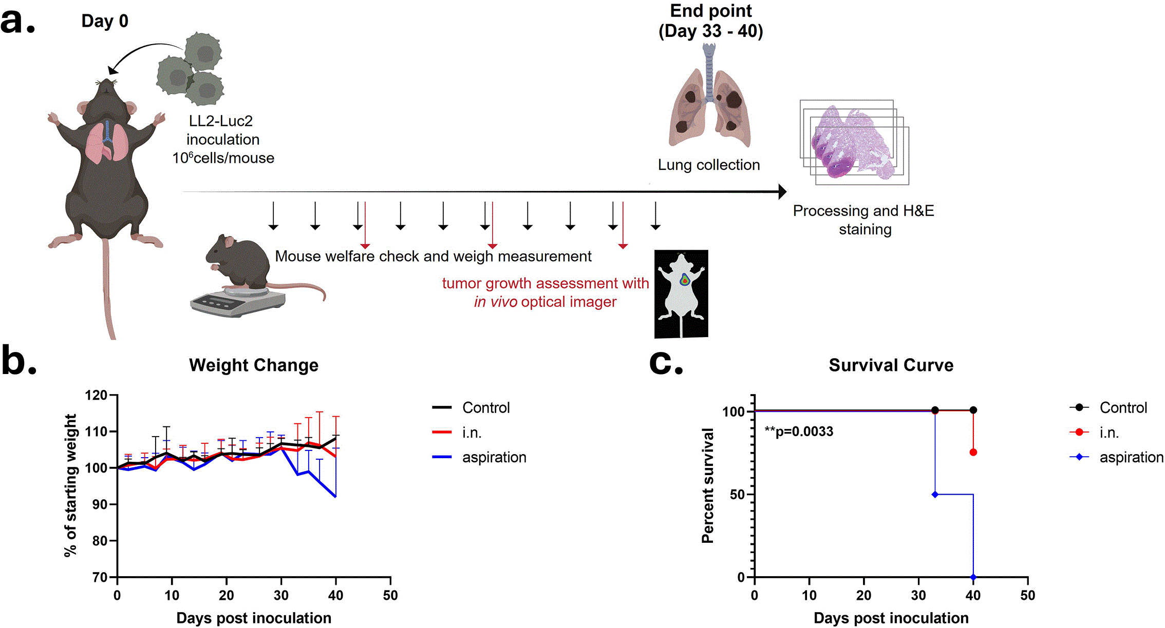
Figure 1. Inoculation of LL2-luc2 tumor cells by aspiration establishes orthotopic lung tumors.
(a) Experimental timeline illustrating tumor cell inoculation, longitudinal monitoring, and endpoint analysis. (b) Body weight change is shown as a percentage of the starting weight for control (n = 5), i.n. (n = 6) and aspiration (n = 6) inoculation and presented descriptively, inferential statistics are not reported. (c) Kaplan–Meier survival curves of control (n = 5), intranasal inoculation (n = 6), and aspiration inoculation (n = 6) groups. Statistical significance was assessed using the Mantel–Cox (log-rank) tests.* p < 0.05; ** p < 0.01; *** p < 0.001.
Without intervention, the health of mice in the aspiration-based inoculation and one mouse from the i.n. tumor engraftment group declined. By day 40 post-inoculation, all mice in the aspiration group reached the humane endpoint (Mantel–Cox log-rank test, p = 0.0033) (Figure 1c), whereas all the mice in the i.n. group, except one, showed no signs of tumor-related distress.
Bioluminescence imaging detects tumor progression with delayed signal emergence
We determined tumor engraftment and subsequent growth by weekly bioluminescence readings. Figure 2a shows a representative image of mice from aspiration-inoculated, i.n. – inoculated and control groups. No consistent tumor-associated BLI signal was observed at days 5 or 12 post-inoculation. Starting on day 19 post-engraftment, we observed a low-level BLI signal that intensified exponentially (Figure 2b, p < 0.0001). On day 33 p.i. mean BLI was 104 e−/s (Figure 2a, b). By day 40 p.i., mean BLI increased to 105 e−/s (Figure 2a,b).
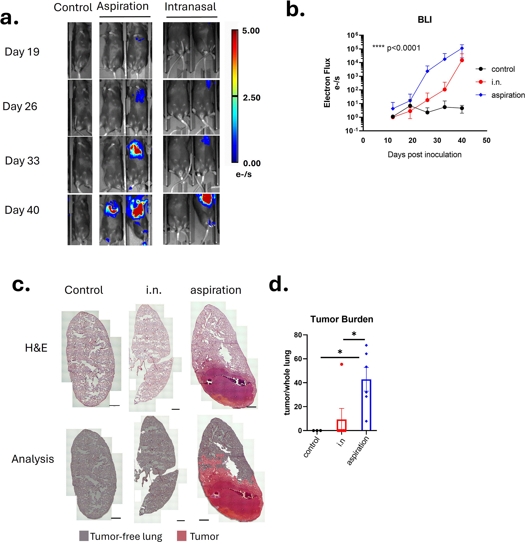
Figure 2. Bioluminescence imaging of tumor progression and pathological assessment of lung tumor burden.
(a) Representative bioluminescence images of the control (n = 5), aspiration inoculation (n = 6), and intranasal inoculation (n = 6) groups, acquired 15 minutes after D-luciferin administration on days 19, 26, 33, and 40 post-inoculation. (b) Bioluminescence signal is expressed as electron flux (e−/s) + 1. Data are shown as mean ± SD on a log10 y-axis. BLI data were analyzed using a mixed-effects model to assess group and time effects. The p-value represents the time × inoculation interaction in the ANOVA mixed-effects models. (c) Representative lung sections stained with hematoxylin and eosin (upper panels) and corresponding tumor versus tumor-free lung region annotations obtained from QuPath analysis (lower panels) Scale bar 1 mm. (d) Tumor burden is quantified as a percentage of tumor area relative to total lung area. Data are shown as mean ± SD. Statistical significance was assessed using the Kruskal–Wallis test. * p < 0.05; ** p < 0.01; *** p < 0.001.
Endpoint histopathology evaluation is a reference standard for orthotopic tumor verification
To verify tumor seeding and quantify tumor burden, the lungs of experimental groups were collected and analyzed with H&E staining (Figures 2c, 2d, and Extended data Figure 1).44 Histopathological examination demonstrated that the tumor is spindle-shaped, with fibrillar cells and anisokaryotic nuclei, eosinophilic cytoplasm, and irregular, hyperchromatic nuclei. Tumor growth is characterized by perivascular seeding, with progression toward areas of necrosis (Extended Data Figure 2).44 Tumor burden was calculated as the percentage of tumor area relative to total lung area using digital pathology (Figure 2c). Using the QuPath software from Bankhead
et al.,28 we trained the classifier to identify tumors and distinguish them from bronchioles, pulmonary veins, and healthy lung tissue (Extended Data Figure 3).44 Once we differentiated healthy lung tissue from the tumor, we calculated the tumor burden (Figure 2d). Aspiration-based inoculation resulted in a significantly elevated tumor burden compared to the i.n. group. In i.n. tumor-inoculated group, only one mouse had successful tumor inoculation, consistent with the BLI signal (Figure 2). While endpoint pathological examination remains the reference standard for tumor verification, we analyzed the correlation between tumor burden and two commonly used indicators for tumor progression: weight loss and bioluminescence. The correlation between tumor burden and weight loss (r = 0.7500, p = 0.066) was stronger than that between tumor burden and bioluminescence (r = 0.5714, p = 0.200), but neither was statistically significant (Extended Data Fig 4).44 Our results suggest that while BLI and weight loss reflect tumor progression, their correlation with quantitative tumor burden indicates that pathological examination remains the most reliable method for tumor assessment.
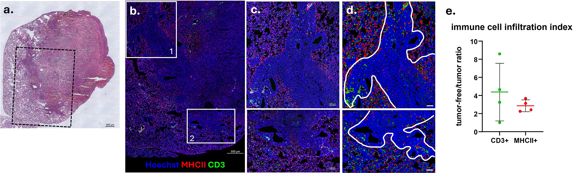
Figure 3. Immune cells located at the tumor borders in an aspiration-inoculated orthotopic lung cancer model.
(a) Hematoxylin and eosin–stained lung section showing tumor localization. Scale bar 500 μm. (b) Consecutive sections were stained for Hoechst, CD3 (AF647), and MHCII (PE). The fluorescence image corresponds to the black dashed rectangle in H&E image at (a). Scale bar 500 μm. (c) Magnified images of the tumor–lung borders, localization of CD3+ and MHCII+ cells. Scale bar 100 μm. (d) QuPath cell annotations illustrating immune marker–positive cells within tumor and tumor-free lung regions. Scale bar 100 μm. (e) The ratio of MHCII+ and CD3+ immune cell densities to the tumor-free and tumor regions is represented as the immune cell infiltration index (n = 4).
LL2-luc2 orthotopic tumors have limited immune cell infiltration
Solid tumors with minimal immune cell infiltration, immune-excluded tumors, are frequently associated with therapeutic resistance.29 Thus, we analyzed immune cell infiltration in our successfully seeded tumors by aspiration. We stained lung sections from the experimental groups with MHC II and CD3 antibodies to identify dendritic cells/B cells and T cells, respectively. Our analysis clearly showed that immune cells were located at the tumor borders, with limited infiltration into the tumor core (Figure 3). Measuring the MHCII+ and CD3+ cells in tumor-free regions over within tumors, described here as the immune cell infiltration index, showed that tumor-free regions were enriched in MHCII+ (2.26–3.68 fold) and CD3+ (1.02–8.59 fold) cells (Figure 3e). Our findings demonstrate that aspiration-based inoculation of the LL2-luc2 cell line establishes tumors with a limited intratumoral immune cell infiltration, providing a valuable approach for studying therapies against treatment-resistant solid tumors.
Discussion
Reliable orthotopic cancer models in immunocompetent mice with minimal technical variability in tumor inoculation are essential for studies of preclinical tumor-microenvironment interaction. In this study, we established and characterized an aspiration-based tumor inoculation method that resulted in a successful tumor engraftment in all inoculated mice and reached a humane endpoint at day 33 to 40 post-inoculation. In contrast, intranasal inoculation resulted in significantly lower inoculation success (1/6), as confirmed by endpoint histopathology. Importantly, we also found that non-invasive tumor seeding via aspiration was associated with limited intratumoral immune cell infiltration, consistent with an immune-excluded-like tumor microenvironment. Altogether, the new method of inoculating lung tumors offers a practical, non-invasive, consistent, and reliable way to study the TME.
Intrathoracic/intrapulmonary injections30–32 are commonly used methods to establish lung tumors. To provide a non-invasive alternative, Tanaka et al.27 adapted the intranasal route that is commonly used in pulmonary infection studies to inoculate Lewis Lung cancer cells (LL2). The study compared the efficacy of inoculation to more traditional engraftment methods, intratracheal and intrapulmonary injection, side by side. The authors reported that i.n. inoculation more closely mimics the progression of primary human lung tumors, which originate from alveolar regions rather than from a random lung parenchymal region.27 Building on this, we investigated whether aspiration, another respiratory delivery route frequently used to deliver biologics directly to the lung, could yield a consistent, reliable orthotopic lung tumor. We included a mock-inoculated control group and an intranasal engraftment group to compare the efficacy of our method with that reported in the current literature. We found that aspiration-based inoculation resulted in a significantly higher tumor incidence (6/6), whereas only 1 mouse in the intranasal group developed tumors (Figures 1 and 2). Survival in the aspiration group declined by day 30 post-inoculation, and all mice reached the humane endpoint by day 40 post-inoculation (Figure 1). LL2 was selected in our studies because LL2-induced tumors closely mimic human NSCLC,33 including the TME,22 have a relatively short doubling time in vitro, and are reported to contain a high proportion of cancer stem cells.34
Tumor aggressiveness is influenced by the inoculation dose.27 In intrapulmonary lung cancer models, the number of inoculated cells ranged from 104 to 5x10⁶ cells/mouse.30–32,35 The non-invasive inoculation routes, like i.n. and aspiration-based delivery are less precise than intrapulmonary inoculation. Tanaka et al. titrated the number of LLC cells. They found that mice receiving 107 cells/mouse died within 25 days post-inoculation, whereas 80% of mice inoculated with 106 cells/mouse survived, and all mice receiving the lowest concentration, 105 cells/mouse, survived.27 Based on these findings, we selected 106 cells/mouse, yielding reproducible tumor establishment under our experimental conditions.
OLCs established by intrapulmonary injections exhibited rapid tumor progression; the bioluminescence signal emerged at day 5 post-inoculation,31 and local tumor growth was detected as early as 2 weeks,32 3 weeks30 post-inoculation. In contrast, in our aspiration model, we detected the BLI signal at 19 days post-inoculation (Figure 2) and small tumor seeds as early as 3 weeks post-inoculation, as shown by histopathology analysis. In previously reported intrapulmonary models, the OLC experimental group without intervention reached the endpoint within 10–20 days post-inoculation. Tumor growth in our model was approximately 2-fold slower, coinciding with the endpoint at 40 days post-inoculation (Figure 1). Overall, this slower progression provides an extended window for investigating tumor–TME interactions.
Monitoring orthotopic tumor growth by bioluminescence imaging signals and CT scans is a valuable tool. Studies report that BLI is often helpful for tracking tumor progression in orthotopic models,36–38 but its correlation with true tumor burden is moderate and context-dependent.4,39–41 A review of preclinical tumor models, either ectopic or orthotopic, that assess tumor growth by BLI found that BLI is strongly correlated with tumor burden in subcutaneous tumors, but this correlation weakens as tumors grow within body cavities.38 Another study assessing the efficacy of BLI combined with MRI imaging of brain tumors in preclinical models found that early tumor progression is better reflected in BLI; however, the BLI vs. tumor burden curve plateaued due to hemorrhage and necrosis within the tumor that masked actual tumor severity, since BLI signals are coming from viable cells.42 Another glioblastoma study focusing on complementary methods to monitor tumor burden reported that BLI alone showed poor correlation with tumor burden due to reduced tumor viability, necrosis, hypoxia, and instability of luciferase constructs within the growing tumor, which could be interpreted as regression.40 Therefore, the tumor burden should be monitored using MRI combined with BLI. Similarly, in the orthotopic colon cancer model, the excised tumor weight correlated with MRI-derived tumor weight (R2 = 0.998), whereas its correlation with BLI was poor (R2 = 0.57).39 Consistent with the literature, the limited linear correlation between bioluminescence signal and histologically quantified tumor burden in our study likely reflects the diffuse and multifocal nature of lung tumors, as well as the known limitations of BLI in capturing total tumor mass in the lung.
Immune exclusion represents a common barrier to effective anti-tumor immunity in solid cancers and remains underrepresented in highly inflamed preclinical models. In our study, we determined that our OLC has limited intratumoral immune cell infiltration; we quantified immune cells localized predominantly at the tumor borders (Figure 3), consistent with previous reports in which GFP-conjugated LLC cells were injected directly into the lung.35 This spatial organization is consistent which demonstrate reduced intratumoral immune infiltration43 compared to ectopic models3,4 and reflects features commonly observed in human solid tumors.
This study has several limitations. The experimental group sizes were modest, and immune profiling was limited to selected markers to assess spatial distribution rather than comprehensive phenotyping. In addition, while BLI enabled longitudinal monitoring, quantitative tumor burden required endpoint histological analysis. Furthermore, the model was established using a single murine lung cancer cell line, and its applicability to other orthotopic lung cancer models remains to be determined.
In conclusion, we describe an aspiration-based syngeneic orthotopic lung cancer model that enables non-invasive and practical tumor establishment in immunocompetent mice. Compared with intranasal delivery, aspiration resulted in a markedly higher tumor inoculation rate and a consistent progression, while avoiding the technical complexity and tissue injury associated with intrapulmonary or intratracheal injection. Importantly, tumors established by aspiration exhibited a limited immune cell infiltration, with immune cells localized predominantly at tumor borders rather than within the tumor core. Together, these features position aspiration-based inoculation as easy to implement for orthotopic lung tumor modeling, particularly well-suited for preclinical studies of immune context- and microenvironment-targeted interventions.
Ethics and consent
All animal experimentation was approved by Izmir Biomedicine and Genome Center’s internal animal experimentation ethics committee (on March 13, 2025, protocol # 2025–007). Animal experiments were conducted in accordance with national law and EU Directive 2010/63/EU. The study did not involve human participants or tissue of human origin thus consent was not applicable.
Data availability statement
All data supporting the findings of this study are available within the paper and its Extended data. Also, the record was deposited in Zenodo: Aspiration-induced orthotopic lung cancer model: a non-invasive method for in vivo tumor microenvironment studies (DOI:10.5281/zenodo.21903877).44
The record contains raw data and statistical analyses, mouse-level information, Extended Data Figure 1, Figure 2, Figure 3 and Figure 4, Extended Data Table 1, Table 2 and the completed ARRIVE 2.0 author checklist.
Extended Data Fig 1. H&E-stained lung sections (upper panel) and QuPath analysis (lower panel) of the samples classified as tumor vs. tumor-free regions. Scale bar 1 mm.
Extended Data Fig 2. Representative images of lung tumors to investigate tumor morphology. Tumor border (scale bar 200 μm) (a), perivascular (within blue line) (scale bar 100 μm) (b), and necrotic (scale bar 200 μm) (c) tumor regions.
Extended Data Fig 3. QuPath analysis flow
Extended Data Fig 4. The association between the terminal bioluminescence signal (a) or weight loss (b) and histologically quantified tumor burden was evaluated using a two-tailed Spearman rank correlation test.
Extended Data Table 1. Mouse welfare scoring system used for animal monitoring and humane endpoint assessment.
Extended Data Table 2. Reagents, suppliers, and catalogue identifiers used in the study.
The files are publicly available under a CC BY 4.0 license.
Acknowledgment
We would like to thank Professor Serap Birincioğlu for her evaluation of tumor H&E samples, core facility specialist Aslı Seren from the optical imaging unit for her assistance with imaging troubleshooting, and Ece Uzun from the histopathology unit for H&E staining of lung sections. We would also like to extend our gratitude to the animal caretakers, Hamide Kaytaz and Hanim Güla, and the vivarium technicians, Burcu Kara, İbrahim Sevim, and Ulaş Saçıntı, for caring for the animals and for their assistance during our time of need.
This publication was created by benefiting from the 2236-B Marie Sklodowska-Curie Actions (MSCA) Cofund Scholarship Programs Contribution Fund Program (TUBITAK project ID: 123C460) and Horizon Europe MSCA Cofund Postdoctoral Program (EU project ID: 101126492). Views and opinions expressed are, however, those of the author(s) only and do not necessarily reflect those of the European Union or TUBITAK. Neither the European Union nor the granting authority can be held responsible for them.
References
- 1.
Lamplugh ZL, Wellhausen N, June CH, et al.:
Microenvironmental regulation of solid tumour resistance to CAR T cell therapy.
Nature Reviews Immunology. Nature Research.
2025; 26: 230–248. PubMed Abstract
| Publisher Full Text
- 2.
Stribbling SM, Beach C, Ryan AJ:
Orthotopic and metastatic tumour models in preclinical cancer research.
Pharmacol. Ther.
2024; 257. Elsevier Inc. PubMed Abstract
| Publisher Full Text
| Free Full Text
- 3.
Snipstad S, Bremnes F, Dehli Haugum M, et al.:
Characterization of immune cell populations in syngeneic murine tumor models.
Cancer Med.
2023; 12: 11589–11601. PubMed Abstract
| Publisher Full Text
| Free Full Text
- 4.
Mao M, Qiu K, Feng L, et al.:
Immunological dynamics in orthotopic compared with subcutaneous murine models of HPV-positive oropharyngeal cancer.
Dis. Model. Mech.
2025; 18: 18. PubMed Abstract
| Publisher Full Text
| Free Full Text
- 5.
Leiter A, Veluswamy RR, Wisnivesky JP:
The global burden of lung cancer: current status and future trends.
Nat. Rev. Clin. Oncol.
2023; 20: 624–639. Springer Nature. Publisher Full Text
- 6.
National Institute of Health Surveillance E and ERP (SEER):
Cancer Stat Facts: Lung and Bronchus Cancer.Reference Source
- 7.
Reck M, Frost N, Peters S, et al.:
Treatment of NSCLC after chemoimmunotherapy — are we making headway?.
Nat. Rev. Clin. Oncol.
2025; 22: 806–830. Springer Nature. Publisher Full Text
- 8.
Sorin M, Rezanejad M, Karimi E, et al.:
Single-cell spatial landscapes of the lung tumour immune microenvironment.
Nature.
2023; 614: 548–554. PubMed Abstract
| Publisher Full Text
| Free Full Text
- 9.
Zhang H, AbdulJabbar K, Moore DA, et al.:
Spatial Positioning of Immune Hotspots Reflects the Interplay between B and T Cells in Lung Squamous Cell Carcinoma.
Cancer Res.
2023; 83: 1410–1425. PubMed Abstract
| Publisher Full Text
| Free Full Text
- 10.
Wang Y, Zhou H, Ju S, et al.:
The solid tumor microenvironment and related targeting strategies: a concise review.
Front. Immunol.
2025; 16. Frontiers Media SA. PubMed Abstract
| Publisher Full Text
| Free Full Text
- 11.
Oser MG, MacPherson D, Oliver TG, et al.:
Genetically-engineered mouse models of small cell lung cancer: the next generation.
Oncogene.
2024; 43: 457–469. Springer Nature. Publisher Full Text
- 12.
Gazdar AF, Hirsch FR, Minna JD:
From mice to men and back: An assessment of preclinical model systems for the study of lung cancers.
J. Thorac. Oncol.
2016; 11: 287–299. Lippincott Williams and Wilkins. Publisher Full Text
- 13.
Woo XY, Srivastava A, Mack PC, et al.:
A Genomically and Clinically Annotated Patient-Derived Xenograft Resource for Preclinical Research in Non-Small Cell Lung Cancer.
Cancer Res.
2022; 82: 4126–4138. PubMed Abstract
| Publisher Full Text
| Free Full Text
- 14.
Huo KG, D’Arcangelo E, Tsao MS:
Patient-derived cell line, xenograft and organoid models in lung cancer therapy.
Transl Lung Cancer Res.
2020; 9: 2214–2232. PubMed Abstract
| Publisher Full Text
| Free Full Text
- 15.
Tanaka R, Yoshinouchi S, Karouji K, et al.:
A mouse model of lung cancer induced via intranasal injection for anticancer drug screening and evaluation of pathology.
FEBS Open Bio.
2023; 13: 51–59. PubMed Abstract
| Publisher Full Text
| Free Full Text
- 16.
Gregório AC, Fonseca NA, Moura V, et al.:
Inoculated cell density as a determinant factor of the growth dynamics and metastatic efficiency of a breast cancer murine model.
PLoS One.
2016; 11: 11. PubMed Abstract
| Publisher Full Text
| Free Full Text
- 17.
Greenlee JD, King MR:
A syngeneic MC38 orthotopic mouse model of colorectal cancer metastasis.
Biol Methods Protoc.
2022; 7: 7. PubMed Abstract
| Publisher Full Text
| Free Full Text
- 18.
Chen C, Neumann J, Kühn F, et al.:
Establishment of an endoscopy-guided minimally invasive orthotopic mouse model of colorectal cancer.
Cancers (Basel).
2020; 12: 1–16. PubMed Abstract
| Publisher Full Text
| Free Full Text
- 19.
Lee SB, Jeon HJ, Hyun H, et al.:
Establishment of an Orthotopic and Metastatic Colorectal Cancer Mouse Model Using a Tissue Adhesive-Based Implantation Method.
Cancers (Basel).
2025; 17: 17. PubMed Abstract
| Publisher Full Text
| Free Full Text
- 20.
Rovers S, Farahmand P, Liu D, et al.:
A Minimally Invasive Transthoracic Injection Technique for Reproducible Intrapleural Delivery in Mice.
Methods Protoc.
2025; 8: 8. PubMed Abstract
| Publisher Full Text
| Free Full Text
- 21.
Xu JJ, Lucero MY, Herndon NL, et al.:
Comparison of a Minimally Invasive Transthoracic Approach and a Surgical Method for Intrapleural Injection of Tumor Cells in Mice.
Comp. Med.
2023; 73: 120–126. PubMed Abstract
| Publisher Full Text
| Free Full Text
- 22.
Yamada-Hara M, Takahashi N, Byun JW, et al.:
In Vivo Bioluminescence Imaging of Tumor Progression in the Lewis Lung Carcinoma Orthotopic Mouse Model: A Comparison Between the Tail Vein Injection and Intranasal Instillation Methods.
Curr Protoc.
2024; 4: e70071. PubMed Abstract
| Publisher Full Text
| Free Full Text
- 23.
Anselmo AC, Gokarn Y, Mitragotri S:
Non-invasive delivery strategies for biologics.
Nat. Rev. Drug Discov.
2018; 19–40. Nature Publishing Group. Publisher Full Text
- 24.
Kinaret P, Ilves M, Fortino V, et al.:
Inhalation and Oropharyngeal Aspiration Exposure to Rod-Like Carbon Nanotubes Induce Similar Airway Inflammation and Biological Responses in Mouse Lungs.
ACS Nano.
2017; 11: 291–303. PubMed Abstract
| Publisher Full Text
- 25.
Shrum B, Anantha RV, Xu SX, et al.:
A robust scoring system to evaluate sepsis severity in an animal model.
BMC. Res. Notes.
2014; 7: 7. PubMed Abstract
| Publisher Full Text
| Free Full Text
- 26.
Özkan M, Eskiocak YC, Wingender G:
Macrophage and dendritic cell subset composition can distinguish endotypes in adjuvant-induced asthma mouse models.
PLoS One.
2021; 16: e0250533–e0250519. PubMed Abstract
| Publisher Full Text
| Free Full Text
- 27.
Tanaka R, Yoshinouchi S, Karouji K, et al.:
A mouse model of lung cancer induced via intranasal injection for anticancer drug screening and evaluation of pathology.
FEBS Open Bio.
2023; 13: 51–59. PubMed Abstract
| Publisher Full Text
| Free Full Text
- 28.
Bankhead P, Loughrey MB, Fernández JA, et al.:
QuPath: Open source software for digital pathology image analysis.
Sci. Rep.
2017; 7: 16878. PubMed Abstract
| Publisher Full Text
| Free Full Text
- 29.
Wu B, Zhang B, Li B, et al.:
Cold and hot tumors: from molecular mechanisms to targeted therapy.
Signal Transduct. Target. Ther.
2024; 9: 274. Springer Nature. PubMed Abstract
| Publisher Full Text
| Free Full Text
- 30.
Gan W, He YM, Hu FL, et al.:
Establishment of an orthotopic model of lung cancer by transthoracic lung puncture using tumor fragments.
J. Thorac. Dis.
2023; 15: 2012–2021. PubMed Abstract
| Publisher Full Text
| Free Full Text
- 31.
Tran Chau V, Liu W, Gerbé de Thoré M, et al.:
Differential therapeutic effects of PARP and ATR inhibition combined with radiotherapy in the treatment of subcutaneous versus orthotopic lung tumour models.
Br. J. Cancer.
2020; 123: 762–771. PubMed Abstract
| Publisher Full Text
| Free Full Text
- 32.
Poczobutt JM, De S, Yadav VK, et al.:
Expression Profiling of Macrophages Reveals Multiple Populations with Distinct Biological Roles in an Immunocompetent Orthotopic Model of Lung Cancer.
J. Immunol.
2016; 196: 2847–2859. PubMed Abstract
| Publisher Full Text
| Free Full Text
- 33.
He Q, Sun C, Pan Y:
Whole-exome sequencing reveals Lewis lung carcinoma is a hypermutated Kras/Nras–mutant cancer with extensive regional mutation clusters in its genome.
Sci. Rep.
2024; 14: 14. PubMed Abstract
| Publisher Full Text
| Free Full Text
- 34.
Wang J, Sun Z, Liu Y, et al.:
Comparison of tumor biology of two distinct cell sub-populations in lung cancer stem cells.2017; 8: 96852–96864. PubMed Abstract
| Publisher Full Text
| Free Full Text
Reference Source
- 35.
Weiss ID, Ella E, Dominsky O, et al.:
In the hunt for therapeutic targets: Mimicking the growth, metastasis, and stromal associations of early-stage lung cancer using a novel orthotopic animal model.
J. Thorac. Oncol.
2015; 10: 46–58. PubMed Abstract
| Publisher Full Text
- 36.
Klerk CPW, Overmeer RM, Niers TMH, et al.:
Validity of bioluminescence measurements for noninvasive in vivo imaging of tumor load in small animals.
BioTechniques.
2007; 43: 7–13, 30. PubMed Abstract
| Publisher Full Text
- 37.
Tuli R, Surmak A, Reyes J, et al.:
Development of a novel preclinical pancreatic cancer research model: Bioluminescence image-guided focal irradiation and tumor monitoring of orthotopic xenografts.
Transl. Oncol.
2012; 5: 77–84. PubMed Abstract
| Publisher Full Text
| Free Full Text
- 38.
Shen YT, Asthana R, Peeters C, et al.:
Potential Limitations of Bioluminescent Xenograft Mouse Models: A Systematic Review.
J Pharm Pharm Sci (www.cspsCanada.org).
2020; 23: 177–199. PubMed Abstract
| Publisher Full Text
Reference Source
- 39.
Ravoori MK, Margalit O, Singh S, et al.:
Magnetic Resonance Imaging and Bioluminescence Imaging for Evaluating Tumor Burden in Orthotopic Colon Cancer.
Sci. Rep.
2019; 9: 6100. PubMed Abstract
| Publisher Full Text
| Free Full Text
- 40.
Le TNT, Lim H, Hamilton AM, et al.:
Characterization of an Orthotopic Rat Model of Glioblastoma Using Multiparametric Magnetic Resonance Imaging and Bioluminescence Imaging.
Tomography.
2018; 4: 55–65. Publisher Full Text
- 41.
Sechrist ZR, Lee G, Schwarz EM, et al.:
Validation of dual energy X-ray absorptiometry for longitudinal quantification of tumor burden in a murine model of pancreatic ductal adenocarcinoma.
PLoS One.
2024; 19: e0292196. PubMed Abstract
| Publisher Full Text
| Free Full Text
- 42.
Jost SC, Collins L, Travers S, et al.:
Measur ing brain tumor growth: Combined bioluminescence imaging-magnetic resonance imaging strategy.
Mol. Imaging.
2009; 8: 245–253. PubMed Abstract
| Publisher Full Text
| Free Full Text
- 43.
Na M, Kang H, Kim N, et al.:
Characterization of an orthotopic mouse transplant model reveals early changes in the tumor microenvironment of lung cancer.
BMB Rep.
2024; 57: 484–489. PubMed Abstract
| Publisher Full Text
| Free Full Text
- 44.
Özkan M, Evcili İ, Güngör B:
Aspiration-induced orthotopic lung cancer model: a non-invasive method for in vivo tumor microenvironment studies. [Dataset].
Zenodo.
2026. Publisher Full Text









