Introduction
Streptococcus suis is an encapsulated, facultative anaerobic, Gram-positive coccus that commonly colonizes the upper respiratory tract of pigs, particularly the tonsils and nasal cavity.1–3 Although it is part of the normal respiratory microbiota of its host, S. suis is also one of the most important bacterial pathogens in swine production. When this pathogen spreads systemically, it can be responsible for a wide range of pathological manifestations, including meningitis, septicemia, arthritis, and endocarditis, leading to significant health, welfare and economic losses.2,4
S. suis displays high phenotypic and genomic diversity, comprising numerous capsular serotypes and genetic lineages that vary in pathogenic potential, with serotype 2 most frequently associated with invasive disease in pigs and zoonotic infections in humans.1,2,5 The clinical outcome of the S. suis infection depends on the virulence of the infecting strain and the host context.3,6,7
Beyond its impact on animal health, S. suis is an emerging zoonotic pathogen, particularly in Asia, where it has been recognized as a major cause of invasive human infection.2,8 This dual role highlights its relevance within a One Health framework, linking animal, human, and environmental health.9
In this context, antimicrobial resistance (AMR) in S. suis has become an increasing concern,4,10 since multidrug-resistant (MDR) strains have increasingly been reported.11 Control of this pathogen in swine production systems has historically relied heavily on antimicrobial use, due to the limited availability of broadly effective vaccines that restricts preventive strategies.4 As a result, selective pressure has contributed to the emergence and dissemination of resistant strains, particularly high rates against tetracyclines, macrolides, and lincosamides.4,10
From a global perspective, AMR in S. suis is part of the broader problem of MDR bacteria, defined as non-susceptibility to multiple antimicrobial categories.12 Under a One Health approach, the use of antimicrobials in animal production, together with the horizontal transfer of resistance genes mediated by mobile genetic elements, facilitates the spread of resistance traits among animals, humans and the environment.9 In this scenario, S. suis not only represents a pathogenic threat, but also acts as a reservoir and vehicle for resistance determinants, reinforcing its epidemiological importance.10,13,14
In light of the growing challenge of antimicrobial resistance, the search for new anti-infective compounds has regained considerable attention, particularly those derived from natural sources.15 Actinomycetes have historically represented one of the most important reservoirs of naturally occurring antimicrobial compounds and continue to be a privileged source of biosynthetic diversity for the discovery of new inhibitory molecules.16–19 In recent years, research efforts have increasingly focused on rare Actinomycetes and other underexplored bacterial taxa, as these microorganisms offer a greater likelihood of producing metabolites with novel chemical scaffolds and less redundant mechanisms of action than those observed in more conventional microbial collections.17,20,21 This shift in focus is largely driven by the need to overcome the persistent issue of rediscovery of known compounds, which has historically limited the efficiency of traditional antimicrobial screening approaches.17,19 Within this framework, the exploration of rare and underrepresented bacteria presents several strategic advantages and the possibility of diversifying current anti-infective development pipelines.17,19,21 On one hand, it expands the accessible chemical space for the identification of bioactive compounds with inhibitory activity against pathogenic microorganisms.22,23 On the other hand, it enables the integration of complementary approaches, such as taxonomic diversity, metabolomic profiles, and dereplication tools, to prioritize extracts and strains with a higher probability of yielding truly novel metabolites.19,24
Despite the recognized potential of Actinomycetes as a source of antimicrobial compounds,19 relatively limited attention has been given to their application against S. suis, particularly in the context of antimicrobial-resistant strains.4 This gap highlights the need for targeted studies aimed at identifying novel bioactive metabolites with activity against this pathogen. Therefore, the aim of this study was to evaluate the antimicrobial activity of Actinomycetes-derived extracts against S. suis, with the objective of identifying novel inhibitory molecules with potential relevance for the control of this clinically and economically important pathogen.
Methods
Bacterial strains and cultivation of bacterial strains
Two S. suis strains representing the two serotypes most commonly associated with clinical cases in Europe were used in this study. The reference strain P1/7 (serotype 2, frequent in pigs and associated with clinical cases in humans) was kindly provided by Dr. Marcelo Gottschalk (Université de Montreal, Canada) and has been previously described.25 The second strain, 895-LCR (serotype 9), originates from CReSA’s collection and was isolated from cerebrospinal fluid obtained from pigs with meningitis. Both strains were initially cultured on chocolate PolyViteX agar plates (bioMérieux, France) and subsequently grown for the assays in Todd-Hewitt Broth (T1438, Sigma-Aldrich, Merck, Germany) supplemented with 2% (w/v) yeast extract (70161, Sigma-Aldrich, Merck, Germany) (THY).26
Antimicrobial susceptibility testing
Antimicrobial susceptibility of S. suis strains was performed by determining the minimum inhibitory concentration (MIC) of selected antibiotics using the broth microdilution method, in accordance with the guidelines of the Clinical and Laboratory Standards Institute (CLSI). As no species-specific clinical breakpoints are available for S. suis, MIC results were interpreted following the breakpoints of the Viridans group streptococci (or Streptococcus pneumoniae when the value was not available for the Viridans group) found in CLSI document M100–27 (2017).
Briefly, serial dilutions appropriate for each antibiotic were prepared from stock solutions at 10 mg/mL and diluted in THY. Stock solutions were prepared in appropriate solvents according to their solubility, including dimethyl sulfoxide (DMSO) (D8418, Sigma-Aldrich, Merck, Germany) when required. To evaluate the potential effect of DMSO on bacterial growth, the solvent was tested separately over a range of concentrations under the same experimental conditions.
For the assay, each well was inoculated with 90 μL of a bacterial suspension adjusted to a final concentration of 1 x 10⁵ CFU/mL and 10 μL of the corresponding antibiotic dilution. Wells containing no antibiotic served as growth controls, while wells containing medium only were used as sterility controls. Plates were incubated at 37 °C for 24 h in a Synergy 2 microplate reader (Biotek), and optical density was measured at 600 nm (OD600). The MIC was defined as the lowest concentration of antibiotic that completely inhibited visible bacterial growth compared to the growth control.
The antibiotics tested included representatives from different antimicrobial classes: penicillin G and cefuroxime (β-lactams), chloramphenicol (amphenicols), clindamycin (lincosamides), erythromycin (macrolides), linezolid (oxazolidinones), levofloxacin (quinolones), rifampicin (ansamycins), tetracycline hydrochloride (tetracyclines), and telithromycin (macrolide-ketolides). All antibiotics were obtained from commercial sources (Table S1; Sigma-Aldrich, Merck, Germany) and prepared according to the manufacturer’s instructions.
Actinomycetes extracts
The NAICONS strain collection, its origin and composition, have been described before.17 Full extracts from randomly selected strains were prepared as described.24 Crude extracts were obtained through standardized high-quality extraction procedures from different Actinomycetes genera (e.g., Streptomyces, Nocardia, Micromonospora, among others). For plate preparation, dried extracts derived from strains of the NAICONS microbial collection (NAICONS, Milan, Italy) were employed. Extract plates were prepared following a protocol adapted from previously described methodologies.24,27
Briefly, a 10 mL culture was processed to generate both whole and fractionated extracts. For crude extracts, 2 mL of culture were mixed with 4 mL of ethanol, incubated under agitation for 1 h at 30 °C and subsequently centrifuged (4000 rpm, 8 min), with the supernatant collected as full extract.
For fractionation, the remaining 8 mL of culture, were separated into mycelium and supernatant. The mycelial pellet was sequentially extracted with ethanol at increasing concentrations (20%, 50% and 100%; 4 mL each), with shaking for 1 h at 30 °C and centrifugation (4000 rpm, 8 min) after each step to obtain mycelium-associated fractions.
In parallel, the supernatant was incubated with a HP20 resin (0.8 mL, 1 h, 30 °C) to adsorb extracellular metabolites. After centrifugation (3000 rpm, 1 min), the resin was sequentially eluted with ethanol solutions of increasing concentration (5%, 30% and 80%) with incubation steps (15 min, 30 °C) and centrifugation (3000 rpm, 1 min) between each elution.
All extracts were dried overnight at room temperature and stored at 4 °C until use.
Screening of Actinomycetes extracts for antimicrobial activity against S. suis
A total of 800 Actinomycetes-derived extracts were screened with S. suis strains P1/7 and 895-LCR. Dried extracts were initially resuspended in 12.5 μL of dimethyl sulfoxide (DMSO) per well in a 96-well plate. Subsequently, 112.5 μL of autoclaved MilliQ water were added to obtain a stock solution containing 10% (v/v) DMSO.
For the antimicrobial assays, 10 μL of each stock solution of full extracts were transferred to a new 96-well plate containing 90 μL of bacterial culture adjusted to 1 x 10⁵ CFU/mL. This resulted in a final volume of 100 μL per well and a final DMSO concentration of 1% (v/v). Assays were performed independently for each Streptococcus suis strain. Each plate included a growth control consisting of 10 μL of 10% DMSO and 90 μL of bacterial culture, and a second control consisting of a chloramphenicol dilution series at known concentrations. Plates were incubated at 37 °C under static conditions and bacterial growth was monitored by measuring OD600 at 0 and 24 h. Growth inhibition was calculated by normalizing the OD600 values to the growth control as indicated in the formula below:
An 80% inhibition threshold was used to define active extracts. This arbitrary cut-off was selected to prioritize extracts showing strong antibacterial activity and to facilitate hit identification. In some cases, OD decreased with incubation time, yielding an inhibition value higher than 100%. For clarity, these values are reported as 100% inhibition.
Selection and fractionation of active extracts
Selected extracts were subjected to fractionation following a previously described HPLC-based approach.27 Briefly, the extracts were separated into fractions by reverse-phase high-performance liquid chromatography (HPLC). Fractionation was performed using a Dionex UltiMate 300 system (ThermoFisher) equipped with a Merck LiChrospher RP-18 reverse-phase column (5 μm, I.D. x L 4.6 x 125 mm, Merck, Darmstadt, Germany) maintained at 50 °C. The mobile phase consisted of 0.05% formic acid in water (phase A) and acetonitrile (phase B), with a flow rate of 1 mL/min. Compounds were eluted using a linear gradient from 10% to 95% acetonitrile over 18 min, followed by a 5 min hold at 95%. UV absorbance was monitored at 230 and 270 nm. For each selected extract, 24 fractions were collected at one-minute intervals. The fractions were then dried using a speed vacuum concentrator and subsequently resuspended in 10% DMSO for biological evaluation using the screening assay described above.
Fractions displaying antimicrobial activity were selected for further analysis by high-resolution mass spectrometry (HRMS) to identify the active compound(s). To help identify the active compounds, adjacent inactive fractions (immediately preceding and following the active fraction) were also included in the LC-HRMS analyses, which were performed as described before,24 using a Vanquish UHPLC system coupled to an Orbitrap Exploris 120 mass spectrometer, equipped with a YMC-Triart ODS column. The mobile phase consisted of 0.1% formic acid in water (A), LC-MS grade acetonitrile (B), and LC-MS grade isopropanol (C), using a gradient program as previously described.23 The flow rate was set to 0.8 mL/min, with an injection volume of 8 μL and a column temperature of 40 °C. During separation, the flow was split, directing 75% to a diode array detector and 25% to the mass spectrometer, which operated with a heated electrospray ionization source in both positive and negative modes. MS/MS data were subsequently analyzed and compared against the Molecules Gateway and external spectral databases to facilitate compound identification.24,28–31
Results
Antimicrobial susceptibility profiles and DMSO concentration optimization in S. suis
Preliminary assays with antibiotics from different classes at a range of concentrations were performed to optimize the screening conditions for S. suis (Figure S1 and S2). These experiments were also used to establish the maximum non-interfering concentration of DMSO for subsequent assays.
The reference strain S. suis P1/7 exhibited high susceptibility to all antibiotics tested (Figure S1;
Table 1), consistent with previous reports.32 In contrast, the clinical isolate 895-LCR displayed an MDR phenotype (Figure S2;
Table 1), showing resistance to erythromycin (macrolides), clindamycin (lincosamides), and tetracycline (tetracyclines), together with higher MIC for penicillin (penicillins), cefuroxime (cephems) and telithromycin (macrolide-ketolides) than P1/7. Other antimicrobial agents, including linezolid, rifampicin, and fluoroquinolones, retained activity against this isolate. This pattern is consistent with resistance profiles commonly described in clinical S. suis isolates32 and suggests the presence of acquired resistance mechanisms.
Table 1. Minimum inhibitory concentration (MIC) of antibiotics for reference Streptococcus suis strain P1/7 and clinical isolate 895-LCR.
| Antibiotics | MIC (μg/mL) |
|---|
| P1/7 |
895-LCR |
|---|
| Penicillin | 0.06 (S) | ≥ 1# |
| Cefuroxime | 0.03 (S) | 0.125 (S) |
| Erythromycin | ≤ 0.06 (S) | > 32 (R) |
| Telithromycin* | ≤ 0.06 (S) | 1 (S) |
| Clindamycin | 0.03 (S) | > 1 (R) |
| Chloramphenicol | 1 (S) | 2 (S) |
| Linezolid | 1 (S) | 1 (S) |
| Levofloxacin | 1 (S) | 1 (S) |
| Rifampicin* | ≤ 0.06 (S) | ≤ 0.06 (S) |
| Tetracycline | 0.25 (S) | 32 (R) |
The effect of DMSO on bacterial growth was evaluated across a range of concentrations in both strains, revealing a dose-dependent inhibitory effect (Figure S1 y S2). High concentrations resulted in marked growth inhibition, which progressively decreased as the solvent concentration was reduced. At 2.5% DMSO, inhibition was minimal, while at lower concentrations it was virtually absent. Based on these results, 1% DMSO was considered suitable for solubilization of extracts and was selected for all subsequent experiments.
Screening of Actinomycetes extracts against S. suis
A direct screening approach was performed to identify extracts with antimicrobial activity against the S. suis strains P1/7 and 895-LCR. The inhibitory activity of the tested extracts showed a broad distribution, ranging from non-detectable effect to strong growth inhibition (Figure S3).
To prioritize candidates for further characterization, extracts displaying high antimicrobial activity were selected using a threshold of >80% growth inhibition. Based on this criterion, 142 extracts were identified as highly active against the reference strain P1/7. A similar analysis performed with the clinical strain 895-LCR resulted in the identification of 123 extracts exceeding the same inhibition threshold. In both cases, highly active extracts formed a clearly defined subset, distinguishable from the majority of extracts showing low or moderate inhibitory effects. From the active extracts, 117 inhibited both strains, while 25 extracts inhibited only P1/7 and 6 inhibited only 895-LCR. These findings highlight the presence of a significant fraction of bioactive compounds within the screened collection and provide a focused set of candidates for subsequent analyses.
Selection and fractionation of active extracts
Out of the 117 active extracts that inhibited both S. suis strains (P1/7 and 895-LCR), 14 extracts did not contain - as listed in the Molecules Gateway24 - known antimicrobial compound that could account for the observed activity. For further analysis, we selected four extracts (from the 14 above) from rare Actinomycetes taxa (73990, 78443, 78450 and 78471), four additional extracts from Streptomyces (72356, 77387, 78495 and 88546) and one extract that contained deferoxamine, a chelating compound (78455).
These selected extracts were subjected to HPLC fractionation, generating 24 fractions per extract, which were subsequently evaluated for antimicrobial activity against the P1/7 strain (
Figure 1). Analysis of the bacterial growth curves in the presence of fractions derived from extracts 78443, 78455, and 88546 showed that antimicrobial activity was restricted to specific fractions, while most fractions did not inhibit bacterial growth and displayed growth curves comparable to the untreated control. For extract 78471, no fractions exhibiting full inhibitory activity were identified, and only one fraction caused a delay in bacterial growth, indicating partial inhibitory effects. Finally, none of the fractions derived from extracts 72356, 73990, 77387, 78450 and 78495 inhibited bacterial growth.
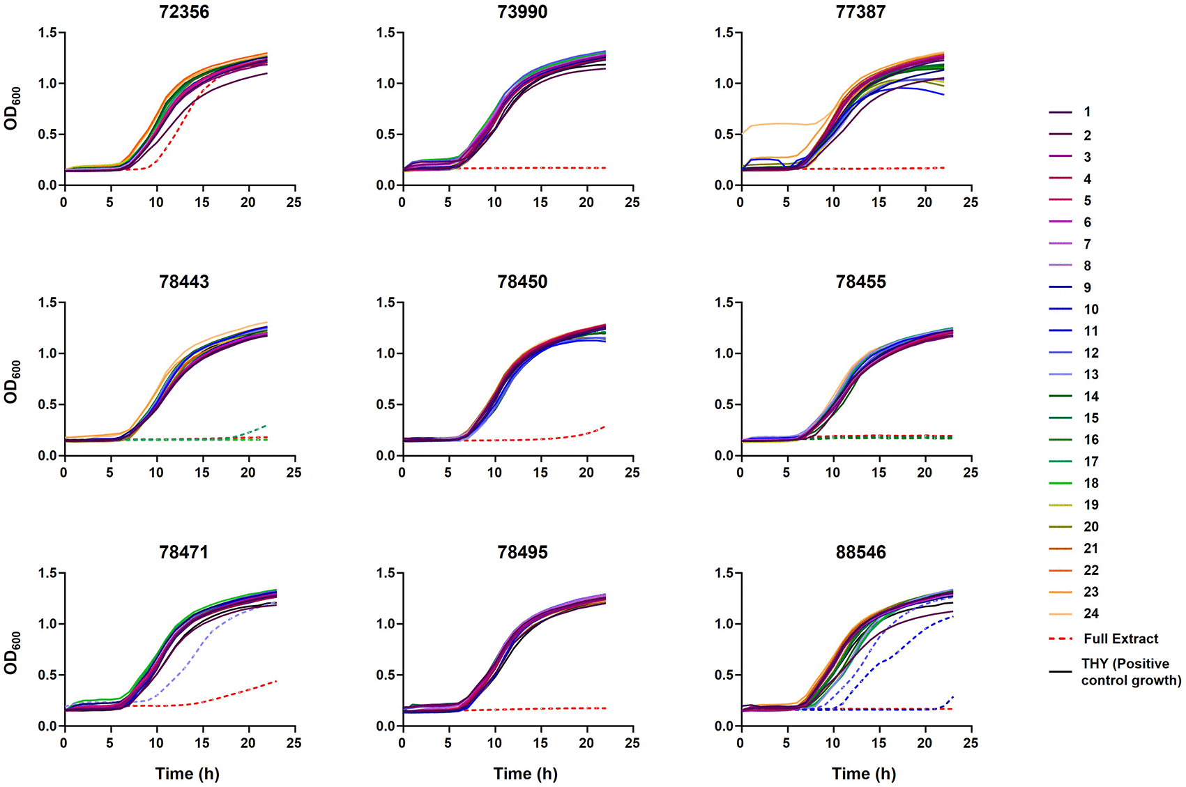
Figure 1. Growth curves of Streptococcus suis strain P1/7 in the presence of extract fractions.
S. suis strain P1/7 was cultured in the presence of the 24 fractions obtained from the selected extracts. Bacterial growth was monitored over 24 hours by measuring optical density at 600 nm (OD600). Dashed lines represent fractions that exhibited partial or complete inhibition of bacterial growth. The following controls were included: untreated P1/7 grown in THY (positive growth control) and P1/7 cultured in the presence of the full extract. The title of each graph corresponds to the extract analyzed.
Overall, only four out of the nine fractionated extracts yielded fractions with detectable antimicrobial activity (
Figure 1), whereas no active fractions were identified for the remaining five extracts. This suggests that, in some cases, the antimicrobial activity observed in the raw extracts may depend on the combined or synergistic effect of multiple components, which could be lost upon fractionation. Alternatively, the active compounds were not eluted from the column under the conditions used, may be present at concentrations below the detection threshold after separation or may exhibit reduced stability during the fractionation process.
Based on the activity profiles obtained for the P1/7 strain, the fractions of the four extracts showing antimicrobial activity were selected for further analysis against the clinical strain 895-LCR. Instead of testing all 24 fractions per extract, a subset of eight fractions per extract was selected, including those displaying complete or partial inhibition, as well as their immediately adjacent fractions, to better delineate the active regions (
Figure 2). In contrast to the results observed for the P1/7 strain, only one of the tested fractions (from extract 88546) resulted in complete growth inhibition in 895-LCR. The remaining fractions did not fully inhibit bacterial growth but instead induced a delay in growth, indicating a partial inhibitory effect. These results suggest that, although the identified fractions retain antimicrobial activity against both strains, their effect is less pronounced in the clinical isolate 895-LCR.
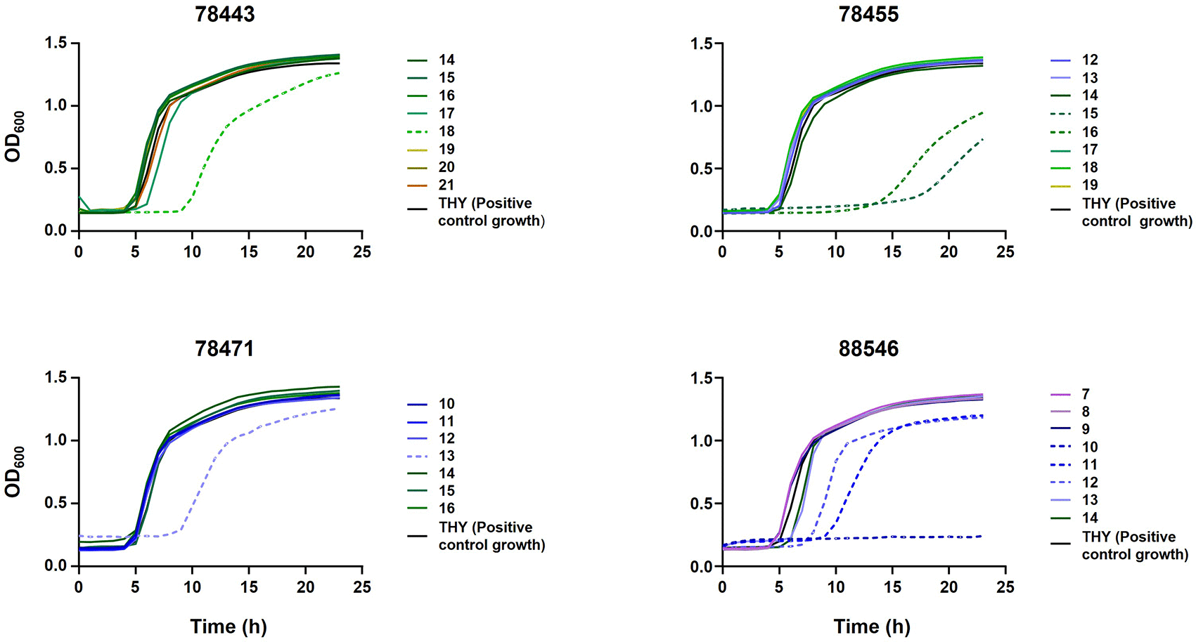
Figure 2. Growth curves of Streptococcus suis strain 895-LCR in the presence of selected extract fractions.
S. suis 895-LCR was cultured in the presence of selected fractions from the four extracts that previously exhibited antimicrobial activity against strain P1/7. For each extract, 7 to 8 fractions were evaluated, including those that had shown complete or partial inhibition of bacterial growth in P1/7, together with their immediately adjacent fractions. Bacterial growth was monitored over 24 hours by measuring optical density at 600 nm (OD600). Dashed lines represent fractions that exhibited partial or complete inhibition of bacterial growth. The following control was included: untreated 895-LCR grown in THY (positive growth control). The title of each graph corresponds to the extract analyzed.
Identification of bioactive compounds by HRMS
A total of eight selected bioactive fractions, together with the corresponding inactive flanking fractions, were subjected to high-resolution mass spectrometry (HRMS) analysis to identify the compounds responsible for the observed antimicrobial activity (
Table 2).
Table 2. Selected extracts for fractionation and further analysis, together with their main characteristics (percentage of inhibition of the growth of S. suis strains P1/7 and 895-LCR, Actinomycetes genus used to produce the extract [producer] and compound identified).
| | % of growth inhibition | |
|---|
| Extract | Producer | P1/7 | 895-LCR | Compound |
|---|
| 73990 | Amycolatopsis
| 100 | 100 | N.D. |
| 78443 | Actinomadura
| 100 | 100 | Verucopeptin |
| 78450 | Micromonospora
| 100 | 100 | N.D. |
| 78455 | Micromonospora
| 100 | 100 | Sch 18640 |
| 78471 | Micromonospora
| 100 | 100 | 8-Deoxy-3-hydroxyrifamycin S |
| 72356 | Streptomyces
| 100 | 100 | N.D. |
| 77387 | Streptomyces
| 100 | 95 | N.D. |
| 78495 | Streptomyces
| 100 | 100 | N.D. |
| 88546 | Streptomyces
| 100 | 100 | Not clear |
For extract 78443, fractions 17 and 18 were analyzed (
Figure 3). LC-MS analysis of these fractions revealed a major ion at m/z 918.515+ with a retention time of 12.91 min, absent in the inactive fractions. The corresponding full mass acquired in negative ionization mode showed the presence of a signal at m/z 894.5194−. The comparison of these data revealed that m/z 918.515 was a sodium adduct, corresponding to a neutral mass of 895.5253 Dalton (Da). This value matches the reported molecular mass of verucopeptin, an antitumor antibiotic isolated from Actinomadura verrucosospora in 1993.33 The obtained MS and MS2 behaviors were consistent with the proposed structure, providing additional support for its identification. Furthermore, taxonomic annotation supports this assignment, since extract 78443 was prepared from a culture of an Actinomadura strain as the original verucopeptin producer (
Figure 3).
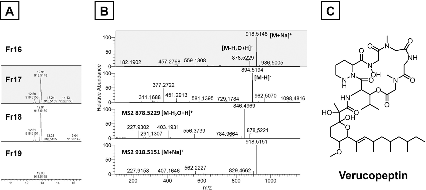
Figure 3. Identification of verucopeptin in extract 78443.
Panel A: Extracted ion chromatograms of m/z 918.5148+ from active (17 and 18) and inactive (16 and 19) fractions from extract 78443. Panel B, from top to down: Positive and negative Full Mass spectra at 12.9 minutes in fraction 18, MS2 spectra of the dehydrated and sodium adducts. Panel C: Chemical structure of verucopeptin.
For extract 78455, fractions 15 and 16 were analyzed (
Figure 4). LC-MS analysis revealed an ion at m/z 772.7280+2 with a retention time of 9.7 min and 841.7505+2 with a retention time of 10.3 min. Based on the observed masses and fragmentation patterns, these compounds are tentatively assigned as Sch 18640 thiopeptide and its congener lacking 2 dehydroalanines at the C-terminal position. These compounds are thiostrepton-type peptides isolated from a Micromonospora arborensis strain in 198134 (
Figure 4).
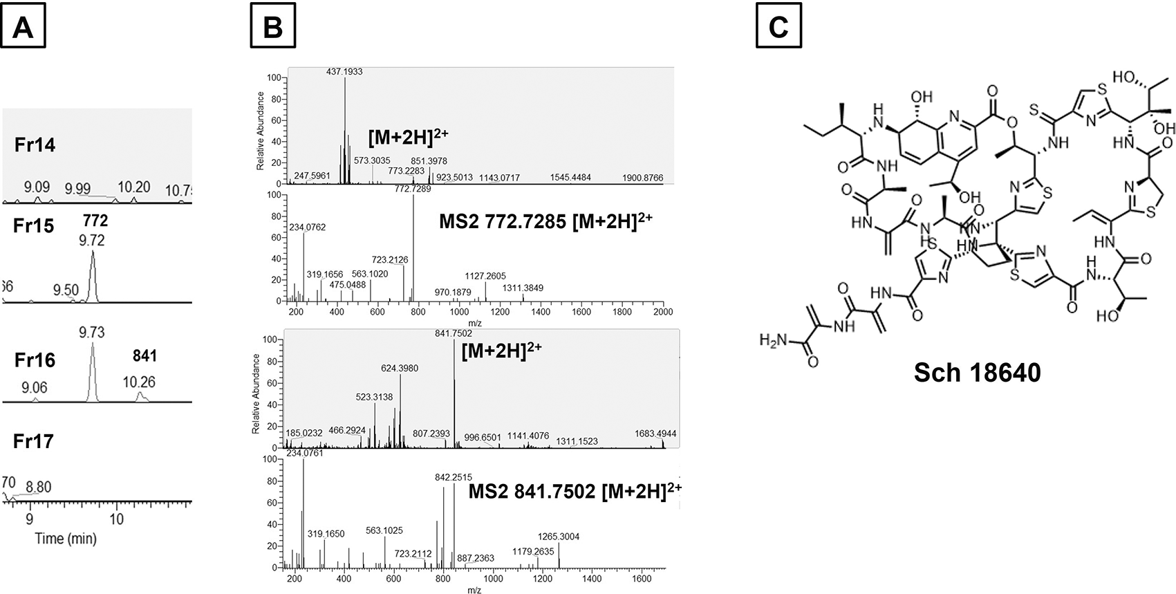
Figure 4. Identification of compound Sch 18640 in extract 78455.
Panel A: Extracted ion chromatograms of m/z 772.72802+ and m/z 841.75022+ from active (15 and 16) and inactive (14 and 17) fractions from extract 78455. Panel B, from top to down: Positive Full Mass spectrum at 9.73 minutes in fraction 16, MS2 spectrum of m/z 772.72852+, positive Full Mass spectrum at 10.26 minutes in fraction 16, MS2 spectrum of m/z 841.75022+. Panel C: Chemical structure of Sch 18640.
For extract 78471, fraction 13 was analyzed (
Figure 5). LC-MS analysis revealed an ion at m/z 694.2877, corresponding to a neutral mass of 695.2942 Da, consistent with the [M-H]− adduct. This mass, fragmentation pattern and retention time matched that of the original standard of 8-deoxy-3-hydroxyrifamycin
S.
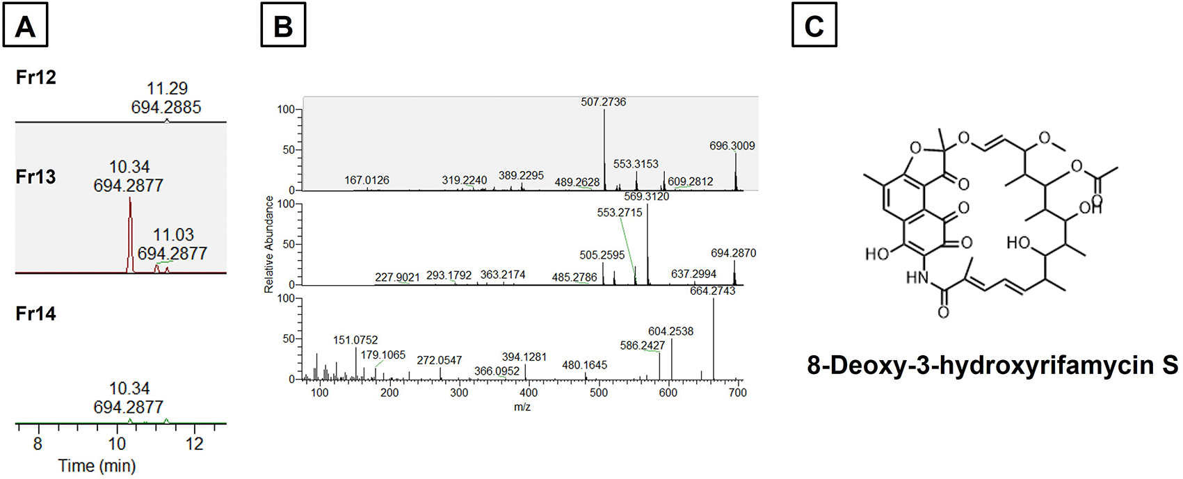
Figure 5. Identification of 8-Deoxy-3-hydroxyrifamycin S in extract 78471.
Panel A: Extracted ion chromatograms of m/z 694.2877− from active (13) and inactive (12 and 14) fractions from extract 78471. Panel B, from top to down: Positive and negative Full Mass spectra at 10.3 minutes in fraction 13, MS2 spectrum of m/z 696.3009+. Panel C: Chemical structure of 8-Deoxy-3-hydroxyrifamycin
S.
For extract 88546, fractions 10, 11 and 12 were analyzed. LC-MS analysis indicated a complex chemical profile, with several detected ions lacking clear database matches, suggesting that the activity may be due to a novel compound.
Discussion
AMR constitutes a major One Health concern, affecting both humans and animals, where interactions between human health, animal health and the environment play a key role in its emergence, maintenance, and dissemination.35–37 In this context, MDR represents a critical challenge, since it restricts therapeutic options, promotes the persistence of resistant pathogens across different reservoirs, and contributes to the spread of resistance determinants among interconnected ecosystems.37,38 In parallel with efforts to reduce antimicrobial use in animal production and human health, and consequently limit the emergence of new resistances,39,40 there is an urgent need to develop or discover new antimicrobials. Within this framework, S. suis represents a particularly relevant model due to its significance as a porcine pathogen and an emerging zoonotic agent.2 The high burden of S. suis in swine production promotes the use of antibiotics in livestock, thereby contributing to the selection of resistant strains and increasing the risk of transmission and infection in humans.10,13,41,42 In this study, we performed a screening of Actinomycetes extracts against S. suis, with particular emphasis on underexplored genera within this phylum and we identified extracts with inhibitory activity that cannot be assigned to already-known compounds. Although based on only two S. suis strains, these promising findings warrant further validation using a broader collection of strains to better reflect the diversity of this bacterial species.
Our results allowed the identification of Sch 18640, a compound with inhibitory activity against S. suis, belonging to the thiopeptide family. Thiopeptides are sulfur-rich natural peptide antibiotics classified as RiPPs (ribosomally synthesized and post-translationally modified peptides), as they originate from a ribosomal precursor peptide that undergoes extensive enzymatic modifications.43–45 Their biological relevance lies in their potent activity against Gram-positive bacteria, mainly through inhibition of protein synthesis. Depending on the specific compound, they can bind to the 50S ribosomal subunit or interfere with the elongation factor EF-Tu.43,44,46 Given that S. suis is a Gram-positive bacterium and therefore falls within the typical target spectrum of thiopeptides, the observed activity is consistent with the known antibacterial profile of this family.43,44 This supports its potential as a promising candidate for further structural and functional characterization, especially to address problems of solubility, bioavailability, and chemical synthesis, which have constrained the clinical development of thiopeptides.43,44
Another relevant finding was that the extract 78455, according to the Molecules Gateway, contained deferoxamine, a chelating compound used in the treatment of iron intoxications. In an attempt to explore the repurposing of this drug as an antibiotic, we tested this extract against S. suis and, in fact, the extract inhibited the growth of the bacterium. However, upon fractionation, this compound was not identified as the agent responsible for the inhibition of S. suis. This apparent discrepancy might be explained by the presence of another active compound in the extract. Deferoxamine (also known as desferrioxamine B) is a hydroxamate siderophore with a high affinity for ferric iron (Fe3+), a property that underlies its clinical use as an iron-chelating agent.47 Indeed, it is an approved drug for the treatment of acute iron intoxication and chronic iron overload.48 This molecule is also of microbiological interest, as it was originally described as a metabolite of Streptomyces pilosus, where it plays a natural role in iron acquisition under conditions of limited metal availability.49 Deferoxamine can also function as an exogenous xenosiderophore in some bacterial species, promoting iron uptake and bacterial growth or even virulence.47,50,51 However, this capability is not universal and depends on the presence of specific uptake systems for siderophore-bound iron.51 In contrast, our results suggest that S. suis does not exploit deferoxamine and neither its presence in the medium adversely affects the bacterium. This is consistent with previous reports indicating that S. suis does not produce siderophores and instead relies on alternative strategies to cope with iron limitation.52 Consequently, deferoxamine does not appear to contribute to the observed antimicrobial activity, which is more likely driven by other compounds present in the extract that were not clearly identified in our analysis.
One of the active metabolites identified in the screening was verucopeptin, produced by Actinomadura, a genus widely recognized as a source of chemically diverse bioactive compounds. Verucopeptin has been identified as a molecule of particular interest,53 primarily for its antitumor activity. This compounds presents inhibitory activity against the vacuolar ATPase (v-ATPase) and research efforts have mainly focused on its chemical structure, biosynthesis, and mechanism of action.33,54–56 Current evidence supports also its action as an antibacterial agent,55 which can explain the activity found against S. suis.
Hydroxyrifamycin S compounds are hydroxylated derivatives of rifamycin S within the rifamycin family, primarily produced by Amycolatopsis mediterranei (formerly Nocardia mediterranei in earlier literature).57–59 It is known that these molecules are generally described as biosynthetic intermediates, derived metabolites, or structural analogues employed to investigate structure-activity relationships, rather than as end products with well-established clinical or industrial applications.60–62 Thus, their relevance lies in providing insight into the diversification of the rifamycin biosynthetic pathway and in elucidating how specific structural modifications influence biological activity.63,64
The specific compound found in this study, 8-deoxy-3-hydroxyrifamycin
S, can be linked to two well-documented features: the occurrence of 3-hydroxyrifamycin S derivatives61 and the existence of an 8-deoxy branch in rifamycin biosynthesis.65–68 Indeed, Traxler & Ghisalba69 explicitly positions this compound within the proposed biosynthetic scheme involving late-stage substitution at the C3 position, supporting its interpretation as a plausible intermediate within this metabolic network. Regarding its activity against S. suis, a mechanistic rationale supports its potential relevance, as members of the rifamycin family are known to target bacterial RNA polymerase.63 Furthermore, structurally related derivatives have demonstrated activity against other bacterial species, such as S. aureus,
60,67 and we determined that our two S. suis strains were susceptible to rifampicin (rifamycin class).
Conclusion
Overall, this study represents a step forward in the search for novel antimicrobials against the zoonotic pathogen S. suis, with potential implications not only for swine health but also for human medicine within a One Health framework. Although further research is needed to fully characterize the identified-compounds efficacy and safety, these findings lay the groundwork for the development of new therapies to combat this worldwide relevant pathogen.
Author roles
Ayelén Perez-Falcèn: Data curation, Investigation, Visualization, Writing – Original Draft Preparation. Marianna Iorio: Conceptualization, Resources, Writing – Review & Editing. Margherita Sosio: Conceptualization, Resources, Writing – Review & Editing. Florencia Correa-Fiz: Conceptualization, Funding Acquisition, Resources, Supervision, Writing – Review & Editing. Stefano Donadio: Conceptualization, Funding Acquisition, Resources, Supervision, Writing – Review & Editing. Virginia Aragèn: Conceptualization, Funding Acquisition, Resources, Supervision, Writing – Review & Editing.
Ethics and consent
Ethical approval and consent were not required.