Introduction
With increased interest in sulphur as a candidate cathode material for high-performance lithium-ion batteries, the characterisation of sulphur to determine structure–property–function relations has become increasingly important in recent years1–3. The characterisation of sulphur by electron microscopy poses a number of interesting challenges. The foremost issue is the high-vacuum environment required for electron microscopy; in the low pressure regime sulphur may undergo sublimation under high vacuum at room temperature, rendering it challenging to image in the transmission electron microscope (TEM)4–6.
In order to prevent sublimation and give the microscopist enough time to complete imaging before the sample disappears entirely, a sample must be held in the solid region of its phase diagram5–7. This can be achieved in a number of ways for samples that can undergo sublimation; by cryogenically cooling the sample to a temperature past the sublimation line, by increasing the pressure surrounding the sample to a level greater than its equilibrium vapor pressure (for sulphur; ~10-7 Torr @ 20°C), or a combination of both4,5,7.
At the typical TEM vacuum pressures (approx. 7 × 10⁻⁸ Torr), sulphur exists in the gas phase4,8. To overcome this issue, cryogenic cooling to below 250 K or increasing pressure around the sample is required to keep sulphur in the solid orthorhombic phase4,5,7.
Both methods have been previously demonstrated by Levin et al.5, showing stable imaging of sulphur using a Cryo-holder for the TEM and using an ambient pressure SEM. Building on this work, a novel method for imaging sulphur in the TEM using a SiNx window gas cell holder is here proposed. With the introduction of commercially available gas cell holders, a sample can be held at a set pressure with any compatible gas connected to the sample holder. In this work, the sample was left open to air at atmospheric pressure for experimental simplicity. Given that the only necessary modification to the system would be the pressure, a set pressure of any non-reactive gas (such as N2 or Ar) would also work if the sample of interest is also sensitive to air. The concept of protectively encapsulating a sample to inhibit degradation in the microscope environment has been used in previous work by Kwon et al. to limit oxidation of copper nanowires, and a conceptually similar solution is demonstrated in this work9. However, the increased thickness added to the sample from the SiNx windows used in this method will reduce the contrast of the image formed, and the field of view and viewable sample area is limited by the size of the viewing window relative to standard TEM grids9. Such compromises are considered acceptable for the sake of sample stability.
Methods
The sulphur particles were observed via field-emission (scanning) transmission electron microscopy ((S)TEM) (Titan, Thermo Fisher Scientific Inc.). The macrostructure and elemental composition of the sulphur particles were analysed using (S)TEM coupled with simultaneous energy dispersive X-ray spectroscopy (EDX; Ametek 30mm2 EDX detector). A vacuum of 7 × 10⁻⁸ Torr and an acceleration voltage of 300 keV were employed for both TEM and STEM imaging during the measurements.
A sample of elemental sulphur was prepared by dispersing the as purchased sulphur powder (325 mesh, 99.5% purity, Alpha Aesar) in isopropanol (IPA) via ultrasonic bath sonication (Fisherbrand 11207 operated at 37kHz)10. The dispersion was deposited on a lacey carbon 400 mesh Cu grid (01896-F, TED PELLA Inc.) by dropcast for observation in vacuum, and allowed to dry in air. Sulphur particles were deposited onto Climate E-chips (P.T.GH.SS.2, DENS Solutions B.V. Delft, Netherlands, shown in Figure 4) in the same facile dropcast manner. Each chip, which consisted of an ~30-nm-thick SiNx membrane, was mounted in a Climate TEM holder (DENS Solutions B.V. Delft, Netherlands.). Here the SiNx membrane acts as sample support while simultaneously isolating the sample from the vacuum environment.
The sample was loaded into the microscope, and a sulphur particle was chosen as the region of interest. The column valves were closed between each consecutive image to reduce any beam-induced artefacts for both standard Cu grid experiment and SiNx window encapsulated experiment.
The encapsulated sample was left open to atmosphere at 1 bar during the experiment.
Results
In order to demonstrate sublimation of sulphur in normal TEM vacuum conditions a sulphur sample was prepared as described above and introduced into the microscope. As can be seen in the bright-field (BF) TEM images in Figure 1, the sulphur particle undergoes sublimation in the column vacuum over a period of time as little as 10 minutes. After this time, the residual material becomes vacuum-stable and does not sublimate further. It is believed to be super-sublimated polymeric sulphur, according to literature sources11,12. This is known to have a much lower vapour pressure than elemental sulphur11 and is comparable to the experiments conducted by Levin et al.5. Examination of the residue by EDX STEM mapping at 300 keV confirms the residue as sulphur as evidenced by the S-Kα peak at 2.3keV (Figure 2).
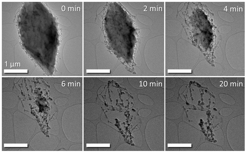
Figure 1. BF TEM time series images of sulphur particle subliming in TEM column vacuum of 7x10⁻⁸ Torr.
The large particle in the region of interest is seen to dramatically shrink over time under vacuum exposure. Column valves were closed between images to prevent beam-induced effects altering the sublimation process. Scale bar is 1 µm in all above images.
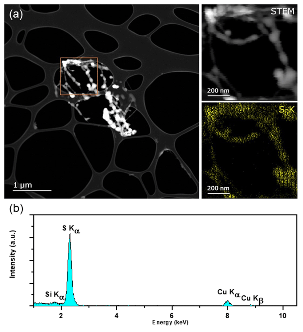
Figure 2.
a) HAADF STEM of residual S post-sublimation acquired on FEI Titan @300keV. Inset shows STEM image of highlighted ROI and EDX maps of S corresponding to the EDX spectrum. b) Integrated spectrum of region of interest, confirming elements listed in a) are present. Maps confirm that the residue is indeed composed of sulphur. (Si peak due to an EDX spectral artefact; an internal fluorescence peak from the silicon window on the silicon drift detector likely. Cu peak is a background signal from the TEM grid.)
In order to investigate the potential of the commercial gas cell sample holder MEMS chips13,14 as a method to image the sulphur without sublimation the sample was deposited on SiNx window DENS Solutions Climate MEMS chips (DENS Solutions B.V. Delft, Netherlands.), inserted into the column, and imaged over a period of 2 hours using the same experimental conditions as the previous experiment (300 keV TEM and STEM with a dwell time of 10ms/pixel and beam current of 0.5nA for EDX analysis). Unlike the previous experiment, the initial state of the particle is maintained in the column vacuum, and no change in particle morphology was observed over the observation period of 2 hours (as seen in Figure 3) in the high-angle annular dark field (HAADF) STEM image. The particle was kept at ambient pressure (1 bar) inside the Climate holder.
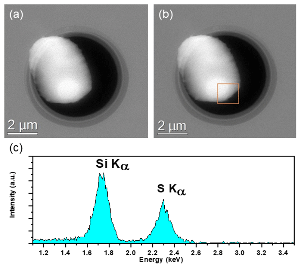
Figure 3.
HAADF STEM images of a sulphur particle before (a) and after (b) a 2-hour exposure inside the TEM column vacuum. The particle is isolated from the column vacuum by the SiNx window of the gas cell chips, thus preventing sublimation of the sample.
(c) Integrated EDX Spectra of highlighted ROI of sulphur particle on SiNx window of the gas cell chips after 2-hour exposure shown in (b) acquired @300 keV in FEI Titan equipped with an Ametek 30mm2 EDX detector. S and Si peaks from the S particle and SiNx window respectively clearly detected.
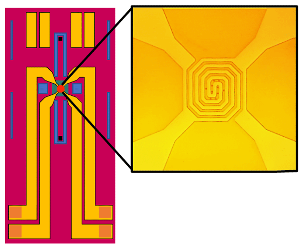
Figure 4. Schematic view of DENS Solutions Climate SiNx window chips.
SiNx windows isolate the gas flow inside the holder from the vacuum of the TEM column. Heating coil visible over SiNx windows.
The EDX spectrum acquired after the 2 hours showing S-Kα and Si-Kα peaks (located at 2.3keV and 1.74keV respectively) in Figure 3c confirms that the particle is indeed sulphur supported on a SiNx window. The suppression of the sublimation clearly demonstrates the viability of the use of windowed in-situ sample holders for imaging and characterisation of high vapour pressure materials in the TEM.
Being an electrically insulating material, sulphur is still very susceptible to radiolytic ionisation damage from the electron beam, and careful consideration must be made to the beam current passing through the sample for both quantitative and qualitative spectroscopy15–18. For example, in order to acquire data for an EDX map of the sulphur particle, higher beam doses (using longer pixel dwell times or a higher beam current) must be used in order to generate enough counts for the detector. This has the effect of damaging the sample heavily due to sulphur’s susceptibility to knock-on damage, and alternate methods must be employed to characterise the elemental composition of the sample with this method15–18. It is still possible to collect spectra to confirm elemental composition of the sample via EDX using a lower dose19,20, as can be seen in Figure 3. In this regard, the cryo-EM method utilised by Levin et al.5 has its benefits over the SiNx window approach that can be found in many in-situ holders. Cryogenic electron microscopy has the secondary benefit of slowing or even inhibiting beam induced artefacts and sublimation artefacts simultaneously, allowing for easier collection of mapping data21. However, cryo-EM may induce a phase change as a result of the change in temperature, depending on the sample being observed, and this method avoids this particular issue7.
Conclusions
The ability to image potentially vacuum sensitive materials for energy storage applications such as sulphur composites without sublimation artefacts creates new possibilities for their characterisation. To control sublimation in TEM analysis, an encapsulation method utilizing a commercial gas cell TEM sample holder is demonstrated. Where before, samples would sublime before imaging could take place, this novel encapsulation method allows for sample stability over the time frames necessary for both imaging, and spectroscopic analysis by EDX, or even EELS with ultra-thin SiNx windows (providing the sample of interest does not contain peaks/edges that would be obfuscated by Si or N peaks/edges). The proposed method is suitable for the TEM analysis of other specimens that may undergo sublimation at TEM vacuum pressures, such as zinc, magnesium, or some polymers to name a few. Such techniques would have been impossible for such materials in the past and presents exciting new opportunities for battery materials researchers.
Data availability
Underlying data
Open Science Framework: Inhibition of vacuum sublimation artefacts for (Scanning) Transmission Electron Microscopy ((S)TEM) of sulphur samples via encapsulation, https://doi.org/10.17605/OSF.IO/Z4MDV22.
This project contains the following underlying data:
- Fig 1_1.dm3
- Fig 1_1.jpg
- Fig 1_1.tif
- Fig 1_2.dm3
- Fig 1_2.jpg
- Fig 1_2.tif
- Fig 1_3.dm3
- Fig 1_3.jpg
- Fig 1_3.tif
- Fig 1_4.dm3
- Fig 1_4.jpg
- Fig 1_4.tif
- Fig 1_5.dm3
- Fig 1_5.jpg
- Fig 1_5.tif
- Fig 1_6.dm3
- Fig 1_6.jpg
- Fig 1_6.tif
- Fig 2_1.tif
- Fig 2_2.tif
- Fig 2_3.tif
- Fig 2_Integrated EDX spectrum.csv
- Fig 3.tif
- Fig 3_1.tif
- Fig 3_Integrated EDX spectrum.csv
- Fig3_1.emi
- Fig3_1_1.ser
- Fig3_2.emi
- Fig3_2_1.ser
- S_window-EDX.txt
- S-EDX.txt
- Sublimated Sulphur Drift corrected EDX spectrum image.emi
- Sublimated Sulphur Drift corrected EDX spectrum image_1.ser
- Sulphur Window Drift corrected EDX spectrum image.emi
- Sulphur Window Drift corrected EDX spectrum image_1.ser
Data are available under the terms of the Creative Commons Zero "No rights reserved" data waiver (CC-By 4.0 Public domain dedication).
Acknowledgments
The authors acknowledge the Advanced Microscopy Laboratory for the provision of their facilities. Microscopy characterization and analysis has been performed at the CRANN Advanced Microscopy Laboratory (AML) (www.tcd.ie/crann/aml/).
Faculty Opinions recommendedReferences
- 1.
Manthiram A, Fu Y, Chung SH, et al.:
Rechargeable lithium-sulfur batteries.
Chem Rev.
2014; 114(23): 11751–11787. PubMed Abstract
| Publisher Full Text
- 2.
Ji X, Lee KT, Nazar LF, et al.:
A highly ordered nanostructured carbon-sulphur cathode for lithium-sulphur batteries.
Nat Mater.
2009; 8(6): 500–506. PubMed Abstract
| Publisher Full Text
- 3.
Cheng XB, Huang JQ, Peng HJ, et al.:
Polysulfide shuttle control: Towards a lithium-sulfur battery with superior capacity performance up to 1000 cycles by matching the sulfur/electrolyte loading.
J Power Sources.
2014; 253: 263–268. Publisher Full Text
- 4.
Ferreira AGM, Lobo LQ:
The low-pressure phase diagram of sulfur.
J Chem Thermodyn.
2011; 43(2): 95–104. Publisher Full Text
- 5.
Levin BDA, Zachman MJ, Werner JG, et al.:
Characterization of Sulfur and Nanostructured Sulfur Battery Cathodes in Electron Microscopy Without Sublimation Artifacts.
Microsc Microanal.
2017; 23(1): 155–162. PubMed Abstract
| Publisher Full Text
- 6.
Raiß C, Peppler K, Janek J, et al.:
Pitfalls in the characterization of sulfur/carbon nanocomposite materials for lithium–sulfur batteries.
Carbon.
2014; 79(1): 245–255. Publisher Full Text
- 7.
Kotz JC, Treichel PM Jr:
Chemistry & Chemical Reactivity. Saunders College Publishing, 1999. Reference Source
- 8.
Williams DB, Barry Carter C:
Transmission Electron Microscopy. 2nd ed. Springer. 2009. Publisher Full Text
- 9.
Kwon Y, An BS, Moon JY, et al.:
Control of oxidation behavior in high vacuum transmission electron microscopy.
Mater Charact.
2021; 172: 110870. Publisher Full Text
- 10.
Nicolosi V, Chhowalla M, Kanatzidis MG, et al.:
Liquid Exfoliation of Layered Materials.
Science.
2013; 340(6139): 1226419. Publisher Full Text
- 11.
Nash DB:
Sulfur in vacuum: Sublimation effects on frozen melts, and applications to Io's surface and torus.
Icarus.
1987; 72(1): 1–34. Publisher Full Text
- 12.
Tobolsky AV:
Polymeric sulfur and related polymers.
J Polym Sci Pol Sym.
1966; 12(1): 71–78. Publisher Full Text
- 13.
Allard LF, Overbury SH, Bigelow WC, et al.:
Novel MEMS-based gas-cell/heating specimen holder provides advanced imaging capabilities for in situ reaction studies.
Microsc Microanal.
2012; 18(4): 656–666. PubMed Abstract
| Publisher Full Text
- 14.
Beker AF, Sun H, Lemang M, et al.:
In situ electrochemistry inside a TEM with controlled mass transport.
Nanoscale.
2020; 12(43): 22192–22201. PubMed Abstract
| Publisher Full Text
- 15.
Brydson R:
Electron Energy Loss Spectroscopy. 1st ed. Taylor and Francis. 2001. Reference Source
- 16.
Pennycook SJ, Nellist PD:
Scanning Transmission Electron Microscopy: Imaging and Analysis. 1st ed. Springer. 2011. Publisher Full Text
- 17.
Hren JJ, Goldstein JI, Joy DC, (Eds.):
Introduction to Analytical Electron Microscopy. PlenumPress. 1979. Publisher Full Text
- 18.
Csencsits R, Gronsky R:
Damage of zeolite Y in the TEM and its effects on TEM images.
Ultramicroscopy.
1987; 23(3–4): 421. Publisher Full Text
- 19.
Leijten ZJWA, Wirix MJM, Strauss M, et al.:
Low-dose (S)TEM elemental analysis of water and oxygen uptake in beam sensitive materials.
Ultramicroscopy.
2020; 208: 112855. PubMed Abstract
| Publisher Full Text
- 20.
Woehl TJ, Jungjohann KL, Evans JE, et al.:
Experimental procedures to mitigate electron beam induced artifacts during in situ fluid imaging of nanomaterials.
Ultramicroscopy.
2013; 127: 53–63. PubMed Abstract
| Publisher Full Text
- 21.
Nogales E:
The development of cryo-EM into a mainstream structural biology technique.
Nat Methods.
2016; 13(1): 24–27. PubMed Abstract
| Publisher Full Text
| Free Full Text
- 22.
Ronan O:
Inhibition of vacuum sublimation artefacts for (Scanning) Transmission Electron Microscopy ((S)TEM) of sulphur samples via encapsulation. 2021. http://www.doi.org/10.17605/OSF.IO/Z4MDV