Introduction
Epidemic and pandemic emergencies require a broad repertoire of prevention and containment strategies as demonstrated during the COVID-19 pandemic.1 Enhanced surveillance represents a key component of an effective response to outbreaks caused by respiratory viruses such as severe acute respiratory syndrome coronavirus 2 (SARS-CoV-2), Influenza A, Influenza B, and respiratory syncytial virus (RSV).2,3 Molecular testing using nucleic acid amplification tests (NAAT) plays a central role in modern respiratory virus detection. Current guidelines recommend the collection of respiratory tract specimens for nucleic acid detection as the standard approach for laboratory diagnosis of acute respiratory viral infections.4–6 For upper respiratory tract sampling, nasopharyngeal swabs (NPS) are still considered the gold standard.7,8 However, NPS alone may be suboptimal for comprehensive detection of respiratory pathogens. Several studies have shown that combining NPS with additional specimen types, such as oropharyngeal swabs (OPS) and/or nasopharyngeal washings (NPW), can improve detection rates.9–11 Nevertheless, the range of non-invasive sampling alternatives remains limited.
The collection of human breath offers a non-invasive alternative to swab-based sampling and has been shown to be effective for COVID-19 diagnosis.12–15 Earlier studies also demonstrated its applicability for detecting other respiratory viruses, including RSV, Influenza and human rhinovirus (HRV).16 During the COVID-19 pandemic, medical face masks proved effective in reducing transmission17–19 and were widely accepted by the public,20 supporting the feasibility of establishing a novel, broadly applicable non-invasive sampling platform.
AveloMask, is a facemask-based breath collection kit that was previously evaluated for the detection of Mycobacterium tuberculosis in exhaled breath aerosol (XBA) samples.21 The system employs a fiber filter to capture XBA, which can be easily released from the mask via a simple push-step and subsequently stored in denaturing buffer that stabilizes the sample for transport, biobanking or downstream analysis (Figure 1). A key limitation of XBA sampling compared to swab-based collection are substantially lower specimen amounts, which can challenge the sensitivity of downstream applications such as nucleic acid extractions and PCR. Indeed, for SARS-CoV-2 detection, viral nucleic acid quantities in breath samples are typically 10-
to 100-fold lower than those obtained from NPS in routine PCR workflows.22,23 Consequently, optimization of extraction and PCR detection methods is critical to achieving adequate sensitivity for mask-based XBA samples.
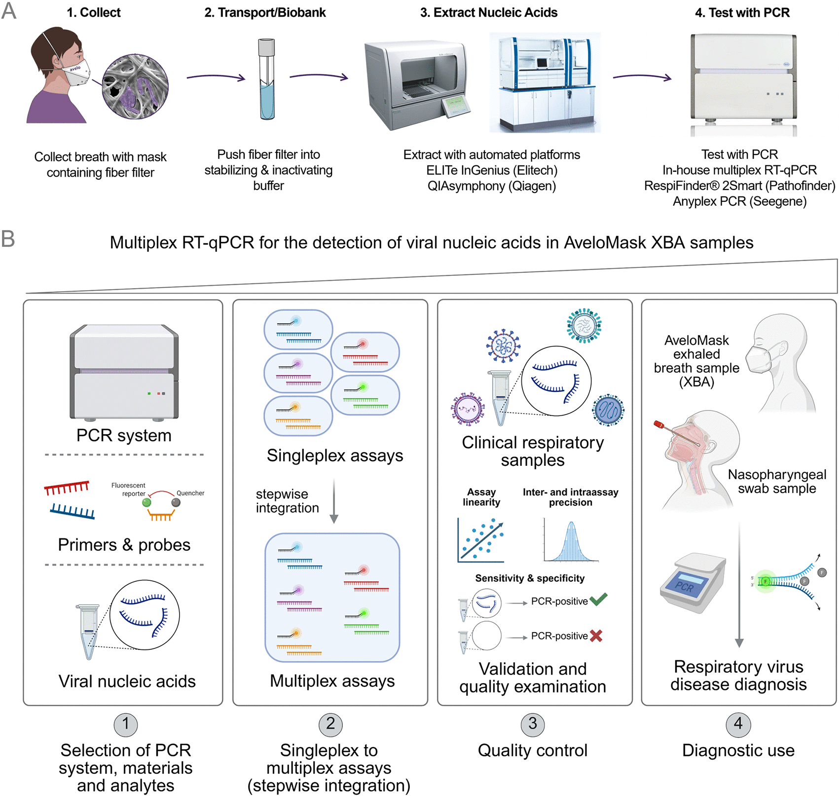
Figure 1. AveloMask analysis workflow and construction of in-house multiplex RT-qPCR assay.
(A) Simplified workflow for collection and analysis of AveloMask XBA samples: 1. Infected individual wears mask for at least 30 min; 2. Fiber filter is incubated in Avelo buffer for stabilization and inactivation, 3. Nucleic acid extraction is performed by ELITech and QIAsymphony extraction platforms, 4. RT-qPCR on eluates generates detection results. (B) Simplified workflow for the setup of the in-house RT-qPCR assay: 1. Selection of PCR system, materials and analytes; 2. Singleplex reactions were merged into multiplex reactions testing primer cross-annealing and overall compatibility of input materials; 3. Viral respiratory clinical samples were selected for quality control assays including linearity testing, intra- and interassay precision assessment as well as test sensitivity and specificity evaluations; 4. Use of the assay in real-world respiratory samples originating from AveloMask or nasopharyngeal swab sampling. Figure part B was created in BioRender. Kelch, M. A. (2026) https://BioRender.com/m8rlcs2.
In this study, we developed a multiplex RT-qPCR assay tailored for AveloMask samples and implemented it on the ELITe InGenius and QIAsymphony extraction platforms. We performed an analytical comparison of our in-house assay with two commercially available multiplex RT-qPCR kits and conducted a preliminary evaluation of the workflow using a limited number of AveloMask-collected XBA samples (Figure 1). The overall objective was to assess and compare extraction and molecular detection workflows for AveloMask samples and establish technical feasibility in initial clinical samples, laying the groundwork for future, larger diagnostic studies of XBA sampling for detection of respiratory viruses.
Methods
Samples, masks and buffer
For validation of the in-house RT-qPCR assay, biobanked patient isolates and cell culture isolates were used (Extended Data Tables 1 and 2). Artificial RNA samples were prepared by spiking 3 mL of Avelo buffer with defined quantities of synthetic RNA standards (see LOD section for concentrations). Unless otherwise indicated, assays were performed using clinical samples from healthy or infected individuals. For workflow comparison, results from XBA and NPS samples collected from seven infected patients, eight healthy controls and one individual recovering from a cold were analyzed.
Table 1. Relative sensitivity (%) and relative specificity (%) of in-house RT-qPCR assay.
| Pathogen | % Sensitivity (Ct < 35)* | % Sensitivity (Ct > 35)* |
% Specificity |
|---|
| Influenza | 100 | 33.3 | 99.6 |
| RSV | 100 | 75.0 | 99.6 |
| SARS-CoV-2 | 100 | 66.6 | 99.2 |
| HRV | 56.5 | 38.1 | 99.1 |
| Total (w/o HRV) | 100 | 73.9 | 98.5 |
Table 2. Limit of detection (LOD) for respiratory viruses across extraction platforms and RT-qPCR assays.
| Respiratory virus | ELITe InGenius [copies/mL] | QIAsymphony [copies/mL] | ELITe InGenius [copies/mL] | QIAsymphony [copies/mL] |
|---|
| In-house RT-qPCR* | RespiFinder 2Smart§ |
|---|
| SARS-CoV-2 | 56 | 95 | ≤ 10 | ≤ 10 |
| Influenza A | 245 | 213 | 13 | 56 |
| Influenza B | 525 | 183 | 27 | 93 |
| RSV | 236 | 511 | 17 | 96 |
AveloMask kits (Avelo AG, Cat. No. AMK01) were used according to manufacturer’s instructions. The Avelo buffer is a guanidine-thiocyanate-based solution that inactivates pathogens and preserves nucleic acids, enabling transport at ambient temperature until further processing or biobanking. Mask filters were collected in Avelo buffer, frozen at −20 °C and processed in batches.
Extraction of nucleic acids
Prior to extraction, samples were spiked with 9 μL MS2 bacteriophage internal control, prepared by 1:1,000 dilution of the stock (0.8 μg/μL; Roche, Cat. No. 10165948001) in AVE buffer (Qiagen, Cat. No. 937055). Extractions were performed using the QIAsymphony DSP Virus/Pathogen Kit (Qiagen, Cat. No. 937055) on the QIAsymphony SP system (Qiagen, Germany) or the ELITe InGenius SP 1000 (ELITech Group, Ref. No. INT033SP1000) on the ELITe InGenius system (ELITech Group, Italy).
Validation samples and clinical XBA samples in Avelo buffer were pulse-vortexed for 10 s, incubated at 70 °C for 15 min, vortexed again for 10 s, and 2 mL was recovered for extraction. This step aims to support inactivation, lysis and elution from the filter. In each run, a positive control (Avelo buffer spiked with all the target viruses) and, as a negative control, unspiked material were included. Nucleic acids were extracted either on the QIAsymphony SP using the Cellfree 1000 V7 default IC protocol (1 mL input, 60 μL elution) or ELITe InGenius SP 1000 (ELITech Group, Italy) in extraction-only mode (1 mL input, 50 μL elution). Extracted nucleic acids were used for RT-qPCR.
Commercial RT-qPCR assays
Seegene Allplex Respiratory Panels 1A, 3, and SARS-CoV-2 assays (Seegene Germany Cat. No. RP10231Z, RP10181Z, RV10248X, respectively) were performed following manufacturer’s instructions. Real-time PCR was conducted on a CFX96 instrument (Bio-Rad, USA) and results were analyzed using Seegene Viewer software (Seegene, Germany).
The RespiFinder 2Smart panel (PathoFinder, Cat No. PF2600-2S) was performed according to manufacturer’s instructions. The assay was run on a LightCycler® 480 II instrument (Roche Diagnostics, Switzerland) using 10 μL extracted RNA/DNA per reaction. Results were interpreted using FastFinder software (PathoFinder, Netherlands).
In-house RT-qPCR assay
We developed a one-step multiplex RT-qPCR assay for influenza A/B, SARS-CoV-2, RSV A/B and HRV using primers and probes selected for high specificity and sensitivity. Lyophilized oligonucleotides (from Integrated DNA technologies, Belgium) were reconstituted to 100 μM, pre-diluted to 20 μM, and stored at −20 °C. Optimal working concentrations were 300 nM for primers and 150 nM for probes. All primer and probe sequences, along with relevant references, are provided (Extended Data
Table 3).
Table 3. RT-qPCR results for AveloMask samples following ELITe InGenius extraction.
| | NPS | AveloMask | AveloMask | AveloMask |
|---|
| Group | ID | Commercial test result | In-house RT-qPCR | RespiFinder2Smart | Seegene Allplex |
|---|
| Infected | 20_250202 | Influenza B (PCR) | Influenza (27.38) | Influenza B | Influenza B (32.95) |
| 04_250722 | SARS (PCR and antigen) | SARS-CoV-2 (35.78) | SARS-CoV-2 | SARS-CoV-2 (33.42) |
| 09_250902 | SARS (PCR and antigen) | SARS-CoV-2 (32.55) | SARS-CoV-2, HRV# | SARS-CoV-2 (30.67) |
| 28_250306 | HRV (PCR) | HRV (21.8) | HRV, Adenovirus | HRV (22.62) |
| 26_241122 | HRV (PCR) | HRV (36.6) | HRV | Neg. |
| 20_241028 | HRV (PCR) | Neg. | HRV | HRV (26.29) |
| 20_241029 | HRV (PCR) | Neg. | HRV, SARS Cov2# | HRV (34.47) |
| Healthy | 08–12 and 14–16 | Neg. | Neg. | Neg.*+ | Neg.* |
| Recovery | 13 | Neg. | Neg. | Neg. | Neg. |
RT-qPCR was performed using Reliance One-Step Multiplex RT-qPCR Supermix (Bio-Rad Laboratories, Cat. No. 12010220) according to manufacturer’s instructions on a CFX96 Touch Real-Time PCR Detection System (Bio-Rad Laboratories, Cat. No. 1855195). Reactions were 20 μL, with 5 μL of RNA input. Cycling consisted of reverse transcription at 50 °C for 10 min, followed by DNA polymerase activation and denaturation at 95 °C for 10 min, and 45 cycles of 95 °C for 10 s (denaturation) and 60 °C for 30 s (annealing/extension). Cycle thresholds were set at fluorophore-specific relative fluorescence units (RFU) to enhance reproducibility across different experiments, VIC: 25 RFU, 6-FAM: 50 RFU, Cy5: 75 RFU, Texas Red: 100 RFU, Cy5.5: 50 RFU. In all experiments, a negative control (extracted negative material or no-template control, respectively) and a positive control, containing all the target viruses, were included.
Assay sensitivity and specificity were evaluated using 273 NPS samples previously tested with the Seegene Allplex Respiratory panels 1–3 and SARS-CoV-2 assay (Seegene Germany, Cat. No. RP10179Z, RP10180Z, RP10181Z, RV10248X). The sample pool contained 133 positive samples for either Influenza, RSV, SARS-CoV-2, HRV or combinations thereof (Extended Data Figure 3). Ct 35 was defined as the cut-off to distinguish strong positives (Ct < 35) from weak positives (Ct > 35), accounting for qPCR threshold uncertainty. Assay cross-reactivity was evaluated against Seegene assays (Extended Data Table 8).
For determination of intra assay precision, each sample was measured in batch consisting of three technical replicates. Replicate reactions originated from the same mastermix. For determination of inter assay precision, each sample was measured in two separate runs on two different days in addition to the intra assay precision setup (Extended Data Figure 1). Validation samples used for assay precision assessments are listed in Extended Data Table 1.
Assay linearity and amplification efficiency were calculated by setting up dilution series of every validation sample in technical duplicates. Each sample was subjected to a 1:10 dilution series with at least four dilution steps. 1:10 dilutions were performed by adding 2 μL RNA to 18 μL of water. Validation samples used for linearity testing are listed in Extended Data Table 2. Example curves, linear regressions and amplification efficiencies are listed in the extended data (Extended Data Figure 2, Extended Data Tables 6 and 7).
For intra assay precision, mean Ct values ± SD is depicted in Extended Data Figure 1, Extended Data Tables 4 and 5. Concentration variations were determined by multiplying SD (Ct) by ln (2):
(1)
Linear regressions were performed by applying method of least squares for a model that considers a semi log data format using a maximum of 1000 iterations and medium stringency.
Regression equations have the following structure in which x represents the dilution factor and y represents the corresponding Ct value:
(2)
Amplification efficiencies were derived from slopes of linear regressions by using the following formula:
(3)
Assay sensitivity was determined by dividing the number of true positives (TP) samples by the sum of true positives (TP) and false negatives (FN):
(4)
Assay specificity was determined by dividing the number of true negative (TN) samples by the sum of true negatives (TN) and false positives (FP):
(5)
In-house RT-qPCR sensitivity and specificity were calculated relative to Seegene Allplex results.
Limit of detection (LOD)
Limits of detection (LODs) were established using synthetic SARS-CoV-2, Influenza A, Influenza B and RSV standards (Zeptometrix HiSS Diagnostics, Freiburg, Germany, Cat. No. NATSARS (COV2)-ST, NATFLUAH1(2009)-STQ, NATFLUB-STQ, NATRSVA-STQ), diluted in Avelo buffer to final concentrations of 800, 400, 200, 100, 50 and 10 copies/mL and extracted using either the QIAsymphony or ELITe InGenius system. Extracted RNA was tested in both the in-house PCR test and the RespiFinder 2Smart panel, with eight or four technical replicates respectively, performed at each concentration. Binary detection results were modeled as a function of RNA concentration using probit regression in R Studio. The LOD for each pathogen was defined as the concentration at which the probability of detection reached 95%, based on the fitted model.
Statistical analysis
Statistical analyses were performed in R (version 4.3.2) using RStudio. Probit regression was used to estimate the concentration corresponding to a 95% probability of detection. No custom software or analysis code was developed for this study.
Ethics statement and informed consent
The research has been performed in accordance with the principles stated in the Declaration of Helsinki. Nasopharyngeal swab samples used for analytical evaluation were pre-existing samples obtained from the research biobank of Frankfurt University Hospital/Goethe University Frankfurt and were used in accordance with the applicable biobank governance, the Ethics Committee of the Faculty of Medicine at Goethe University Frankfurt am Main has granted a waiver (dated July 25th 2019) stating that there is no obligation to provide counselling in the context of a retrospective analysis of anonymised residual samples for scientific purposes.
For the collection of AveloMask samples from a small number of adult members of staff/the study team, formal ethical approval was not obtained. Prior to sample collection, the study team consulted the relevant institutional ethics committee at Goethe University Frankfurt, which advised that formal ethical approval was not required for this small number of voluntary samples. All participants provided written informed consent for collection and research use of their samples. All samples and associated data were pseudonymized prior to analysis.
The statutes of the Ethics Committee of the Faculty of Medicine at Goethe University Frankfurt am Main are available online (unimedizin-ffm.de/fileadmin/redakteure/Klinikum/Ethik-Kommission/2025/Satzung_EK_FB16_GU_20250311.pdf).
Results
In-house RT-qPCR assay shows high sensitivity and specificity for SARS-CoV-2, Influenza and RSV relative to the Seegene Allplex assay
To detect relevant respiratory viruses in clinical breath samples, we developed a multiplex RT-qPCR assay targeting SARS-CoV-2, influenza A/B and RSV A/B, and included HRV as a marker for general infection incidence. Sensitivity relative to the Seegene Allplex assay for influenza, RSV and SARS-CoV-2 was 100% for samples with Ct < 35 and 74% for samples with Ct > 35 in the reference assay. The assay’s relative specificity for SARS-CoV-2, influenza A/B and RSV was 98.5% with 4 false positive results for SARS-CoV-2 (Ct 38.41, Ct 37.66), Influenza (Ct 38.28), and RSV (Ct 36.12). For HRV the assay showed lower relative sensitivity of 57% for samples with Ct < 35 and 38% for samples with Ct > 35. We detected two false positive samples for HRV (Ct 37.58, Ct 38.57) (
Table 1). Intra- and inter-assay standard deviations were consistently below 0.5 Ct for all tested samples (Extended Data Figure 1). Maximum concentration variation was ~22% (~0.32 Ct) for intra-assay comparison (Extended Data Table 4) and ~ 33% (~0.47 Ct) for inter-assay comparison (Extended Data Table 5), with overall 7/10 samples showing <10% concentration variation. Regression analyses yielded coefficients >0.98 (Extended Data Figure 2, Extended Data Table 6). Analysis of dilution series demonstrated detection of viral nucleic acids with average slopes between −3.1 and − 3.6, corresponding to amplification efficiencies of 90–110% (Extended Data Table 7). No sample cross-reactivity was observed, and the assay reliably detected common viral subtypes, including RSV-A and RSV-B, as well as multiple Influenza A strains (Extended Data Table 8).
Comparison of ELITech and QIAsymphony extraction systems yield similar limits of detection (LOD) for respiratory viruses
We compared the nucleic acid extraction performance of the ELITe InGenius and QIAsymphony platforms for clinically relevant respiratory viruses, including SARS-CoV-2, Influenza A, Influenza B, and RSV. Synthetic RNA standards at defined concentrations were spiked into Avelo buffer and subjected to extraction, followed by analysis using both in-house RT-qPCR assay and commercial RespiFinder 2Smart panel. Across all target viruses and extraction platforms, LOD values for the in-house RT-qPCR ranged from 56 to 525 copies/mL. In contrast, the RespiFinder 2Smart assay demonstrated overall lower LODs, ranging from ≤10 to 96 copies/mL (
Table 2). Based on an Avelo Buffer volume of 3 mL and an extraction input volume of 1 mL, these LODs correspond to approximately 168–1,575 viral copies on the mask filter for detection by the in-house RT-qPCR assay and 30–288 copies for detection by the RespiFinder 2Smart panel.
ELITech and QIAsymphony extractions detect viral nucleic acids in AveloMask samples from infected individuals
Shifting the focus from artificial samples that were necessary to develop workflows, we attempted to assess feasibility with real-world XBA samples. We selected a set of seven AveloMask samples from patients with respiratory symptoms, including cough, fever, chills and rhinorrhea, eight samples from healthy individuals as negative controls, and one sample from an individual recovering from a respiratory infection. For each patient, paired NPS and AveloMask samples were collected for direct comparison (
Tables 3 and 4). Using the in-house RT-qPCR, results were fully concordant with the NPS reference results in 5/7 infected individuals across both extraction platforms. However, HRV was not detected in two samples (20_241028 and 20_241029) although it was tested positive by comparator assays. The RespiFinder 2Smart assay in combination with QIAsymphony extraction showed complete concordance with NPS results (7/7). In contrast, when combined with ELITe InGenius extraction, the RespiFinder 2Smart assay yielded additional detections not supported by comparator results, including HRV in sample 09_250902 and SARS-CoV-2 in sample 20_241029. Sample 13 originates from an individual who, at the time of sampling, was recovering from cold symptoms and was therefore classified separately. The sample tested positive in the QIAsymphony + RespiFinder 2smart workflow, while it tested negative in the others. The PCR no-template control was negative, indicating no contamination was detected during PCR setup or amplification. The Allplex assay detected 6/7 infected samples with both extraction platforms but failed to detect HRV in sample 26_241122. Comparison of extraction platforms using the Allplex assay revealed a mean Ct difference of approximately 2.7 cycles (pValue 0.0056, paired t-test), with slightly lower Ct values observed for ELITe InGenius extraction. A similar trend was observed with the in-house RT-qPCR assays.
Table 4. RT-qPCR results for AveloMask samples following QIAsymphony extraction.
| | NPS | AveloMask | AveloMask | AveloMask |
|---|
| Group | ID | Commercial test result | In-house RT-qPCR | RespiFinder2Smart | Seegene Allplex |
|---|
| Infected | 20_250202 | Influenza B (PCR) | Influenza (31.65) | Influenza B | Influenza B (35.41) |
| 04_250722 | SARS (PCR and antigen) | SARS-CoV-2 (36.76) | SARS-CoV-2 | SARS-CoV-2 (35.30) |
| 09_250902 | SARS (PCR and antigen) | SARS-CoV-2 (35.37) | SARS-CoV-2 | SARS-CoV-2 (32.67) |
| 28_250306 | HRV (PCR) | HRV (24.75) | HRV, Adenovirus | HRV (25.74) |
| 26_241122 | HRV (PCR) | HRV (42.08) | HRV | Neg. |
| 20_241028 | HRV (PCR) | Neg. | HRV | HRV (27.75) |
| 20_241029 | HRV (PCR) | Neg. | HRV | HRV (39.89) |
| Healthy | 08–12 and 14–16 | Neg. | Neg. | Neg. | Neg. |
| Recovery | 13 | Neg. | Neg. | HRV | Neg. |
Discussion
The aim of this proof-of-concept study was to evaluate whether exhaled breath aerosol (XBA) samples collected with AveloMask can be integrated into routine molecular workflows for respiratory virus detection, and to assess the impact of the extraction platform and downstream PCR panel on analytical performance. This workflow prioritization objective addresses the central challenge of XBA-based testing, namely low viral loads in breath samples, which make workflow sensitivity critical.
Overall, our findings demonstrate that AveloMask-derived XBA samples seem compatible with established automated extraction systems and downstream multiplex PCR assays. This represents a technical advance, indicating that mask-based breath sampling can potentially be integrated into routine diagnostic workflows. These findings support the idea of XBA sampling becoming a non-invasive approach for respiratory virus detection.
A key finding is that the choice of PCR assay had a greater impact on analytical sensitivity than the extraction platform. RespiFinder 2Smart yielded lower LODs (≤96 copies/ml) compared to the in-house RT-qPCR assay (≤ 525 copies/mL), consistent with previous reports.24–26 This suggests that, in XBA testing, PCR assay selection is a major determinant of performance and may outweigh differences between extraction systems. Notably, the LODs for RespiFinder 2Smart were lower than manufacturer specifications, likely due to differences in template material and limited replicate numbers. These estimates should therefore be considered preliminary (“pre-LOD”) rather than definitive and larger studies are needed to determine clinical relevance. A limitation of our LOD estimates is RNA spiking as an imperfect surrogate for XBA sampling, usually including exhaled aerosol collection and filter membrane release. To overcome this missing real-world representation, we sought to analyze mask samples directly.
In contrast to the PCR assay differences, the two extraction platforms performed comparably. Neither ELITe InGenius nor QIAsymphony was consistently superior across targets using synthetic RNA standards. Comparable performance across different nucleic acid extraction methods has also been reported for respiratory virus detection in other analytical settings.27,28 In clinical XBA samples, ELITe InGenius yielded slightly lower Ct values (~2.7 cycles) on average in both the in-house and Allplex assays. However, this did not improve concordance with swab comparator results, indicating that Ct values alone are not sufficient to assess workflow performance.
Among the tested combinations, most favorable results were observed for QIAsymphony extraction followed by RespiFinder 2Smart analysis. This workflow combined low LODs with high concordance in the small XBA sample data set. While QIAsymphony extraction has previously been validated with the RespiFinder 2Smart assay for NPS (RespiFinder 2Smart instructions for use), our data indicate compatibility with XBA samples. In contrast, the same RespiFinder 2Smart with ELITe InGenius extraction generated additional detections not confirmed by the swab comparator results. These may reflect low viral loads detected due to higher sensitivity, or false-positives due to non-specific amplification or cross-reactivity. Differences in assay design, including oligo target regions, may further contribute to discordant results. In addition, stochastic effects near the LOD, as well as pre-analytical factors such as sample handling, RNA stability, or contamination, may influence detection outcomes. Importantly, given the absence of a true gold standard, it cannot be excluded that some of these additional detections represent true infections not identified by the comparator assay. These discordant findings warrant further investigation, including evaluation of potential contamination.
The in-house RT-qPCR panel offers the advantage of an open and cost-effective workflow that can be broadly implemented for research and development purposes. Its performance in comparison to Seegene Allplex was robust for SARS-CoV-2, influenza, and RSV. However, HRV detection remained limited, likely due to the extensive genetic diversity of HRV, which complicates primer and probe design,29,30 compounded by low viral loads and multiplex conditions. This limitation highlights the need for further assay optimization, particularly for HRV coverage. While the assay is currently not suitable for HRV detection, it underlines the suitability for SARS-CoV-2, influenza, and RSV detection.
Another caveat is the use of the Seegene Allplex assay as the reference method producing relative sensitivity and specificity estimates and therefore no conclusions on absolute clinical performance can be made. Additional limitations include the small clinical sample size, restricted pathogen range, and unresolved discordant results. Strengths include the comparison of two automated extraction platforms and three downstream PCR approaches, as well as the combined use of synthetic standards and clinical XBA samples. In conclusion, this proof-of-concept study suggests that AveloMask-derived XBA samples may be integrated into standard molecular diagnostic workflows for respiratory virus detection. Whereas no conclusions on clinical relevance can be drawn due to small sample size of n = 7, the combination of QIAsymphony and RespiFinder 2Smart demonstrated the most favorable performance. However, this requires further confirmation and validation as in a larger ongoing study. Our findings support further prospective evaluation of optimized XBA-based workflows for respiratory virus diagnostics and surveillance.
Consent
Written informed consent was obtained from all participants for the collection and research use of their samples and associated data.
Data and software availability
Underlying and extended data
Participant-level data underlying
Table 1 cannot be made publicly available because the applicable participant consent does not permit public sharing of pseudonymized individual-level data. The ethics committee did not provide a separate determination regarding data sharing; access is therefore governed by the applicable participant consent and institutional data-protection requirements. Requests for access to these restricted data should be addressed to Prof. Annemarie Berger (annemarie.berger@em.uni-frankfurt.de), specifying the research purpose and proposed analyses. Requests will be considered in accordance with the applicable consent and institutional data-protection requirements.
All other data supporting the findings of this study are available directly within the paper, including participant-level data in
Tables 3 and 4, or in the extended data. Extended data for this study are available on Zenodo (https://doi.org/10.5281/zenodo.22710012).31
Data are available under the terms of the Creative Commons Attribution 4.0 International license (CC-BY 4.0).
Acknowledgements
We thank Frankfurt Virus Solutions of the Goethe University & University Hospital Frankfurt for providing us virus isolates and associated protocols (for further information contact the corresponding author or virussolutions@unimedizin-ffm.de).
We thank the clinical and laboratory staff involved in sample collection, processing, and data management.
In addition to the authors named above, the B-Path consortium includes the following members: University Hospital Heidelberg, Heidelberg, Germany: Maria del Mar Castro Noriega, Ralitza Dekova, Shannon McMahon, Jonas Wachinger, Agrin Putri, Abdulkadir Civan, Carrie Anderson, Magdalena Pajonk, Maximilian Schirmer, Zhizhao Jiang, Parnian Karimi, Kevin Yuan and Frank Tobian. IRCCS San Raffaele Scientific Institute, Milan, Italy: Roberta Monardo, Caterina Franco, Francesca Saluzzo and Mariam Omrani. Vita-Salute San Raffaele University, Milan, Italy: Nicola Clementi. Desmond Tutu Health Foundation, Cape Town, South Africa: Robin Wood, Juliet Joubert, Ryan Dinkele, Ronnett Seldon, Andiswa Vazi, Zeenat Hoosen, Tricia Nhlapho and Andrea McKerry. Avelo AG, Schlieren, Switzerland: Katie Tiseo. Institutul de Pneumoftiziologie Marius Nasta, Romania: Elmira Ibraim, Alexandru Stoichita, Mihaela Simion, Madalina Burecu, Alexandru Daniel Radu, Lita Maricica, Camelia Nita, Simona Mustata, Teodora Onitoiu and Daniela Chirila.
References
- 1.
Agyapon-Ntra K, McSharry PE:
A global analysis of the effectiveness of policy responses to COVID-19.
Sci. Rep.
2023; 13(1): 5629. PubMed Abstract
| Publisher Full Text
| Free Full Text
- 2.
Cascini F, Hoxhaj I, Zace D, et al.:
How health systems approached respiratory viral pandemics over time: a systematic review.
BMJ Glob. Health.
2020; 5(12): e003677. PubMed Abstract
| Publisher Full Text
| Free Full Text
- 3.
Hamza IA, Mao K, Gao C, et al.:
Elements of Viral Outbreak Preparedness: Lessons, Strategies, and Future Directions.
Viruses.
2026; 18(1): 50. PubMed Abstract
| Publisher Full Text
| Free Full Text
- 4.
Evans SE, Jennerich AL, Azar MM, et al.:
Nucleic Acid-based Testing for Noninfluenza Viral Pathogens in Adults with Suspected Community-acquired Pneumonia. An Official American Thoracic Society Clinical Practice Guideline.
Am. J. Respir. Crit. Care Med.
2021; 203(9): 1070–1087. PubMed Abstract
| Publisher Full Text
| Free Full Text
- 5.
Gulholm T, Basile K, Kok J, et al.:
Laboratory diagnosis of severe acute respiratory syndrome coronavirus 2.
Pathology.
2020; 52(7): 745–753. PubMed Abstract
| Publisher Full Text
| Free Full Text
- 6.
Hanson KE, Azar MM, Banerjee R, et al.:
Molecular Testing for Acute Respiratory Tract Infections: Clinical and Diagnostic Recommendations From the IDSA's Diagnostics Committee.
Clin. Infect. Dis.
2020; 71(10): 2744–2751. PubMed Abstract
| Publisher Full Text
| Free Full Text
- 7.
Larsen KD, Jensen MM, Homoe AS, et al.:
Head-to-Head Comparison of Nasopharyngeal, Oropharyngeal and Nasal Swabs for SARS-CoV-2 Molecular Testing.
Diagnostics (Basel).
2023; 13(2): 283. PubMed Abstract
| Publisher Full Text
| Free Full Text
- 8.
Tsang NNY, So HC, Ng KY, et al.:
Diagnostic performance of different sampling approaches for SARS-CoV-2 RT-PCR testing: a systematic review and meta-analysis.
Lancet Infect. Dis.
2021; 21(9): 1233–1245. PubMed Abstract
| Publisher Full Text
| Free Full Text
- 9.
Begier E, Aliabadi N, Ramirez JA, et al.:
Detection by Nasopharyngeal Swabs Alone Underestimates Respiratory Syncytial Virus-Related Hospitalization Incidence in Adults: The Multispecimen Study's Final Analysis.
J. Infect. Dis.
2025; 232(1): e126–e136. PubMed Abstract
| Publisher Full Text
| Free Full Text
- 10.
Kim C, Ahmed JA, Eidex RB, et al.:
Comparison of nasopharyngeal and oropharyngeal swabs for the diagnosis of eight respiratory viruses by real-time reverse transcription-PCR assays.
PLoS One.
2011; 6(6): e21610. PubMed Abstract
| Publisher Full Text
| Free Full Text
- 11.
Lieberman D, Lieberman D, Shimoni A, et al.:
Identification of respiratory viruses in adults: nasopharyngeal versus oropharyngeal sampling.
J. Clin. Microbiol.
2009; 47(11): 3439–3443. PubMed Abstract
| Publisher Full Text
| Free Full Text
- 12.
Bellarmino N, Cantoro R, Castelluzzo M, et al.:
COVID-19 detection from exhaled breath.
Sci. Rep.
2024; 14(1): 23245. PubMed Abstract
| Publisher Full Text
| Free Full Text
- 13.
De Almeida MB, Aharonov-Nadborny R, Gabbai E, et al.:
Clinical trial and detection of SARS-CoV-2 by a commercial breath analysis test based on Terahertz technology.
PLoS One.
2022; 17(9): e0273506. PubMed Abstract
| Publisher Full Text
| Free Full Text
- 14.
Leding C, Skov J, Uhrbrand K, et al.:
Detection of SARS-CoV-2 in exhaled breath from non-hospitalized COVID-19-infected individuals.
Sci. Rep.
2022; 12(1): 11151. PubMed Abstract
| Publisher Full Text
| Free Full Text
- 15.
Sawano M, Takeshita K, Ohno H, et al.:
SARS-CoV-2 RNA load and detection rate in exhaled breath condensate collected from COVID-19 patients infected with Delta variant.
J. Breath Res.
2022; 16(3): 036006. PubMed Abstract
| Publisher Full Text
- 16.
Houspie L, De Coster S, Keyaerts E, et al.:
Exhaled breath condensate sampling is not a new method for detection of respiratory viruses.
Virol. J.
2011; 8: 98. PubMed Abstract
| Publisher Full Text
| Free Full Text
- 17.
Chen C, Zhou W, Qi J, et al.:
Adherence to mask-wearing and its impact on the incidence and deaths of viral respiratory infectious diseases: a systematic review, meta-analysis and modelling study.
BMJ Glob. Health.
2025; 10(2): e017087. PubMed Abstract
| Publisher Full Text
| Free Full Text
- 18.
Hemmer CJ, Hufert F, Siewert S, et al.:
Protection From COVID-19-The Efficacy of Face Masks.
Dtsch. Arztebl. Int.
2021; 118(5): 59–65. PubMed Abstract
| Publisher Full Text
| Free Full Text
- 19.
Tran TQ, Mostafa EM, Tawfik GM, et al.:
Efficacy of face masks against respiratory infectious diseases: a systematic review and network analysis of randomized-controlled trials.
J. Breath Res.
2021; 15(4): 047102. PubMed Abstract
| Publisher Full Text
- 20.
Carbon CC:
About the Acceptance of Wearing Face Masks in Times of a Pandemic.
Iperception.
2021; 12(3): 20416695211021114. Publisher Full Text
- 21.
Risch P, Broger T, Booi Z, et al.:
AveloMask, a novel breath aerosol collection kit for airborne Mycobacterium tuberculosis: a proof-of-principle assessment.
J. Clin. Microbiol.
2025; 63(9): e0054625. PubMed Abstract
| Publisher Full Text
| Free Full Text
- 22.
Kim HS, Lee H, Park J, et al.:
Collection and detection of SARS-CoV-2 in exhaled breath using face mask.
PLoS One.
2022; 17(8): e0270765. Publisher Full Text
- 23.
Smolinska A, Jessop DS, Pappan KL, et al.:
The SARS-CoV-2 viral load in COVID-19 patients is lower on face mask filters than on nasopharyngeal swabs.
Sci. Rep.
2021; 11(1): 13476. PubMed Abstract
| Publisher Full Text
| Free Full Text
- 24.
Choudhary ML, Anand SP, Tikhe SA, et al.:
Comparison of the conventional multiplex RT-PCR, real time RT-PCR and Luminex xTAG(R) RVP fast assay for the detection of respiratory viruses.
J. Med. Virol.
2016; 88(1): 51–57. PubMed Abstract
| Publisher Full Text
| Free Full Text
- 25.
Gadsby NJ, Hardie A, Claas EC, et al.:
Comparison of the Luminex Respiratory Virus Panel fast assay with in-house real-time PCR for respiratory viral infection diagnosis.
J. Clin. Microbiol.
2010; 48(6): 2213–2216. PubMed Abstract
| Publisher Full Text
| Free Full Text
- 26.
Xu A, Xie L, Zhong L, et al.:
Development of a multiplex real-time RT-PCR assay for simultaneous detection of 18 respiratory viruses.
Front. Cell. Infect. Microbiol.
2025; 15: 1727780. PubMed Abstract
| Publisher Full Text
| Free Full Text
- 27.
Campos KR, Sacchi CT, Goncalves CR, et al.:
COVID-19 laboratory diagnosis: comparative analysis of different RNA extraction methods for SARS-CoV-2 detection by two amplification protocols.
Rev. Inst. Med. Trop. Sao Paulo.
2021; 63: e52. Publisher Full Text
- 28.
Sabatier M, Bal A, Destras G, et al.:
Comparison of Nucleic Acid Extraction Methods for a Viral Metagenomics Analysis of Respiratory Viruses.
Microorganisms.
2020; 8(10). PubMed Abstract
| Publisher Full Text
| Free Full Text
- 29.
Silva ER, Watanabe AS, Carraro E, et al.:
Rhinovirus detection using different PCR-based strategies.
Braz. J. Microbiol.
2012; 43(2): 739–743. PubMed Abstract
| Publisher Full Text
| Free Full Text
- 30.
Lu X, Holloway B, Dare RK, et al.:
Real-time reverse transcription-PCR assay for comprehensive detection of human rhinoviruses.
J. Clin. Microbiol.
2008; 46(2): 533–539. PubMed Abstract
| Publisher Full Text
| Free Full Text
- 31.
Kelch M, Santhosh Kumar H, Broger T, et al.;
Molecular Detection of Respiratory Viruses from Breath Samples: Evaluation of Nucleic Acid Extraction Systems and PCR Panels. [Dataset].
Zenodo.
2026. Publisher Full Text







