Plain language summary
Parkin promotes activity of the cell cleaning machinery by targeting specific damaged proteins and organelles for their degradation, which allows cells, tissues and whole organisms to cope with stress and aging. Individuals carrying genetic mutations in the gene encoding Parkin develop Parkinson’s disease at a young age likely due to disrupted processing of the neuronal protein alpha-Synuclein. This phenomenon is evolutionarily conserved because animal models, such as mice, fish or flies, exhibit progressive alpha-Synuclein pathology upon Parkin inactivation. Here, we generated a worm model with fluorescently labeled Parkin, which, when accumulated in the cells, worsened that pathology associated with Parkinson’s disease. This study shows that mutations that cause excessive Parkin accumulation in the roundworm C. elegans resemble some characteristics of aging-related diseases and could therefore explain the cellular pathology of some Parkin-related diseases, such as Parkinson's disease or cancer.
Introduction
Various cellular responses to stress conditions that can promote either their survival, or death through apoptosis, have evolved to maintain the health of the whole organism. Parkin is an evolutionarily conserved E3-ubiquitin ligase with a broad role in cell stress by targeting various proteins for degradation through ubiquitin-proteasomal system1. Several loss-of-function mutations in the human Parkin RING-in-between-RING (RBR) E3 Ubiquitin Protein Ligase (PRKN) gene, which encodes Parkin, have been associated with the autosomal recessive juvenile form of Parkinson’s disease (PD), a devastating disease due to loss of dopaminergic neurons in the midbrain Substantia Nigra2. Since disease-causing mutations are spread across all exons, and may thus affect several aspects of Parkin biology, including autoinhibition, translocation, interaction or ligase activity and its regulation, it raises the possibility of distinct pathogenetic mechanisms induced by various Parkin mutants that occur in different domains of the protein3. Indeed, some mutations, such as R275W, appear to have gain-of-function neurotoxic properties with dominant inheritance highlighting the fact that sometimes heterozygous carriers may develop PD4.
Parkin is expressed in the cytosol and possesses several regulatory and functional domains. The N-terminal part of the protein encodes an autoinhibitory ubiquitin-like domain (Ubl), that structurally resembles ubiquitin (Ub), which is followed by four zinc-coordinating Really Interesting New Gene (RING)-like domains (RING0, RING1, IBR and RING2) that mediate ubiquitin ligase activity5. How Parkin is regulated for selective protein targeting is not well understood. Parkin is autoinhibited under basal conditions and requires activation that has been described for its role in mitophagy (autophagy of the damaged mitochondria)6. Phosphatase And Tensin Homolog Induced Putative Kinase 1 (PINK1), a sensor of the depolarized mitochondria, is a direct activator of Parkin3. Insufficiently imported PINK1 into mitochondria phosphorylates both the Ubl domain of Parkin as well as Ubiquitin at the equivalent residue Serine 657. Both phosphorylation events trigger a binding switch between the Ubl domain and Ub that releases autoinhibition through opening of the E2-binding site in RING1 and the catalytic site in RING28. Activated Parkin is then recruited onto the outer mitochondrial membrane where it mediates ubiquitination of proteins to trigger mitophagy9.
Functional C. elegans Parkin ortholog, that structurally resembles all Parkin domains, including regulatory sites targeted by PINK1 and phospho-Ubiquitin, is encoded by the pdr-1 gene10 and regulates accumulation of mitochondrial DNA mutations, mitochondrial fusions as well as the mitochondrial untranslated protein response11–13. Next to PDR-1’s conserved role in mitochondrial quality control, it also targets the small guanine triphosphatases (GTPase) CED-10 (Cell Death Abnormality) to regulate cytoskeletal rearrangements preventing apoptotic cell engulfment14.
The activation of the Parkin towards non-mitochondrial targets, such as synaptic endophilin and synaptojanin15,16, endo-lysosomal GTPase Rab7 (Rat Sarcoma Virus-associated binding)17 or alpha-Synuclein18 are unclear. Several phosphorylation sites, S-nitrosylation and S-sulfhydration have been detected though the effect of these modification needs further studies19,20. Here, we used the genetics of C. elegans to seek regulators of C. elegans Parkin ortholog PDR-1. We generated a new functional mCherry reporter of PDR-1 and identified regulators of its abundance. We show that abundant PDR-1 reporter impacts autophagy-lysosomal dynamics and alters processing of exogenously expressed human alpha-Synuclein in C. elegans.
Methods
C. elegans strains
Animals were maintained under standard procedure with nematode growth media (NGM) plates unless otherwise stated. The C. elegans construct for dmaIs48 transgene was generated by Invitrogen Gateway recombination cloning technology. Specifically, we cloned the entire pdr-1 gene, which was amplified by direct polymerase chain reaction (PCR) using 5’-agGGAAGTGGCTCGAGTATGTCTGATGAAATCTCTATA-3’ and 5’-GGCCGATGCGGAGCTCTTAATTAAACCAATGGTCCCATT-3’ primers, into pDEST-mCherry vector in frame to mCherry reporter gene with unc-54 3'-untranslated region (3'UTR). The pDEST-mCherry::pdr-1-unc-54 3'-UTR vector was subsequently recombined with pENTRY vector carrying rpl-28 promoter sequence. Transgenic strains were generated by germline transformation using microinjection technique. Transgenic construct was injected at 50 ng/μl into the Bristol strain N2 (20 individual nematodes were used) and stable extrachromosomal lines of mCherry positive animals were established. Extrachromosomal array was subsequently integrated by ultraviolet (UV) irradiation (5 individual nematodes were used) and the strain carrying dmaIs48 was 2x outcrossed.
The strains used were as follow: wild isolate N2, xmSi(mai-2p::mai-2::GFP::mai-2 3’-UTR), xmSi;pdr-1(gk448), xmSi;drp-1(tm1108), xmSi;eat-3(ad426), xmSi;dmaIs48(rpl-28p::mCherry::pdr-1);pdr-1(gk448), dmaIs48;xmSi, dmaIs48;xmSi; rde-1(dma341), dmaIs48;adIs2122(lgg-1p::GFP::lgg-1), dmaIs48;adIs2122; rde-1(dma341), dmaIs58(rpl-28p::lmp-1::GFP), dmaIs48;dmaIs58, dmaIs48;dmaIs58;rde-1(dma341), uonEx1(alpha-Synuclein::YFP), dmaIs48;uonEx1(alpha-Synuclein::YFP), LB138 (him-8(e1489);uaDf5/+), uaDf5/+;pdr-1(gk448); uaDf5/+;dmaIs48, uaDf5/+;dmaIs48;pdr1(gk448), uaDf5/+;dmaIs48; rde-1(ne300), uaDf5/+;dmaIs48; rde-1(ne300);pdr-1(gk448).
Imaging and image analysis
Animals were mounted onto a 2% agarose pad containing 10 mM sodium azide and imaged with an EVOS FL auto digital microscope for epifluorescence imaging or a confocal LeicaSP8-X confocal laser scanning microscope within 2–5 minutes. For anoxia stress assay, animals were placed into hypoxia incubator chamber (StemCell, 27310) with constant nitrogen flow delivery to achieve nearly 0% oxygen for 6 or 24 hours prior the imaging21. For Carbonyl cyanide p-trifluoro-methoxyphenyl hydrazone (FCCP) assay, animals were exposed to 10uM FCCP 1 hour before imaging. ImageJ (Fiji) was used for the quantification of the fluorescent signal and image processing. Identical setting and conditions were used to compare experimental groups with controls. For the quantification of alpha-Synuclein inclusions, we measured size of the fluorescent foci in the body wall muscle cells from the confocal images using Fiji. At least three images representing each condition from three independent biological replicates were analyzed.
Assessment of mtDNA heteroplasmy
The mtDNA heteroplasmy was assessed by quantitative PCR using previously established protocol with a few modifications22. Specifically, 20 L4 animals of each biological group were collected and washed in M9 buffer (Na2HPO4 5.8 g/L, KH2PO4 3 g/L, NaCl 0.5 g/L and NH4Cl 1 g/L) and total DNA was extracted by nematode lysis using Proteinase K at 65C for 60 minutes followed by incubation for 10 minutes at 95C. Quantitative PCR was performed on a CFX96 qPCR/Real-Time PCR Module w/ C1000 Touch Thermal Cycler (Bio-Rad) using All-in-One™ qPCR Mix (GeneCopoeia) and the following set of primers to detect truncated mtDNA (5’TGAGACTTTTAATTATTTACATCCC and 5’CAGTGCATTGACCTAGTCATC), wt mtDNA (5’TGAGACTTTTAATTATTTACATCCC and 5’CAATTTTGCGTGCTATTCC) or total mtDNA (5’CTTTAGGTGGGTTGACAGG and 5’GTAACACCCGTGAAAATCC). The threshold cycle (CT) values were obtained from CFX Manager software (Bio-Rad). The average CT (threshold cycle) of triplicate and or duplicate values obtained for each biological group of mtDNA molecules was normalized using the 2−ΔΔCT method23, relative to the average CT values obtained for the total mtDNA and/or wt mtDNA molecules from the same sample. At least three independent biological experiments were used to determine the normalized CT values of each strain.
Forward genetic screen
Forward genetic screen for altered pdr-1 reporter mutants after ethyl methanesulfonate (EMS)-induced random mutagenesis was performed as described previously24,25. To screen for mutations that alter expression pattern of the mCherry::PDR-1 reporter, we mutagenized 120 L4 animals carrying the dmaIs48; xmSi[mai-2::GFP] with 50 mM EMS in M9 for four hours at 20°C with constant rotation. Worms were subsequently washed in M9 and placed on new NGM plates. We observed the F2 progeny for phenotype using a fluorescence dissecting microscope (Nikon SMZ800N). Animals with constitutively bright mCherry fluorescence were isolated and subsequently sequenced by whole-genome sequencing to obtain lists of candidate genes. Whole genome sequencing was carried out at the UCSF Genomics Facility. DNA sequences from each mutant were analyzed using the CloudMap pipeline Unmapped Mutant Workflow26 available on the open-source Galaxy web-based platform27. Ribonucleic acid interference (RNAi) directed against genes with putative causal mutations was performed to confirm phenocopying.
RNA interference
Feeding RNAi was performed as previously described28. Five gravid animals carrying dmaIs48 transgene were placed on NGM media containing ampicillin 25 μg/ml and 1mM Isopropyl β-d-1-thiogalactopyranoside (IPTG) and seeded with bacteria producing the desired double stranded RNA (dsRNA). Progeny were subsequently grown at 23°C and screened for the phenotype in young adult stage. To induce mild RNAi against ant-1.1, ubq-2 and ubl-1, that induced larval arrest in the screened progeny under standard procedure, L1 and L2 animals were placed on RNAi plates and screened for the phenotype in young adult stage. Visual examination of the animals was done on fluorescent stereoscope (Nikon SMZ800N). The bacterial clones were obtained from C. elegans RNAi collection - Ahringer (Source Bioscience, 3318).
Western blot analysis
Animals were lysed in the Laemmli sample buffer (Bio-Rad, 1610747) supplemented with the reducing agent β-mercaptoethanol, followed by boiling the samples for 10 min. The worm lysates were separated on 4–15% sodium dodecyl sulfate-polyacrylamide gel electrophoresis (SDS PAGE) (Bio-Rad, 4561086). The proteins were transferred to a nitrocellulose membrane (Bio-Rad, 1620167) and subsequently detected by anti-mCherry rat monoclonal antibody (ThermoFisher Scientific, M11217 1:1000), anti-Synuclein Mouse monoclonal antibody (Abnova, MAB5383 1:2000) and re-incubated with anti-histone H3 rabbit polyclonal antibody (Abcam, ab1791 1:2000) as a loading control.
Co-Immunoprecipitation (Co-IP) Analyses
Whole-animal extracts (dmaIs48; xmSi(mai-2::GFP); rde-1(dma341)) were prepared by sonication in M9 buffer (22 mM KH2PO4, 42 mM Na2HPO4, 86 mM NaCl) with protease/phosphatase inhibitor mixture (Sigma-Aldrich, 11836153001 1x). Centrifugation-cleared animal extracts were precleaned by control magnetic beads (bmab-20, ChromoTek) for 30 mins at 4°C, and followed by immunoprecipitation with RFP-trap magnetic beads (ChromoTek, rtma-10) at room temperature for one hour. The RFP-trap beads were subsequently washed five times with M9 buffer. Co-immunoprecipitated proteins were eluted by heating at 70 °C in Laemmli buffer (Bio-Rad, 1610747) for 15 min. Eluates were then subjected to SDS-PAGE and stained with Coomassie Blue to visualize the bands for subsequent in-gel digestion.
Protein identification using liquid chromatography coupled with tandem mass spectrometry (LC-MS/MS)
LC-MS/MS analysis following an in-gel digestion was performed by International Research Resource Center in Biomolecular Mass Spectrometry and Proteomics at University of California San Francisco (UCSF). The in-gel digestion was carried out using their standard sample preparation procedure, available here. The proteins in each gel band were reduced, alkylated, and finally digested overnight with 100 ng of Trypsin (Promega, v511c). The resultant peptide mixture was desalted with µC18-ZipTips (Millipore, ZTC18M960), speed vacuum dried, suspended in 0.1% formic acid, and analyzed on a Velos Pro Elite Orbitrap Mass Spectrometer. The mass spectrometric data was obtained in a data-dependent acquisition mode. The data was then converted into peak lists with PAVA29, a software developed by International Research Resource Center in Biomolecular Mass Spectrometry and Proteomics. The raw data can be also processed by software package MaxQuant. Using ProteinProspector search engine (v5.20.1), the peak lists were searched against the SwissProt C. elegans database and the proteins detected by the LC-MS/MS were subsequently identified.
Results
C. elegans Parkin reporter colocalizes with autophagy-lysosomal compartments
To study regulatory pathways of the Parkin we first investigated the mitochondrial morphology in pdr-1 mutant animals using mitochondrial reporter xmSi encoding Mitochondrial ATPase Inhibitor fused to Green Fluorescent Protein (MAI-2::GFP)30. We focused on the hypodermal tissue in adult animals, the largest multinucleated syncytial cell in C. elegans. We found that pdr-1 deficiency leads to elongation of the mitochondrial pattern as seen previously using different mitochondrial reporters12,13. We compared such morphology with other known mediators of the mitochondrial dynamics. First, we examined the mitochondrial morphology in the genetic mutants of drp-1 (Dynamin-Related Protein) encoding the ortholog of DRP1 whose activity is required for the mitofission and eat-3 (Eating: abnormal pharyngeal pumping) encoding ortholog of Optic atrophy 1 (OPA1) whose activity is required for mitofusion. As expected, while drp-1 mutants exhibited large disorganized mitochondrial stains, the eat-3 mutants exhibited fragmented mitochondrial pattern. Next, we investigated mitochondrial morphology in animals subjected to anoxic conditions and mitochondrial oxidative phosphorylation uncoupler FCCP (Carbonyl cyanide p-trifluoro-methoxyphenyl hydrazone). Interestingly, animals exposed to six hours of anoxia (near to 0% oxygen) exhibited donut-like shapes while animals exposed to 24 hours of anoxia exhibited hypermitofusion characterized by the enlarged mitochondria of various shapes. On the other hand, the FCCP triggered fragmented round shape morphology, that differs from the mitochondrial fragmentation in eat-3 mutants, highlighting mitochondrial shape under disrupted membrane potential. These data show that induced mitochondrial elongations due to loss of pdr-1/Parkin differ from the anoxia-induced mitochondrial fusions as well as from disrupted fragmentation in drp-1 mutants (Figure 1A).
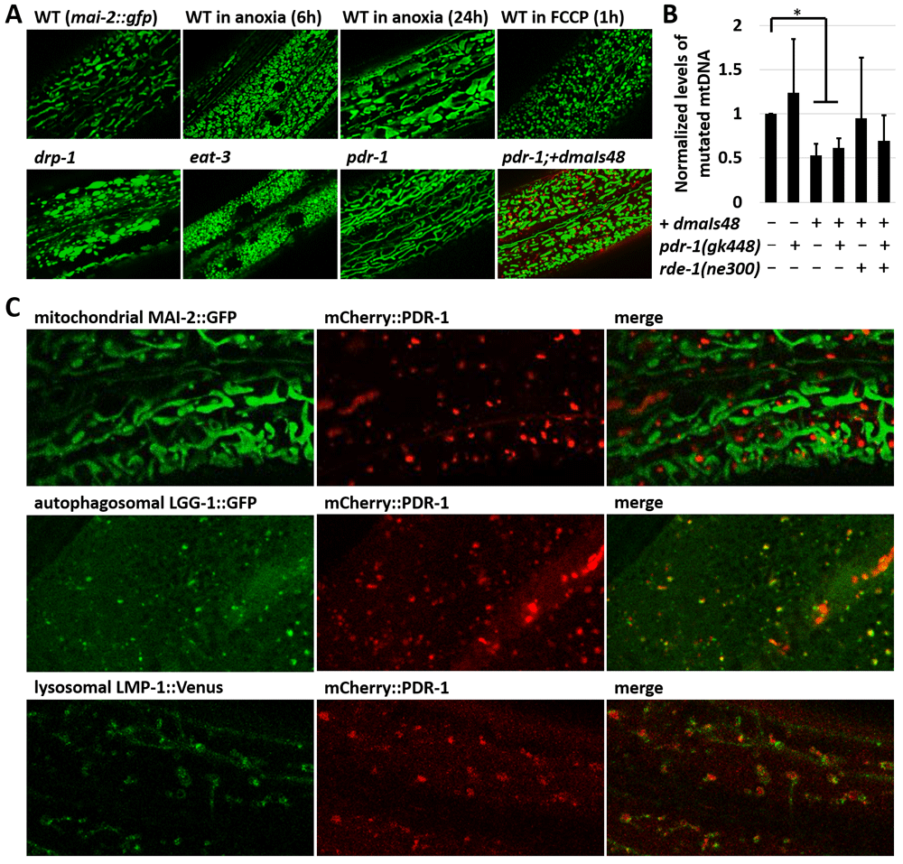
Figure 1. The mCherry reporter of C. elegans Parkin (PDR-1) is enriched in the autophagy-lysosomal system.
A. Exemplar green fluorescent protein (GFP) fluorescence confocal images showing mitochondrial morphology in the hypoderm of xmSi(mai-2::GFP) animals under several environmental and genetic stresses, including drp-1(tm1108), eat-3(ad426) and pdr-1(gk448). The PDR-1 reporter expressed from dmaIs48 transgene restores mitochondrial pattern in pdr-1(gk448) mutants. B. Normalized levels of mutated mtDNA in indicated strains carrying uaDf5/+. Data are presented as mean ± standard deviation (SD) with p-values calculated by Wilcoxon Mann–Whitney rank sum test to compare two independent populations, * p<0.05. C. Confocal GFP and mCherry fluorescence images showing expression pattern of the mCherry::PDR-1 together with mitochondrial reporter xmSi(mai-2::GFP), autophagosomal reporter adIs2122(lgg-1::GFP) and lysosomal reporter dmaIs58(lmp-1::Venus) in the hypoderm. The PDR-1 reporter colocalizes with the autophagosomal and lysosomal reporters.
We used the pdr-1 phenotype to validate the newly generated mCherry reporter of PDR-1. We cloned the entire pdr-1 gene in frame behind the mCherry reporter under ubiquitous promoter rpl-28. Using confocal microscopy we found that the newly generated transgenic strain carrying dmaIs48(rpl-28p::mCherry::pdr-1) exhibits bright fluorescent foci of mCherry::PDR-1 that do not colocalize with GFP reporter for mitochondria MAI-2::GFP, though still reduced mitochondrial fusions in pdr-1 mutants (Figure 1A, 1B). We further investigated the functionality of the mCherry::PDR-1 towards mitochondrial biology by exploring the heteroplasmy of mutated mtDNA using LB138 strain carrying uaDf5/+, a heteroplasmic ~ 3 kb mtDNA truncation22,31,32. We determined that the mCherry::PDR-1 expression significantly reduced levels of truncated mtDNA in WT as well as pdr-1 mutants (Figure 1B).
Since mCherry::PDR-1 appears to be a functional Parkin reporter towards mitochondrial biology, we investigated whether the mCherry::PDR-1 reporter recruits onto mitochondria upon exposure to FCCP by following the PDR-1 subcellular localization for 30 minutes after the exposure to FCCP. Although the FCCP altered the mitochondrial pattern that formed round shape morphology, we did not observe recruitment of the mCherry::PDR-1 onto mitochondria despite its dynamic expression pattern in both standard and stressed conditions; see Figure 1A, and Supplementary Video 1 and 2 in Extended data33. To determine the subcellular localization of the mCherry::PDR-1 foci we crossed the dmaIs48 transgene with the GFP reporter for autophagosome adIs2122(lgg-1p::GFP::lgg-1)34 and GFP(Venus) reporter for lysosome dmaIs58(rpl-28p::lmp-1::Venus)35. The confocal microscopy showed that the mCherry foci colocalize with both (LC3, GABARAP and GATE-16 family) and LMP-1 (LAMP (lysosome-associated membrane protein) homolog) reporters indicating that mCherry::PDR-1 is targeted to autophagosome-lysosome compartments (Figure 1C). The full data associated with the results are available in Underlying data33.
Endogenous RNAi pathway regulates expression of mCherry::PDR-1
Because of the striking mCherry::PDR-1 expression pattern in dmaIs48 animals we aimed to identify novel regulators of the PDR-1 subcellular expression by unbiased approaches. We performed a forward genetic screen using EMS mutagenesis to isolate mutants with an altered expression pattern. From a screen of approximately 100,000 haploid genomes of the dmaIs48; xmSi(mai-2::GFP) strain we have isolated 10 independent mutants with 100% penetrance of markedly increased red fluorescent signal while the GFP signal was not altered (Table 1, Figure 2A). Confocal microscopy showed that the mutants exhibit increased fluorescent signal in the cytosol as well as increased number and size of the foci (Figure 2B) and revealed accumulated mCherry::PDR-1 reporter across the tissues with a strong signal in intestine and hypoderm (Figure 2C). Interestingly, the expression pattern of accumulated mCherry::PDR-1 in the EMS-derived mutants exhibited reduced dynamics in the hypodermal tissue; see Supplementary Video 3 in Extended data33. Western-Blot analysis using an anti-mCherry antibody showed increased PDR-1 reporter levels in the mutants accompanied with partial processing of the PDR-1 reporter, which is indicated by detection of the shorter variants, compared to parental strain (Figure 2D). Whole genome sequencing of the isolated mutants revealed that 4 mutants carry the protein-changing mutations in the rrf-1 gene. The rrf-1 encodes RNA-directed RNA polymerase and is a component of the WAGO 22G RNAs RNAi pathway that generate siRNA for transcriptional silencing of exogenous RNAs36. Notably, all the remaining isolated mutants carry at least one protein-changing mutation in the genes of the RNAi pathway, such as rde-1, rde-10, ego-1, mut-2 and mut-16 (Table 1). To verify causality of these mutations we employed RNAi against rrf-1, rde-1 and mut-16 using parental strain and, indeed, we observed that all three RNAi conditions increased reporter levels and thus phenocopied isolated mutants. These data show that increased levels of the mCherry::PDR-1 in the isolated EMS-derived mutants are due to loss of transgene silencing activity at the post-transcriptional level. To evaluate functionality of the abundant mCherry::PDR-1 we used loss-of-function allele rde-1(ne300) to induce expression of mCherry::PDR-1 and assessed the heteroplasmy of truncated mtDNA. We determined that the overexpressed mCherry::PDR-1 did not significantly reduce levels of truncated mtDNA neither in WT nor pdr-1 mutants suggesting that the abundant mCherry::PDR-1 might be activity silenced and may induce artificial cellular responses (Figure 1A). The full data associated with the results are available in Underlying data33.
Table 1. Ethyl methanesulfonate (EMS)-derived mutants with abundant Parkin reporter mCherry::PDR-1.
| mutation | chr. | penetrance | Gene | Protein change | Annotation |
|---|
| dma334 | I | 100% | rde-10 | splicing | RNA interference defective protein |
| dma330 | I | 100% | mut-2 | W164STOP | Mutator |
| dma337 | I | 100% | rrf-1 | Q52STOP | RNA-dependent RNA polymerase |
| dma338 | I | 100% | rrf-1 | A944T | RNA-dependent RNA polymerase |
| dma339 | I | 100% | rrf-1 | G648R | RNA-dependent RNA polymerase |
| dma330 | I | 100% | rrf-1 | R1256STOP | RNA-dependent RNA polymerase |
| dma327 | I | 100% | ego-1 | D1305N | RNA-dependent RNA polymerase |
| dma333 | I | 100% | mut-16 | Q808STOP | Mutator |
| dma341 | V | 100% | rde-1 | P963L | Component of RNA-induced silencing complex |
| dma325 | V | 100% | rde-1 | splicing | Component of RNA-induced silencing complex |
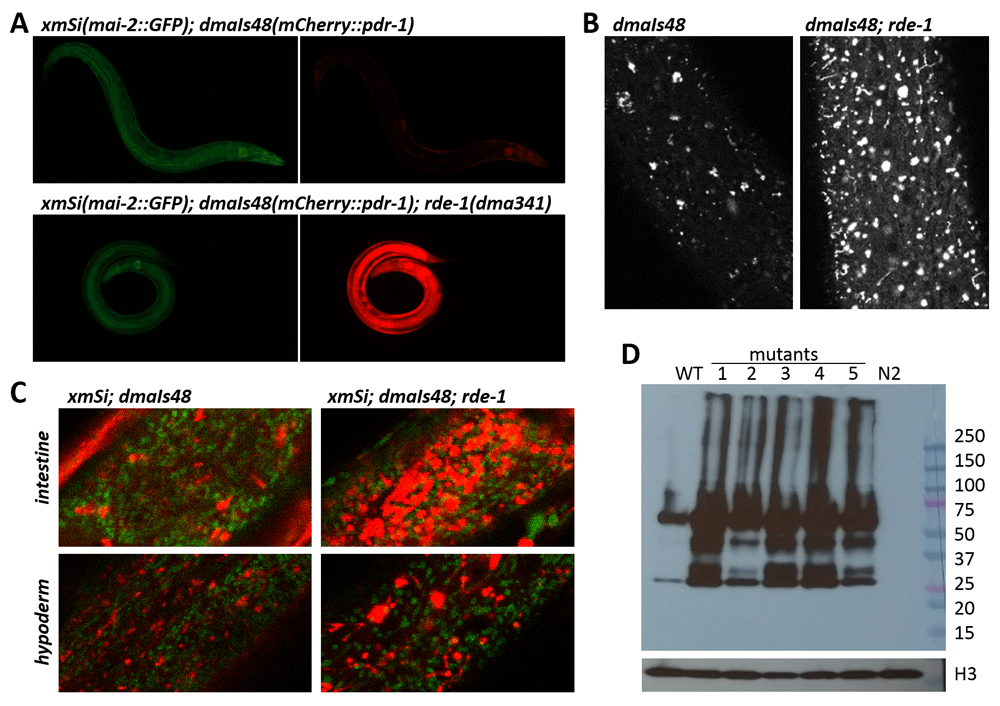
Figure 2 . The abundant C. elegans Parkin (PDR-1) reporter in ethyl methanesulfonate (EMS)-derived mutants.
A. Exemplar green fluorescent protein (GFP) (left) and mCherry (right) fluorescence images showing mCherry::PDR-1 and MAI-2::GFP expression in WT and rde-1(dma341) mutant. Only mCherry::PDR-1 expression is increased in rde-1 mutants. B. Fluorescence confocal images of the hypoderm showing the expression pattern of mCherry::PDR-1 in WT and rde-1(dma341) mutant. C. Confocal view of merged MAI-2::GFP and mCherry::PDR-1 fluorescence in intestine (upper) and hypoderm (lower) of WT and rde-1(dma341) mutant. D. The mCherry::PDR-1 protein levels determined by sodium dodecyl sulfate-polyacrylamide gel electrophoresis (SDS PAGE) followed by western blot analysis using anti-mCherry Ab and anti-histone H3 Ab. C. elegans protein lysates from various genetic backgrounds were analyzed. 50 µg protein samples were loaded per lane. WT represents parental strain carrying dmaIs48(mCherry::pdr-1). Analyzed mutants with dmaIs48 are as follows: 1. rde-1(dma325), 2. ego-1(dma327), 3. rrf-1(dma339), 4. rde-1(dma341), 5. mut-2(dma340). N2 represents wild isolate N2.
Hybrid ubiquitins and mitochondrial ANT and ATPase regulate mCherry::PDR-1 levels
Although unbiased mutagenesis revealed regulators of the transgenic mCherry::PDR-1 expression, novel alleles regulating PDR-1 function have not been isolated. Therefore, we employed Co-immunoprecipitation (CoIP) using RFP-trap to identify putative interacting proteins with the mCherry::PDR-1 reporter. We used crude extract of both parental strain dmaIs48 as well as isolated mutant dmaIs48; rde-1(dma341) as the baits. One-dimensional sodium dodecyl sulfate-polyacrylamide gel electrophoresis (SDS PAGE) revealed five bands of protein pull down that did not differ in size between parental and mutant samples (Figure 3A). We analyzed these bands by gel digest followed by liquid chromatography-tandem mass spectrometry (GeLC-MS/MS) analysis and identified putative interactors with the highest score for adenosine triphosphate (ATP)-Citrate Lyase ACLY-1/hACLY, Heat Shock 70kDa HSP-1/hHSPA1 and hHSPA8, 14-3-3-like proteins PAR-5/hYWHAZ and FTT-2/hYWHAZ and several Ribosomal Proteins. Notably, subunits of the Vacuolar ATPase (VHA-8, VHA-12, VHA-13), and several mitochondrial proteins, such as mitochondrial Adenine Nucleotide Translocator ANT-1.1/hANT, ATP synthase subunits ATP-1/hATP5F1A and ATP-2 hATP5F1B, Voltage Dependent Anion Channel VDAC-1/hVDAC1, or Heat Shock Protein HSP-60/hHSPD1, have been also identified; see Supplementary Table 1 in Extended data33.
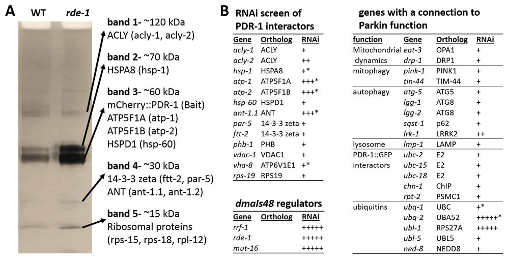
Figure 3. Protein interactor and RNAi screen identify regulators of C. elegans Parkin (PDR-1) abundance.
A. Sodium dodecyl sulfate-polyacrylamide gel electrophoresis (SDS PAGE) followed by Coomassie staining of mCherry::PDR-1 pull down. Red fluorescent protein (RFP)-trap was used for Co-immunoprecipitation of protein extracts from dmaIs48(mCherry::pdr-1) and dmaIs48;rde-1(dma341) animals. Mass spectrometry (MS) analysis of individual bands was used to identify putative interactors. Proteins with the highest peptide coverage are indicated. B. Ribonucleic acid interference (RNAi) screen of indicated genes for abundant mCherry::PDR-1.The “plus” symbol indicates mCherry fluorescence intensity and the “star” symbol indicates larval arrest phenotype based on visual examination.
Next, we performed reverse genetic screen by RNAi against putative PDR-1 regulators. First, we have systematically screened the genes encoding the identified PDR-1 interactors for altered expression pattern of the mCherry::PDR-1 reporter. We found that mCherry::PDR-1 abundance was enhanced by RNAi against mitochondrial ant-1.1, atp-1 and atp-2 accompanying with developmental arrest at early stages of larva development. Because ANT has been recently identified driver of mitophagy trough Parkin activity37, we hypothesized that impaired mitophagy may lead to Parkin accumulation. However, the RNAi against pink-1/hPINK1 and tin-44/hTIM44 (Transport to Inner Mitochondrial Membrane (yeast TIM)), the components of the ANT/Parkin-mediated mitophagy did not increase the expression of mCherry::PDR-1. We have employed RNAi against other genes with a connection to Parkin function, including previously identified PDR-1::GFP interactors10 or genes encoding ubiquitin, and found that inactivation of hybrid ubiquitin genes ubl-1 (Ubiquitin-like) and ubq-2 (ubiquitin) increased the abundance of mCherry::PDR-1, accompanying with developmental arrest at early stages of larva development. These data suggest that accumulation of the PDR-1 reporter is not related to impaired initiation of mitophagy but rather due to impaired mitochondrial function, ubiquitination or proteasomal degradation. The full data associated with the results are available in Underlying data33.
Accumulated mCherry::PDR-1 disrupts lysosomal dynamics
Since increased expression of the PDR-1 reporter in the rde-1 mutants lost its dynamic pattern in the hypoderm we asked whether its subcellular localization differ compared to the parental strain. We crossed the mCherry::pdr-1 reporter with autophagy lgg-1::GFP and hypodermal lmp-1::Venus reporters and found that accumulated PDR-1:mCherry in the rde-1 mutants does not colocalize with LGG-1:GFP reporter, though the ant-1.1 and ubl-1 knockdowns upon mild RNAi still showed partial colocalization (Figure 4A). Interestingly, all conditions, including rde-1(dma341), RNAi ant-1.1 and RNAi ubl-1, induced expression of both mCherry::PDR-1 and LMP-1::Venus reporters when expressed together, and colocalized in the lysosomal-like structures. Notably, the dynamics of the PDR-1 reporter in ubl-1 knockdowns was interrupted (Supplementary Video 4, Extended data33) compared to control and the LMP-1::Venus expression was increased at the plasma membrane where mCherry::PDR-1 has not been detected (Figure 4A). Because the lmp-1::Venus transgene also uses the same ubiquitous promotor rpl-28 as mCherry::PDR-1, we asked whether accumulated LMP-1::Venus in animals with accumulated mCherry::PDR-1 is dependent on the co-expressed mCherry::pdr-1. We employed RNAi against mut-16 and ubq-2, the most striking RNAi conditions that induce mCherry::PDR-1 accumulation, in the animals carrying only the lmp-1::Venus transgene. We found that expression of LMP-1::Venus was not induced and retained its dynamics in respective knockdowns indicating that significant accumulation of the LMP-1::Venus in mut-16 and ubq-2 knockdowns is dependent on the co-expression of the mCherry::PDR-1 (Figure 5A and 5B). These data indicate that lysosomal dynamics are disrupted due to the abundant mCherry::PDR-1.The full data associated with the results are available in Underlying data33.
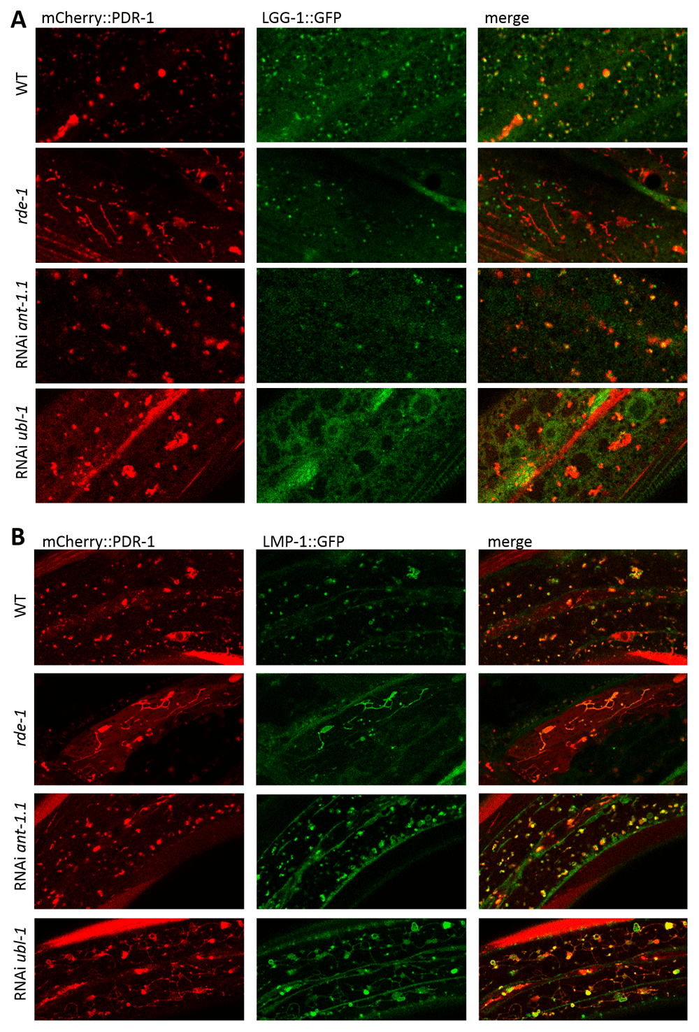
Figure 4. Abundant C. elegans Parkin (PDR-1) reporter alters lysosomal morphology.
A. Enlarged view of confocal green fluorescent protein (GFP) and mCherry fluorescence images showing expression pattern of the autophagosomal reporter adIs2122(lgg-1::GFP) and PDR-1 reporter dmaIs48(mCherry::pdr-1) in the hypoderm of indicated mutants/knockdowns. B. Enlarged view of confocal GFP and mCherry fluorescence images showing lysosomal reporter dmaIs58(lmp-1::Venus) and PDR-1 reporter dmaIs48(mCherry::pdr-1) in the hypoderm of indicated mutants/knockdowns.
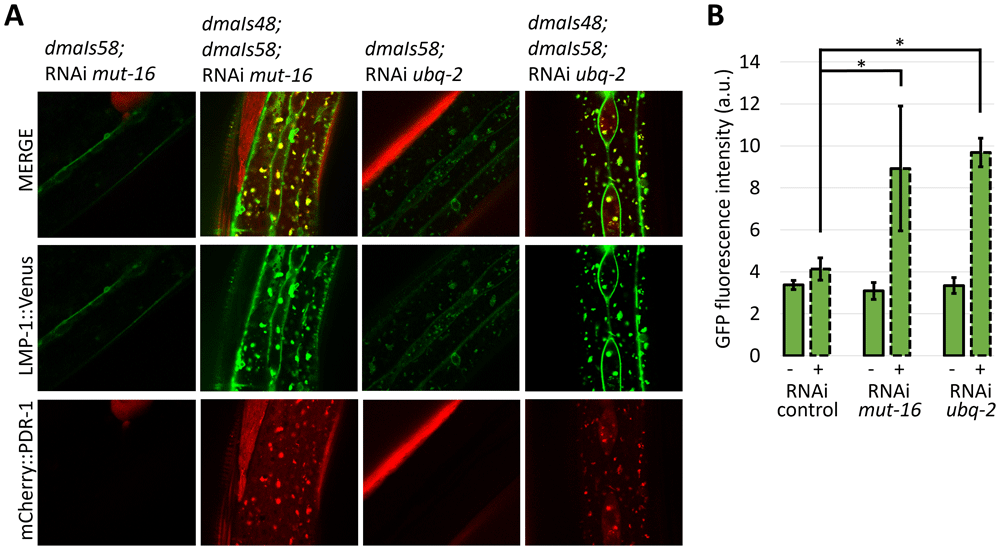
Figure 5. Abundant C. elegans Parkin (PDR-1) reporter increases expression of lysosomal reporter LMP-1. A.
Confocal green fluorescent protein (GFP) and mCherry fluorescence images showing lysosomal reporter dmaIs58(lmp-1::Venus) in the absence and presence of the PDR-1 reporter dmaIs48(mCherry::pdr-1) upon various ribonucleic acid interference (RNAi) assay. B. Quantification of the GFP fluorescence intensity detected in the widefield image of animals carrying dmaIs58(lmp-1::Venus) in the absence (-) and presence (+) of the dmaIs48(mCherry::pdr-1) upon various RNAi assay. 3 animals from independent biological replicates were analysed. Data are presented as mean ± standard deviation (SD) with p-values calculated by one-way analysis of variance (ANOVA) with Bonferroni correction, * p<0.05.
The mCherry::PDR-1 alters formation of alpha-Synuclein inclusions
We then questioned whether disrupted autophagy-lysosomal dynamics in animals with accumulated mCherry::PDR-1 in cytosol and lysosomes alters proteostasis in the C. elegans model of the Parkinson’s disease. We used a previously well-established GFP-based reporter of alpha-Synuclein aggregation in body wall muscle cells uonEx1(unc-54p::alpha-Synuclein::YFP) and with confocal microscopy quantified the size of the YFP fluorescent foci in the presence and absence of the overexpressed mCherry::PDR-1. First, we determined that the size of the foci (inclusions of aggregated signal) was reduced in the young adult animals co-expressing mCherry::PDR-1. In addition, fluorescent microscopy together with western blot analysis revealed that the expression levels of the alpha-Synuclein::YFP were reduced in the presence of the mCherry::PDR-1 (Figure 6). Next, we asked whether animals with accumulated mCherry::PDR-1, such as mut-16 and ubq-2 knockdowns, would further reduce expression of the alpha-Synuclein::YFP. We found that RNAi against mut-16 reduced expression levels of alpha-Synuclein::YFP in the absence as well as in the presence of the mCherry::PDR-1 accompanied with reduction of the YFP foci. These data suggest that reduction of formed alpha-Synuclein::YFP foci is a result of its reduced expression levels. Interestingly, the RNAi against ubq-2 increased expression levels of alpha-Synuclein::YFP in the absence of the mCherry::PDR-1, while the ubq-2 knockdowns with accumulated mCherry::PDR-1 exhibited significantly increased formation of the alpha-Synuclein::YFP foci despite its reduced expression levels (Figure 6). These data indicate that accumulated mCherry::PDR-1 due to inactivated ubq-2 alters cellular processing of exogenously expressed alpha-Synuclein. The full data associated with the results are available in Underlying data33.
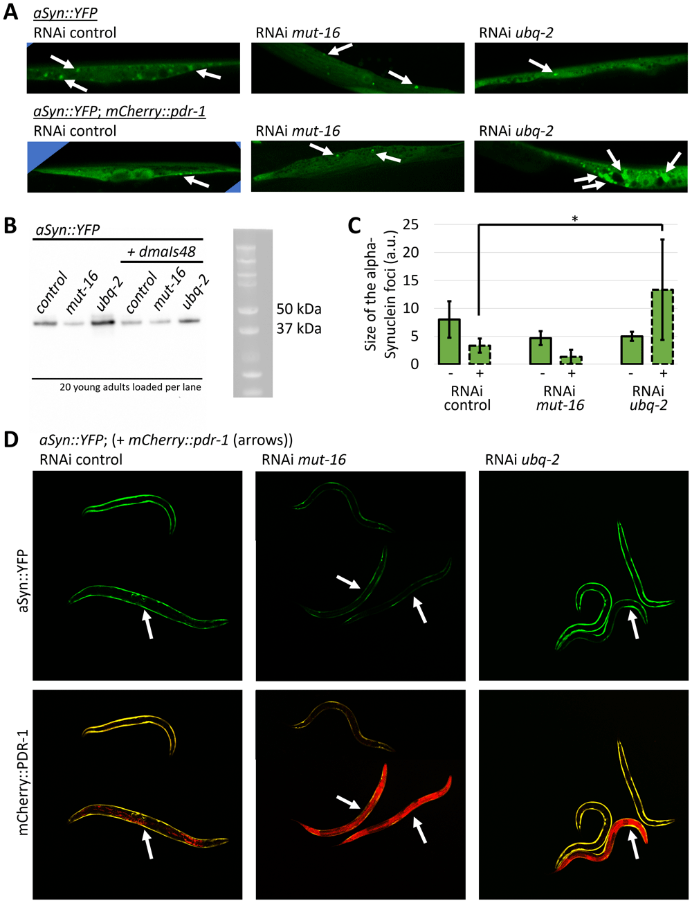
Figure 6. The C. elegans Parkin (PDR-1) alters alpha-Synuclein processing.
A. Representative confocal green fluorescent protein (GFP) fluorescence images of the body wall muscle cells in animals carrying uonEx1(alpha-Synuclein::YFP) in the absence and presence of the dmaIs48(mCherry::pdr-1) upon various ribonucleic acid interference (RNAi) assay. B The alpha-Synuclein::YFP protein levels as determined by sodium dodecyl sulfate-polyacrylamide gel electrophoresis (SDS PAGE) followed by western blot using anti-alpha-Synuclein Ab (Abnova MAB5383). Twenty young adult animals collected from three biological replicates upon various RNAi were loaded per lane. C. Size quantification of detected fluorescent foci in body wall muscle cells expressing alpha-Synuclein::YFP in the absence (-) and presence (+) of the dmaIs48(mCherry::pdr-1) upon various RNAi assay. 3 body wall muscle cells from independent biological replicates were analysed. D. Exemplar GFP and mCherry fluorescence images of animals carrying uonEx1(alpha-Synuclein::YFP) in the absence and presence of the dmaIs48(mCherry::pdr-1) upon indicated RNAi assay. Arrows indicate animals carrying dmaIs48(mCherry::pdr-1) which exhibit reduced GFP fluorescence associated with expression levels of alpha-Synuclein reporter. Data are presented as mean ± standard deviation (SD) with p-values calculated by one-way analysis of variance (ANOVA) with Bonferroni correction, * p<0.05.
Discussion
Parkin has an active role during proteotoxic as well as mitochondrial stress. Understanding the regulatory mechanisms distinguishing specific Parkin targets may explain its role in Parkinson’s disease, tumorigenesis, and other Parkin-associated diseases. Here we present a new functional mCherry reporter of C. elegans Parkin (PDR-1) that exhibits autophagy-lysosomal targeting and disrupts the dynamic movements of this system upon its intracellular accumulation. We demonstrate that the mCherry::pdr-1 transgene reduced mitochondrial fusion in pdr-1 mutants and reduced levels of mutated mtDNA, indicating that the mCherry::PDR-1 reporter retains some functionality towards mitochondrial biology. On the other hand, we cannot exclude the possibility that mCherry tag at the N-terminal autoinhibitory Ubl domain may alter its regulation, targeting and/or subcellular localization. In addition, differently expressed mCherry::pdr-1 compared to endogenous pdr-1 gene due to activity of the rpl-28 promoter as well as overexpressed mCherry::pdr-1 may induce artificial responses that need to be evaluated in future studies.
Our preliminary protein interactor screen revealed several putative PDR-1 targets, such as VDAC-1 or HSP-1 which are the orthologs of previously identified human Parkin interactors VDAC1 and HSPA838,39. Interestingly, several identified interactors, such as ANT-1.1/hANT and ATP-1/hATP5F1A, are components of the inner mitochondrial membrane, and therefore we suspect that they are targeted by Parkin during the process of mitochondrial turnover, when the outer mitochondrial membrane is ruptured. Congruently, a similar assay with human Parkin identified several mitochondrial proteins whose direct physical interaction is proposed to be taken part during mitophagy after disruption of the mitochondrial membranes40. In fact, Parkin-mediated mitophagy requires both the proteasome system for outer- and autophagy system for inner-mitochondrial membrane rupture41.
From RNAi screen directed against putative PDR-1 interactors we identified hybrid ubiquitin genes ubq-2 and ubl-1 or mitochondrial ant-1.1, atp-1 and atp-2 as regulators of the PDR-1 abundance. How exactly these genes maintain mCherry::PDR-1 levels is not clear. Both ubq-2/hUBA52 and ubl-1/hRPS27A encode a single copy of the ubiquitin domain which can be cleaved from the ribosomal subunits L40 and S27a, respectively42,43. It is therefore possible that UBL-1 and UBQ-2 may serve as the ubiquitin substrates for the ATP-dependent PDR-1 auto-ubiquitination. The western blot analysis of overexpressed PDR-1 reporter indicated its partial processing of the mCherry::PDR-1 which supports this hypothesis. Notably, we also found that inactivation of the ubq-2 increased levels of the exogenously expressed alpha-Synuclein reporter in body wall muscle cells while the co-expression of the mCherry::pdr-1 reduced alpha-Synuclein levels indicating a role of UBQ-2 along with expressed PDR-1 reporter in the regulation of alpha-Synuclein expression levels. Congruently, beneficial effect of Parkin on alpha-Synuclein toxicity has been demonstrated in midbrain cell cultures44 as well as in a Drosophila model of Parkinson’s disease45. Parkin can mediate K63-linked poly-ubiquitination of alpha-Synuclein to trigger its degradation through increased aggregation46,47. Whether UBQ-2/hUBA52 serves as a ubiquitin substrate for the PDR-1/hParkin ubiquitin ligase activity targeting alpha-Synuclein merits further studies.
Intriguingly, the abundant PDR-1 reporter due to inactivated ubq-2 altered also alpha-Synuclein processing characterized by enlarged fluorescent foci. Since the RNAi of mitochondrial and ubiquitin genes, which induce abundance of the PDR-1 reporter, results in the developmental arrest we have not been able to investigate whether the enlarged aggregated alpha-Synuclein signal may be beneficial and less toxic than diffused alpha-Synuclein, as observed in other model systems48. Nonetheless, we hypothesize that abundant PDR-1 may sequester intracellular ubiquitin and thus prevent its binding to alternative E3 ubiquitin ligases. Depleted ubiquitin would then result in insufficient ubiquitination of damaged proteins with consequent impairment of the lysosomal dynamics and proteostasis. Interestingly, the abundant PDR-1 along with the affected lysosomal system due to inactivated mut-16 did not alter alpha-Synuclein processing. We believe that the reduced formation of the aggregated alpha-Synuclein signal in mut-16 knockdowns is due to reduced expression levels of alpha-Synuclein reporter in these animals.
Interestingly, lysosomal disfunction with altered lysosomal morphology, that resemble lysosomal morphology in C. elegans with abundant PDR-1, has been observed also in the Parkin-deficient cell line49,50 indicating that both inactivated as well as abundant Parkin may affect lysosomal morphology and function. Taken together, since the enlarged inclusions of alpha-Synuclein in our model system resemble Parkinson’s disease pathology, we propose that an affected lysosomal system upon PDR-1 abundance may be one of the possible pathological mechanisms of mutated Parkin.
Ethics and consent
In this project we utilize a roundworm Caenorhabditis elegans as a genetic model. These microscopic worms are invertebrate and, therefore, they do not fall within the scope of EU Directive 2010/63/EU on the protection of animals used for scientific purposes. No ethical approval was required for this research.
Data availability
Underlying data
Zenodo: Caenorhabditis elegans Parkin: regulators of its abundance and role in autophagy-lysosomal dynamics. https://doi.org/10.5281/zenodo.594006533.
This project contains the following underlying data:
- List of Underlying Data.docx (contains description of individual underlying data files in each folder).
- Figure 1.zip (The mCherry reporter of C. elegans Parkin (PDR-1) is enriched in the autophagy-lysosomal system).
- Figure 2.zip (The abundant PDR-1 reporter in ethyl methanesulfonate (EMS)-derived mutants).
- Figure 3.zip (Protein interactor and RNAi screen identify regulators of PDR-1 abundance).
- Figure 4.zip (Abundant PDR-1 reporter alters lysosomal morphology).
- Figure 5.zip (Abundant PDR-1 reporter increases expression of lysosomal reporter LMP-1).
- Figure 6.zip (The PDR-1 alters alpha-Synuclein processing).
Acknowledgments
We thank R. E. Navarro lab and Caenorhabditis Genetics Center for C. elegans strains. Mass Spectrometry was provided by the Mass Spectrometry Resource at UCSF (A.L. Burlingame, Director) supported by the Dr. Miriam and Sheldon G. Adelson Medical Research Foundation (AMRF) and the NIH-NIGMS. R.V. was supported by International Mobility Program of the Autonomous Province of Bolzano.
Faculty Opinions recommendedReferences
- 1.
Shimura H, Hattori N, Kubo S, et al.:
Familial Parkinson disease gene product, parkin, is a ubiquitin-protein ligase.
Nat Genet.
2000; 25(3): 302–5. PubMed Abstract
| Publisher Full Text
- 2.
Nuytemans K, Theuns J, Cruts M, et al.:
Genetic etiology of Parkinson disease associated with mutations in the SNCA, PARK2, PINK1, PARK7, and LRRK2 genes: a mutation update.
Hum Mutat.
2010; 31(7): 763–80. PubMed Abstract
| Publisher Full Text
| Free Full Text
- 3.
Narendra D, Walker JE, Youle R:
Mitochondrial Quality Control Mediated by PINK1 and Parkin: Links to Parkinsonism.
Cold Spring Harb Perspect Biol.
2012; 4(11): a011338. PubMed Abstract
| Publisher Full Text
| Free Full Text
- 4.
Cookson MR, Lockhart PJ, McLendon C, et al.:
RING finger 1 mutations in Parkin produce altered localization of the protein.
Hum Mol Genet.
2003; 12(22): 2957–65. PubMed Abstract
| Publisher Full Text
- 5.
Seirafi M, Kozlov G, Gehring K:
Parkin structure and function.
FEBS J.
2015; 282(11): 2076–88. PubMed Abstract
| Publisher Full Text
| Free Full Text
- 6.
Narendra D, Tanaka A, Suen DF, et al.:
Parkin is recruited selectively to impaired mitochondria and promotes their autophagy.
J Cell Biol.
2008; 183(5): 795–803. PubMed Abstract
| Publisher Full Text
| Free Full Text
- 7.
Kazlauskaite A, Martínez-Torres RJ, Wilkie S, et al.:
Binding to serine 65-phosphorylated ubiquitin primes Parkin for optimal PINK1-dependent phosphorylation and activation.
EMBO Rep.
2015; 16(8): 939–54. PubMed Abstract
| Publisher Full Text
| Free Full Text
- 8.
Gladkova C, Maslen SL, Skehel JM, et al.:
Mechanism of parkin activation by PINK1.
Nature.
2018; 559(7714): 410–4. PubMed Abstract
| Publisher Full Text
| Free Full Text
- 9.
Koyano F, Okatsu K, Kosako H, et al.:
Ubiquitin is phosphorylated by PINK1 to activate parkin.
Nature.
2014; 510(7503): 162–6. PubMed Abstract
| Publisher Full Text
- 10.
Springer W, Hoppe T, Schmidt E, et al.:
A Caenorhabditis elegans Parkin mutant with altered solubility couples α-synuclein aggregation to proteotoxic stress.
Hum Mol Genet.
2005; 14(22): 3407–23. PubMed Abstract
| Publisher Full Text
- 11.
Ahier A, Dai CY, Kirmes I, et al.:
PINK1 and parkin shape the organism-wide distribution of a deleterious mitochondrial genome.
Cell Rep.
2021; 35(9): 109203. PubMed Abstract
| Publisher Full Text
- 12.
Luz AL, Rooney JP, Kubik LL, et al.:
Mitochondrial Morphology and Fundamental Parameters of the Mitochondrial Respiratory Chain Are Altered in Caenorhabditis elegans Strains Deficient in Mitochondrial Dynamics and Homeostasis Processes.
PLoS One.
2015; 10(6): e0130940. PubMed Abstract
| Publisher Full Text
| Free Full Text
- 13.
Cooper JF, Machiela E, Dues DJ, et al.:
Activation of the mitochondrial unfolded protein response promotes longevity and dopamine neuron survival in Parkinson’s disease models.
Sci Rep.
2017; 7(1): 16441. PubMed Abstract
| Publisher Full Text
| Free Full Text
- 14.
Cabello J, Sämann J, Gómez-Orte E, et al.:
PDR-1/hParkin negatively regulates the phagocytosis of apoptotic cell corpses in Caenorhabditis elegans.
Cell Death Dis.
2014; 5(3): e1120. PubMed Abstract
| Publisher Full Text
| Free Full Text
- 15.
Trempe JF, Chen CXQ, Grenier K, et al.:
SH3 Domains from a Subset of BAR Proteins Define a Ubl-Binding Domain and Implicate Parkin in Synaptic Ubiquitination.
Mol Cell.
2009; 36(6): 1034–47. PubMed Abstract
| Publisher Full Text
- 16.
Cao M, Milosevic I, Giovedi S, et al.:
Upregulation of Parkin in Endophilin Mutant Mice.
J Neurosci.
2014; 34(49): 16544–9. PubMed Abstract
| Publisher Full Text
| Free Full Text
- 17.
Song P, Trajkovic K, Tsunemi T, et al.:
Parkin Modulates Endosomal Organization and Function of the Endo-Lysosomal Pathway.
J Neurosci.
2016; 36(8): 2425–37. PubMed Abstract
| Publisher Full Text
| Free Full Text
- 18.
Shimura H, Schlossmacher MG, Hattori N, et al.:
Ubiquitination of a new form of α-synuclein by parkin from human brain: implications for Parkinson's disease.
Science.
2001; 293(5528): 263–9. PubMed Abstract
| Publisher Full Text
- 19.
Chung KKK, Thomas B, Li X, et al.:
S-Nitrosylation of Parkin Regulates Ubiquitination and Compromises Parkin’s Protective Function.
Science.
2004; 304(5675): 1328–31. PubMed Abstract
| Publisher Full Text
- 20.
Vandiver MS, Paul BD, Xu R, et al.:
Sulfhydration mediates neuroprotective actions of parkin.
Nat Commun.
2013; 4(1): 1626. PubMed Abstract
| Publisher Full Text
| Free Full Text
- 21.
Vozdek R, Long Y, Ma DK:
The receptor tyrosine kinase HIR-1 coordinates HIF-independent responses to hypoxia and extracellular matrix injury.
Sci Signal.
2018; 11(550): eaat0138. PubMed Abstract
| Publisher Full Text
- 22.
Valenci I, Yonai L, Bar-Yaacov D, et al.:
Parkin modulates heteroplasmy of truncated mtDNA in Caenorhabditis elegans.
Mitochondrion.
2015; 20: 64–70. PubMed Abstract
| Publisher Full Text
- 23.
Livak KJ, Schmittgen TD:
Analysis of relative gene expression data using real-time quantitative PCR and the 2(-Delta Delta C(T)) Method.
Method.
2001; 25(4): 402–8. PubMed Abstract
| Publisher Full Text
- 24.
Brenner S:
The genetics of Caenorhabditis elegans.
Genetics.
1974; 77(1): 71–94. PubMed Abstract
| Publisher Full Text
| Free Full Text
- 25.
Ma DK, Vozdek R, Bhatla N, et al.:
CYSL-1 Interacts with the O2-Sensing Hydroxylase EGL-9 to Promote H2S-Modulated Hypoxia-Induced Behavioral Plasticity in C. elegans.
Neuron.
2012; 73(5): 925–40. PubMed Abstract
| Publisher Full Text
| Free Full Text
- 26.
Minevich G, Park DS, Blankenberg D, et al.:
CloudMap: A Cloud-Based Pipeline for Analysis of Mutant Genome Sequences.
Genetics.
2012; 192(4): 1249–69. PubMed Abstract
| Publisher Full Text
| Free Full Text
- 27.
Blankenberg D, Kuster GV, Coraor N, et al.:
Galaxy: A Web-Based Genome Analysis Tool for Experimentalists.
Curr Protoc Mol Biol.
2010; 89(1): Unit 19.10.1-21. PubMed Abstract
| Publisher Full Text
| Free Full Text
- 28.
Kamath RS, Ahringer J:
Genome-wide RNAi screening in Caenorhabditis elegans.
Methods.
2003; 30(4): 313–21. PubMed Abstract
| Publisher Full Text
- 29.
Guan S, Price JC, Prusiner SB, et al.:
A data processing pipeline for mammalian proteome dynamics studies using stable isotope metabolic labeling.
Mol Cell Proteomics.
2011; 10(12): M111.010728. PubMed Abstract
| Publisher Full Text
| Free Full Text
- 30.
Fernández-Cárdenas LP, Villanueva-Chimal E, Salinas LS, et al.:
Caenorhabditis elegans ATPase inhibitor factor 1 (IF1) MAI-2 preserves the mitochondrial membrane potential (Δψm) and is important to induce germ cell apoptosis.
PLoS One.
2017; 12(8): e0181984. PubMed Abstract
| Publisher Full Text
| Free Full Text
- 31.
Lin YF, Schulz AM, Pellegrino MW, et al.:
Maintenance and propagation of a deleterious mitochondrial genome by the mitochondrial unfolded protein response.
Nature.
2016; 533(7603): 416–419. PubMed Abstract
| Publisher Full Text
| Free Full Text
- 32.
Gitschlag BL, Kirby CS, Samuels DC, et al.:
Homeostatic Responses Regulate Selfish Mitochondrial Genome Dynamics in C. elegans.
Cell Metab.
2016; 24(1): 91–103. PubMed Abstract
| Publisher Full Text
| Free Full Text
- 33.
Vozdek R, Wang B, Li KH, et al.:
Caenorhabditis elegans Parkin: regulators of its abundance and role in autophagy-lysosomal dynamics.
Zenodo.
2021. http://www.doi.org/10.5281/zenodo.5940065
- 34.
Kang C, You Y, Avery L:
Dual roles of autophagy in the survival of Caenorhabditis elegans during starvation.
Genes Dev.
2007; 21(17): 2161–71. PubMed Abstract
| Publisher Full Text
| Free Full Text
- 35.
Luo S, Wang X, Bai M, et al.:
The conserved autoimmune-disease risk gene TMEM39A regulates lysosome dynamics.
Proc Natl Acad Sci U S A.
2021; 118(6): e2011379118. PubMed Abstract
| Publisher Full Text
| Free Full Text
- 36.
Billi AC, Fischer SEJ, Kim JK:
Endogenous RNAi pathways in C. elegans.
WormBook.
2014; 1–49. PubMed Abstract
| Publisher Full Text
| Free Full Text
- 37.
Hoshino A, Wang W, Wada S, et al.:
The ADP/ATP translocase drives mitophagy independent of nucleotide exchange.
Nature.
2019; 575(7782): 375–9. PubMed Abstract
| Publisher Full Text
| Free Full Text
- 38.
Geisler S, Holmström KM, Skujat D, et al.:
PINK1/Parkin-mediated mitophagy is dependent on VDAC1 and p62/SQSTM1.
Nat Cell Biol.
2010; 12(2): 119–31. PubMed Abstract
| Publisher Full Text
- 39.
Imai Y, Soda M, Hatakeyama S, et al.:
CHIP Is Associated with Parkin, a Gene Responsible for Familial Parkinson’s Disease, and Enhances Its Ubiquitin Ligase Activity.
Mol Cell.
2002; 10(1): 55–67. PubMed Abstract
| Publisher Full Text
- 40.
Zanon A, Rakovic A, Blankenburg H, et al.:
Profiling of Parkin-binding partners using tandem affinity purification.
PLoS One.
2013; 8(11): e78648. PubMed Abstract
| Publisher Full Text
| Free Full Text
- 41.
Wei Y, Chiang WC, Sumpter R Jr, et al.:
Prohibitin 2 Is an Inner Mitochondrial Membrane Mitophagy Receptor.
Cell.
2017; 168(1–2): 224–238.e10. PubMed Abstract
| Publisher Full Text
| Free Full Text
- 42.
Komander D, Clague MJ, Urbé S:
Breaking the chains: structure and function of the deubiquitinases.
Nat Rev Mol Cell Biol.
2009; 10(8): 550–63. PubMed Abstract
| Publisher Full Text
- 43.
Kobayashi M, Oshima S, Maeyashiki C, et al.:
The ubiquitin hybrid gene UBA52 regulates ubiquitination of ribosome and sustains embryonic development.
Sci Rep.
2016; 6(1): 36780. PubMed Abstract
| Publisher Full Text
| Free Full Text
- 44.
Petrucelli L, O’Farrell C, Lockhart PJ, et al.:
Parkin protects against the toxicity associated with mutant α-Synuclein: proteasome dysfunction selectively affects catecholaminergic neurons.
Neuron.
2002; 36(6): 1007–19. PubMed Abstract
| Publisher Full Text
- 45.
Rana A, Rera M, Walker DW:
Parkin overexpression during aging reduces proteotoxicity, alters mitochondrial dynamics, and extends lifespan.
Proc Natl Acad Sci U S A.
2013; 110(21): 8638–43. PubMed Abstract
| Publisher Full Text
| Free Full Text
- 46.
Olzmann JA, Li L, Chudaev MV, et al.:
Parkin-mediated K63-linked polyubiquitination targets misfolded DJ-1 to aggresomes via binding to HDAC6.
J Cell Biol.
2007; 178(6): 1025–38. PubMed Abstract
| Publisher Full Text
| Free Full Text
- 47.
Tan JMM, Wong ESP, Kirkpatrick DS, et al.:
Lysine 63-linked ubiquitination promotes the formation and autophagic clearance of protein inclusions associated with neurodegenerative diseases.
Hum Mol Genet.
2008; 17(3): 431–9. PubMed Abstract
| Publisher Full Text
- 48.
Winner B, Jappelli R, Maji SK, et al.:
In vivo demonstration that α-Synuclein oligomers are toxic.
Proc Natl Acad Sci U S A.
2011; 108(10): 4194–9. PubMed Abstract
| Publisher Full Text
| Free Full Text
- 49.
Guerra F, Girolimetti G, Beli R, et al.:
Synergistic Effect of Mitochondrial and Lysosomal Dysfunction in Parkinson’s Disease.
Cells.
2019; 8(5): 452. PubMed Abstract
| Publisher Full Text
| Free Full Text
- 50.
Okarmus J, Bogetofte H, Schmidt SI, et al.:
Lysosomal perturbations in human dopaminergic neurons derived from induced pluripotent stem cells with PARK2 mutation.
Sci Rep.
2020; 10(1): 10278. PubMed Abstract
| Publisher Full Text
| Free Full Text