Introduction
Nowadays neurodegenerative research has increasingly moved beyond a brain-centred approach to adopt a multisystemic strategy that emphasizes the role of the microbiota and its interactions with the immune, metabolic, and other systems (Clarke et al., 2013; Cryan et al., 2019; Erny et al., 2015; Lyte, 2014). Indeed, the action of the microbiota is not restricted to the gut (Garcia-Rios et al., 2019; Garcia-Rios et al., 2017; Santos-Marcos et al., 2019b), as its action extends to the central nervous system via the gut-brain (GB) axis (Frost et al., 2014).
Current literature estimates that bacteria in the human gut are comparable to the number of host cells, with approximately 232 M genes (Sender et al., 2016; Tierney et al., 2019). The phyla Firmicutes and Bacteroidetes represent circa 75% of the gut bacteria, contributing to host homeostasis (Eckburg et al., 2005). The gut microbiota (GM) is responsible for the interaction between the host and some environmental factors such as diet and drugs (David et al., 2014; Vich Vila et al., 2020). Furthermore, it is involved in the development of some organs such as the enteric nervous system (ENS), and is highly relevant in the smooth running of the host's innate and adaptative immune system and metabolism, boosting the metabolic capacity of humans (Collins et al., 2014; Dabke et al., 2019; Haro et al., 2016a; Haro et al., 2017; Haro et al., 2016b; Zheng et al., 2020). Moreover, several studies have demonstrated the important crosstalk between the central nervous system (CNS) and the GM (Diaz Heijtz et al., 2011; Sharon et al., 2016). This interaction is bidirectional and has a crucial impact on neurodegenerative disorders such as Alzheimer’s Disease (AD) (Ma et al., 2019).
The gut microbiota-brain axis role in neurodegenerative diseases topic has been previously reviewed (Anis et al., 2022; Gubert et al., 2020; Hashim & Makpol, 2022; Korf et al., 2022; Love et al., 2022; Menees et al., 2022; Needham et al., 2020; Wang et al., 2021), however the implications according to the sex has not been focused. This review describes the more relevant aspects of neurodegenerative diseases associated with ageing, their relationship with GM alterations, and how sex may influence the incidence and progression of these diseases.
Gut-brain axis
The bidirectional mechanism that links the GM and the brain is referred to as the GB axis and is an ongoing dialogue between the GI tract and the CNS, which includes the brain, the spinal cord, the hypothalamic pituitary adrenal (HPA) axis and the autonomic nervous system (ANS), (Carabotti et al., 2015). There is evidence indicating that the GM influences the GB axis at different time points, from early life to neurodegeneration, and from the lumen to the CNS (Figure 1). Consequently, it is also called the brain-gut-microbiota axis (Dinan & Cryan, 2017).
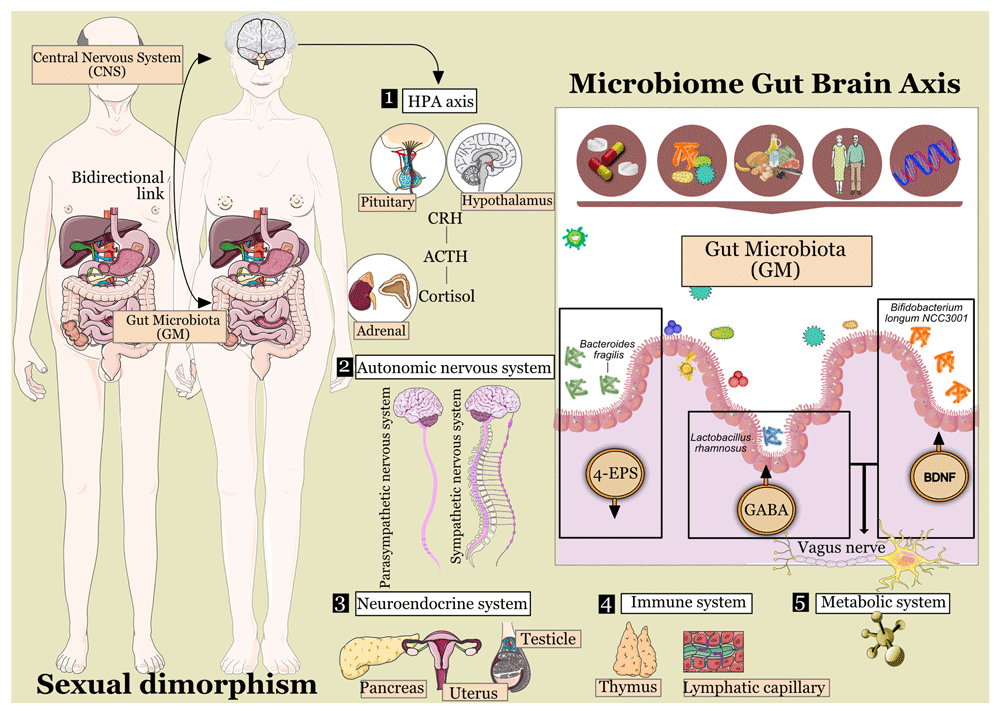
Figure 1. The gut-microbiota-brain axis.
The bidirectional link communicating the gut microbiota and the brain is called ‘The gut-microbiota-brain axis’ (GMB axis). Different pathways contribute to the GMB axis, including the autonomic nervous system, the hypothalamic-pituitary-adrenal (HPA) axis, the neuroendocrine system, the metabolic pathways, and the immune system. Some examples of gut microbiota involved in the modulation of the GMB axis include Bacteroides fragilis, which reduces the levels of 4-EPS (which impacts anxiety-like behaviour and communication skills in rodent models), Lactobacillus rhamnosus (which increases GABA and affects stress responsiveness and anxiety and depressive-like behaviour), and Bifidobacterium longum NCC3001, which increases BDNF decreasing anxiety-like and depressive behaviour. LEGEND: HPA: hypothalamic-pituitary-adrenal; CRH, corticotropin receptor hormone; ACTH, adrenocorticotrophic hormone (Bravo et al., 2011; Hsiao et al., 2013; Kelly et al., 2017; Morais et al., 2021; Pinto-Sanchez et al., 2017).
In addition to the potential influence of the gut microbiota on the gut-brain axis, the gut-brain axis is also regulated by neural, endocrine, immune, and metabolic signalling (Quigley, 2017). The ANS (comprising both, the sympathetic and parasympathetic limbs) controls both; i) the afferent signals going from the lumen to the enteric, spinal, and vagal pathways towards CNS, and ii) the efferent signals going from the CNS to the GI tract (Carabotti et al., 2015). Complementary, the HPA axis is responsible for the host's adaptative response to stressors of any type (Tsigos & Chrousos, 2002). This system produces corticotropin-releasing factor (CRF) in the hypothalamus as a response to pro-inflammatory cytokines and/or environmental stress. The CRF promotes the production of adrenocorticotropic hormone in the pituitary gland which triggers the synthesis of cortisol in the adrenal glands. This last hormone is the main stress-related hormone that influences the organism systemically, including the brain (Carabotti et al., 2015).
The mechanism that orchestrates the GB axis's good functioning includes, following the classification from Morais and colleagues (2021), indirect and direct communications through i) neuronal pathways, ii) chemical signalling, and iii) the immune system (Morais et al., 2021).
The most obvious link between the gut and the brain is the neuronal pathways that physically connect both sites (Hsiao et al., 2013; Kelly et al., 2017; Pinto-Sanchez et al., 2017). The vagus nerve is considered the main modulatory component in the communication between the muscle and mucosal layers of the GI tract with the CNS (Bravo et al., 2011). Indeed, early development of the GI tract is influenced by the GM, and the brainstem (Wang & Powley, 2007; Yoo & Mazmanian, 2017). Luminal volume and chemical signals (such as neurotransmitters, metabolites, and hormones) activate mechanoreceptors, and chemoreceptors, respectively. These stimuli promote the transit of signals from the peripheral end of the vagus nerve to the CNS (Han et al., 2018; Kaelberer et al., 2018; Wang & Powley, 2007). Finally, some GM products such as cell wall components or short-chain fatty acids (SCFA) (for example, butyric acid, propionic acid, and acetic acid) can influence neuronal pathways, modulating gut motility (Golubeva et al., 2017; Mao et al., 2013; Muller et al., 2020). SCFA has been associated with sympathetic nervous system regulation, and serotonin release, as well as with the learning process and memory (Grider & Piland, 2007; Stefanko et al., 2009; Vecsey et al., 2007).
The chemical communication between the GM and the brain includes both, direct and indirect signalling. For example, epigenetic, neuroplasticity and gene expression are directly regulated by SCFA, produced by the gut microbiota through fermentation of dietary fibre, influencing both, the disease, and behaviour of the host. And indirectly through chemical signalling of the neuroendocrine system modulating for example the production of endocrine signals in the enteroendocrine cells such as the glucagon-like peptide 1, which is implied in the sensation of hunger and appetite (Aresti Sanz & El Aidy, 2019; Dalile et al., 2019). Furthermore, microorganisms such as Bifidobacterium, Bacteroides, Escherichia spp., and Parabacteroides are known to generate neurotransmitters which can then trigger the synthesis of neurotransmitters by the host (Strandwitz et al., 2019; Yano et al., 2015).
Besides, the GB axis is also influenced by the immune system, since both, the GM and the CNS are intimately connected with the immune system, from the early development of the individual to the correct functioning of it during life (Zheng et al., 2020). In addition, modulations in the permeability of the blood-brain (BB) barrier induced by the GM were observed in mice, where germ-free animals had increased permeability than those with healthy microbiota (Braniste et al., 2014; Daneman, 2012). This was partially attributed to the decreased production of tight-junction proteins. Moreover, the GM plays a relevant role in shaping the peripheral immune system and is essential for healthy brain development, maturation, and activation of microglia, cells of the nervous tissue with phagocytic and support capacity (Abdel-Haq et al., 2019; Luck et al., 2020; Morais et al., 2021).
Finally, the brain through the GB axis can influence the GM by modulating the mucosal environment, gut permeability, and immune function (Carabotti et al., 2015; Demaude et al., 2006). The modulations in the mucosa are driven by changes in the secretion of acid, mucus, and bicarbonates, the mucosal immune response as well as alterations in the intestinal fluid production; all these changes affect the metabolic niches linked with the mucosa and consequently influence the microbiota living in those habitats (Macfarlane & Dillon, 2007). Stress is considered the main trigger of this process and influences permeability by promoting the production of interferon-γ and reducing the expression of mRNA ZO-2, which then increases permeability (Demaude et al., 2006). The increased permeability allows microbial antigen penetration in the epithelium, which incites the immune response. Besides, the immune response is also stimulated by the ANS, regulating the activity and quantity of mast cells, which produces a decompensation between tryptase and histamine (Morais et al., 2021). This is a general view of the other side of the bidirectional effects of the gut-brain axis; however, most of the processes reported describing the effect of the brain on the gut are related to stress-related conditions, and we are focused on ageing-related diseases in this review. Several studies, both, human trials, and pre-clinical models, have proven the implications of the GM profile in the modulation of diseases associated with the GB axis (Jiang et al., 2015; Sampson et al., 2016). There is evidence showing the implications of GM composition in neurodegenerative diseases, behaviour, and neurodevelopment (Choi et al., 2018; Jiang et al., 2015). This review will focus on neurodegenerative diseases associated with ageing, and how they might be influenced by sex.
The role of gut microbiota in ageing-related conditions
Several mental health disorders, including Lewy body disease (LBD), amyotrophic lateral sclerosis (ALS), Huntington's disease (HD), Alzheimer’s disease (AD), Parkinson’s disease (PD), and mild cognitive impairment (MCI), have been associated with the gut-brain (GB) axis, with the microbiome playing a role in their development (Figure 2). This review focuses on age-related conditions, which are increasingly common in our ageing population (Hou et al., 2019). Life expectancy is expected to increase by 4.4 years for both men and women by 2040 (Foreman et al., 2018). Despite changes in life expectancy, the chronological ceiling of our health remains constant, implying that geriatric diseases, such as dementia or Parkinson's, will affect patients for an extended number of years. This future health scenario encourages us to seek personalized preventive strategies to minimize the negative impact that this phenomenon will have on the quality of life of the ageing population, as well as on social services infrastructures. In this sense, the understanding of the GB axis in neurodegenerative diseases could assist in the development of new therapeutical approaches.
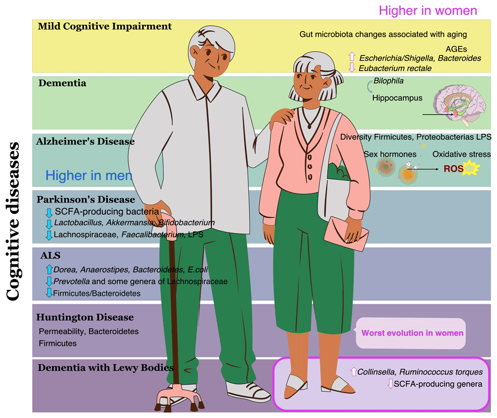
Figure 2. Cognitive diseases prevalence in men and women.
Ageing-associated cognitive diseases such as mild cognitive impairment (MCI), dementia, Alzheimer's disease, Parkinson's disease, Huntington's disease (HD), Amyotrophic Lateral Sclerosis (ALS), and Lewy Body Disease (LBD) (Hou et al., 2019) have different prevalence’s among the sexes (Migliore et al., 2021; Ullah et al., 2019).
Mild Cognitive Impairment (MCI)
MCI is a syndrome that worsens mental abilities in ageing populations. These include awareness, memory, and language skills. This condition has a prevalence of 12–18% in individuals over 60 years old and is considered the anteroom of dementia (Petersen, 2004). This multifactorial disease is determined by both, physiological and environmental factors that predispose a person toward a frailty stage. Some of the physiological aspects involve an increase in oxidative processes that negatively affect compounds such as lipids, proteins, and deoxyribonucleic acid, and unbalance the pro-oxidant and antioxidant factors and mitochondrial function (Revel et al., 2015). Indeed, advanced glycation end-products such as methylglyoxal and N(6)-carboxymethyl have been previously positively associated with brain atrophy and vascular dementia respectively (Fuijkschot et al., 2016; Srikanth et al., 2013). On the other hand, some environmental factors predisposing a segment of the population towards MCI are smoking, dietary habits (due to implications in obesity and hypertension), educational attainment, and cognitive inactivity (Perquin et al., 2012). The gut microbiota has also been linked to mental conditions such as PD and AD as a potential environmental factor (Kowalski & Mulak, 2019; Li et al., 2019).
The role of GM in MCI is not completely understood yet and it is difficult to find studies that focus only on MCI disease and not on dementia or AD. The principal traits associated with MCI are an increase in inflammation caused by metabolites linked with GM and metabolic modulations driven by GM products. For example, Cattaneo and colleagues showed in a study with 73 patients that the microbiome from faecal samples of MCI patients who presented brain amyloidosis had smaller levels of E. rectale and higher levels of Escherichia/Shigella species compared with the healthy controls. Interestingly, the patients who did not present with brain amyloidosis had a profile closer to the one presented by healthy controls. The authors of this study associated this alteration with a peripheral inflammatory state in patients who had brain amyloidosis and cognitive impairment (Cattaneo et al., 2017). Similarly, other studies showed that alterations in vascular inflammation (Tang et al., 2017), as well as in the neuro-inflammatory system (Wu et al., 2017), are driven by gut microbiota metabolites and linked with cognitive decline, which can lead to MCI. A different study in a Japanese population concluded that the prevalence of Bacteroides was independently associated with MCI in patients who did not have dementia and presented with hippocampal atrophy (Saji et al., 2020).
Currently, it seems that preliminary research on MCI indicates that cognitive functions such as memory issues are influenced by bacterial lipopolysaccharide (LPS), which is a component of the bacterial membrane considered a pro-inflammatory neurotoxin (Zhao et al., 2017). LPS has also been identified in brain lysates from the hippocampus and the temporal lobe of the neocortex in patients with AD (Zhao et al., 2017). Modulations in cognitive behaviour involving a decrease in spatial memory were also described in ageing mice, which can be considered a pre-clinical model for MCI. For example, Scott and colleagues described issues in spatial memory and higher anxiety-like behaviours in ageing mice correlating with increased gut permeability, general inflammation, and predominance of Clostridium, Porphyromonadacea, Odoribacter, and Butyricimonas among others (Scott et al., 2017). Memory issues were also described in pet dogs, where fewer Fusobacteria were identified in older individuals, while dogs with better memory scores presented a lower level of Actinobacteria (Kubinyi et al., 2020).
Dementia
Dementia is a general term employed for several progressive diseases involving difficulties in carrying out everyday activities, remembering details, thinking, or making decisions. AD is the most common type of dementia, but other types also include vascular dementia, Lewy body dementia, and mixed dementia (CDC, 2022). Dementia is not part of healthy ageing; however, nowadays around 55 million people have dementia. Current predictions indicate that the number of people with dementia will increase to 152.8 million cases in 2050 (GBD 2019 Dementia Forecasting Collaborators, 2022).
Inadequate nutrition and obesity have been previously associated with cognitive impairment and dementia. Current literature indicates that modulations in GBA and inflammation could play a role in the onset and development of the disease, however, the mechanisms behind this remain largely unknown (Solas et al., 2017). Some authors have indicated that the GB axis, as well as insulin resistance and central inflammation, are some of the mechanisms behind the onset and development of the disease (Anstey et al., 2011; Gorelick, 2010; Solas et al., 2017). Preliminary studies in elderly people suggest a link between cognitive flexibility and diet. The same association was observed in preclinical models with mice where a high-fat diet (or a high-sucrose diet) modified the GM profile and the cognitive ability of the individual, including reduced synaptic plasticity and increased vulnerability to anxiety-like conduct (Liu et al., 2015; Magnusson et al., 2015). Conversely, a preclinical model with rats that followed a healthy diet in adolescence grew in adults with improved cognitive ability and high microbiota diversity (Rogers et al., 2016). The causal effect of the microbiome was also evidenced in mouse models where gut re-colonization was associated with cognitive difficulties (Rogers et al., 2016). Indeed, mice experiments showed that B. wadsworthia bacteria had a detrimental impact on cognitive impairment by negatively modulating the hippocampus, which is the area in the brain controlling learning and memory (Olson et al., 2021). Similarly, the levels of metabolites derived from the microbiome, such as faecal lactic acid, were negatively associated with the risk of dementia. This highlights the need to consider the microbial profile as another component that could predispose the patient toward dementia (Saji et al., 2020).
Alzheimer's disease
Alzheimer's disease (AD) is a neurodegenerative condition that affects approximately 50 million people worldwide and accounts for over 70% of all dementia and cognitive decline cases, particularly in older adults (above 65 years) (Prince et al., 2013). Women are at a higher risk of developing AD, making it crucial to understand the underlying sexual differences that contribute to this disparity. Notably, about two-thirds of individuals diagnosed with AD dementia are women (Beam et al., 2018). Currently, AD is the seventh leading cause of death globally, with the risk of developing the disease increasing with age (Tomaskova et al., 2016). From a clinical standpoint, AD is defined by profound impairments in cognitive, memory, and motor functions, resulting in a significant deterioration in behavioural, mental, and functional capabilities that impact daily life and overall well-being (Tiwari et al., 2019). While the precise origins of AD remain uncertain, it is recognized as a multifactorial disorder with several contributing elements. Ageing is a major risk factor that notably elevates the likelihood of developing the disease. Additionally, genetic mutations, particularly those affecting the amyloid precursor protein (APP) gene, are essential in familial AD. These mutations lead to the accumulation of neurodegenerative amyloid-beta protein, which is a key pathological feature of the disease (Potter et al., 2016).
Current research has shown that environmental factors such as dysbiotic gut microbiome are associated with AD in both human trials and preclinical models. This dysbiosis allows endotoxins such as lipopolysaccharide (LPS) to cross the blood-brain barrier, triggering neuroinflammation. Additionally, the gut microbiome in AD patients influences neuroinflammation through bacterial metabolites (TMAO and SCFA) and the secretion of pro-inflammatory cytokines like TNF-β, IL-1β, and IL-6 (Giau et al., 2018). Furthermore, dysbiosis also increases bacteria that promote inflammation (e.g., Escherichia/Shigella, Verrucomicrobia, Pseudomonas aeruginosa, and Proteobacteria) and decreases bacteria with anti-inflammatory properties (e.g., B. fragilis, E. hallii, F. prausnitzii, E. rectale, and Bifidobacterium), contributing to beta-amyloid plaque formation (Cattaneo et al., 2017) and AD-related features such as insulin resistance and oxidative stress (Liu et al., 2020a). Studies also showed that consequently reduced butyrate levels, a result of dysbiosis, may impair mitochondrial function, leading to increased reactive oxygen species (ROS) production (Mottawea et al., 2016).
Compared to healthy subjects, AD patients have lower Firmicutes levels and higher Proteobacteria levels (Liu et al., 2019). Elevated LPS levels have also been observed in the blood plasma, neocortex, and hippocampus of AD patients (Zhang et al., 2009) (Zhao et al., 2017). Moreover, gut dysbiosis may affect the availability of tryptophan and serotonin—precursors of critical neurotransmitters like dopamine and norepinephrine—which are essential for proper brain (Doifode et al., 2021; Morris et al., 2017). Therefore, these alterations in the gut microbiota could contribute to the pathogenesis of AD by promoting neuroinflammation, disrupting neurotransmitter synthesis, and impairing neuronal function.
Importantly, functional amyloids have been detected in various microbial species, including S. cerevisiae and the Enterobacteriaceae family. These microbial amyloids can be identified by Toll-like receptor 2 (TLR-2) on microglial cells in the enteric nervous system, which triggers the activation of nuclear factor kappa B and TNF-α, thereby enhancing β-secretase activity and leading to the formation of beta-amyloid (Liu et al., 2012). BACE1 (beta-site amyloid precursor protein cleaving enzyme 1) is crucial for producing all forms of amyloid-β (Aβ), including Aβ42, which aggregates into toxic forms associated with Alzheimer’s disease (Fuster-Matanzo et al., 2013). This association, along with BACE1’s role in creating harmful amyloid-β species, suggests that beta-amyloid plaques might function as antimicrobial peptide aggregates, indicating a malfunction in immune defence mechanisms. Further research is needed to clarify how microbial amyloids affect amyloid-β aggregation in vivo and to evaluate whether targeting microbial amyloids or modulating gut-brain axis interactions could present new strategies for preventing or mitigating the progression of AD.
In addition, animal models provide further insight into the microbiota's role in AD. Studies in Drosophila (Clark et al., 2015) and mice (Conley et al., 2016; Thevaranjan et al., 2018) demonstrated that age-related changes in gut microbiota increase intestinal permeability and systemic inflammation. Notably, microbial amyloids from S. cerevisiae and E. coli have been shown to induce amyloidosis and amyloid aggregation in mouse models, suggesting that microbe-induced amyloids may exhibit prion-like properties (Lundmark et al., 2005). In another study, curli from S. typhimurium promoted IL-22, IL-17α, and IL-6 production in the intestinal epithelium, though the response was diminished in TLR-2-deficient mice (Nishimori et al., 2012). Furthermore, LPS injections in rats increased the expression of pro-inflammatory cytokines IL-1β, IL-6, and TNF-α in the brain, along with LPS-associated beta-amyloid plaque deposition (Wang et al., 2018). Microbial amyloids, combined with the effects of increased intestinal permeability and inflammation, may act as a trigger for amyloid aggregation in the brain, suggesting that gut-derived microbial products might play a central role in AD progression. These findings underscore the need for further research to explore the specific pathways through which gut microbiota and microbial amyloids influence neuroinflammation and amyloidosis. Understanding these mechanisms could lead to novel therapeutic approaches targeting the gut-brain axis to mitigate or prevent Alzheimer's disease progression.
Parkinson's disease
PD occurs with an incidence rate of 10–50 cases per 100,000 people annually and a prevalence rate of 100–300 cases per 100,000 people. It is the second most prevalent neurodegenerative disorder after AD and is 1.5 times more common in men than in women (Elbaz et al., 2016). Additionally, due to the ageing global population, the number of PD cases is projected to double by 2030 (Elbaz et al., 2016). PD is a progressive neurodegenerative condition that primarily impacts dopaminergic neurons in the brain, resulting in reduced dopamine levels and motor dysfunction (Hoehn & Yahr, 1967). This latter includes resting tremors, bradykinesia, rigidity, and gait abnormalities, as well as non-motor symptoms such as hyposmia, sleep disorders, depression, and GI symptoms (Kalia & Lang, 2015). Conversely, to AD, women often develop a more benign PD phenotype (Georgiev et al., 2017). In addition, PD is positively associated with an increased risk for progressive cognitive decline from MCI to dementia (Emre, 2003). It has been described that MCI affects over one-fourth of PD patients, and dementia eventually affects over 80% of patients with 20-year survival (Schapira et al., 2017).
Researchers have suggested that gut microbiota might play a significant role in influencing neurodegenerative diseases, with accumulating evidence linking gut microbes to the symptoms and mechanisms of Parkinson’s disease (PD) (Romano et al., 2021). PD is now recognized as a multisystem disorder affecting both central and peripheral nervous systems. Gastrointestinal issues such as gastroparesis and constipation are often observed years before the onset of motor symptoms. Changes in gut microbiota composition have been studied in relation to the development of PD (Cersosimo & Benarroch, 2012). Animal research has demonstrated that gut microbiota may impact PD by influencing α-synucleinopathy and neuroinflammation (Keshavarzian et al., 2020; Sampson et al., 2016). Inflammation is a well-established characteristic of PD, with elevated levels of inflammatory molecules found in the brain, cerebrospinal fluid (Chen et al., 2018; McGeer et al., 1988; Nagatsu et al., 2000), and blood (Chen et al., 2008; Lin et al., 2019; Qin et al., 2016) of PD patients. Although the specific cytokines, chemokines, and inflammatory markers vary, IL-6, TNF, IL-1β, CRP, IL-10, CCL5, and IL-2 are frequently dysregulated in the peripheral blood of PD patients (Qin et al., 2016). These observations suggest that inflammation might originate from a compromised intestinal barrier, which could lead to increased systemic exposure to inflammatory microbial products like LPS, a component of bacterial cell walls (Brown, 2019; Houser & Tansey, 2017).
It has recently been described alterations such as an enrichment of the genera Lactobacillus, Akkermansia, and Bifidobacterium and depletion of bacteria belonging to the Lachnospiraceae family and the Faecalibacterium genus, associated with PD (Romano et al., 2021). Indeed, an increase in the mucin-degrading Akkermansia and a depletion in short-chain fatty acid (SCFA)-producing bacteria are seen to be the features of the gut microbiota associated with PD (Hirayama & Ohno, 2021). Taken together, these alterations drive an impairment of the intestinal barrier integrity with the subsequent exposure to LPS, which leads to abnormal aggregation of α-synuclein fibrils (Hirayama & Ohno, 2021). This increased intestinal permeability is also underscored by decreased serum LPS binding protein in PD (Hasegawa et al., 2015; Unger et al., 2016). Indeed, reductions in faecal SCFA and intestinal inflammatory responses are characteristic of PD, which are related to the microbiota and disease onset. Interestingly, some of these relationships in PD are sex-dependent (Aho et al., 2021).
In summary, the interplay between gut microbiota, intestinal barrier integrity, and neuroinflammation plays a crucial role in the pathophysiology of Parkinson's disease, highlighting the need for further research to explore microbiota-targeted interventions as potential therapeutic strategies for managing and potentially mitigating disease progression.
Amyotrophic lateral sclerosis
Amyotrophic lateral sclerosis (ALS) is a progressive neurodegenerative disease characterized by the loss of motor neurons in the brain and spinal cord, leading to muscle weakness and eventually death. Although ALS is relatively uncommon, with an incidence rate of 1.1–3.5 per 100,000 person-years, it has a significant impact on public health due to its high levels of morbidity and mortality (Abhinav et al., 2007; Al-Chalabi & Hardiman, 2013). In addition to motor symptoms, ALS can also present with non-motor features, such as cognitive and behavioural disturbances (McCombe et al., 2017; Xu et al., 2017). Approximately 10–15% of cases are classified as familial ALS (FALS), while the majority are sporadic ALS (SALS). Sporadic ALS is more common in men than in women, with a male-to-female ratio of 2.6:1 (Uenal et al., 2014). Family inheritance in FALS is mostly autosomal dominant, and more than 30 genes have been implicated (Al-Chalabi & Hardiman, 2013). Major genes include the DNA binding protein TAR (TARDBP/TDP-43), superoxide dismutase 1 (SOD1), the FUS RN binding protein (FUS/TLS), or the C9orf72-SMCR8 complex subunit of the most recently discovered gene (C9orf72) with repeat expansions of ALS-causing hexanucleotides (Renton et al., 2014). Also, environmental neurotoxins could contribute to the development of ALS: for example, β-methylamino-L-alanine (BMAA) and formaldehyde (Banack & Cox, 2003; Murch et al., 2004).
In addition, gut microbiota (GM)'s role in physiopathology has been increasingly emphasized. GM in mice and humans shares similar characteristics at the gender level and could therefore show significant similarities from a functional point of view. For example, in SOD1G93A mice, a commonly used transgenic model, the faeces exhibited reduced levels of butyrate-producing bacteria (Wu et al., 2015). Additionally, another study found that mice with mutations in the C9orf72 gene had a reduction in immunostimulant bacteria. In this study, GM transplantation was demonstrated to protect against systemic inflammation and GM-mediated autoimmunity, enhancing the connection between GM and ALS at the preclinical level (Burberry et al., 2020). Moreover, a study by Kira et al. confirmed that oral administration of L-carnitine can delay the onset and progression of ALS and extend the lifespan of SOD1G93A mice (Kira et al., 2006).
On the one hand, the efficacy of carnitine therapy revealed that N-oxide-trimethylamine (TMAO) from GM and its associated compounds could have a role in the initiation and progression of ALS. In this way, several reports have established similarities in GM composition, among people who cohabit due to similarities in environment and diet. Therefore, the spouses of ALS patients provided a natural control group, thus establishing the null hypothesis, effectively excluding any differences due to environment, diet, and physical intimacy (Chen et al., 2020). The researchers could see that the TMAO metabolic pathway of GM is changed in both ALS patients and their spouses, compared to healthy controls, suggesting that alterations in GM occurred before illness onset. With this type of evidence, we can suspect a strong interrelation between GM and ALS development. In addition, negative correlations between upper motor neuron (UMN) involvement and concentrations of TMAO and other metabolites could suggest that inhibition of this metabolic pathway could lead to a better prognosis in ALS patients (Chen et al., 2020). Furthermore, significant differences in tight junctions and adhesion proteins have been identified in post-mortem examinations of CNS tissues from ALS patients, indicating that these changes allow molecules to pass through the blood-CNS barrier that normally would not (Wu et al., 2015).
On the other hand, LPS could potentially contribute to the pathogenesis of the disease through the promotion of neuroinflammation and/or weakening of the blood-brain barrier and has been found in elevated concentrations in the plasma of patients with SALS (Wright et al., 2018). Plasma LPS levels have also been significantly and positively associated with monocyte/macrophage activation in blood, while negatively associated with IL-10 expression from monocytes, supporting the idea that microbial products can promote systemic inflammation in ALS (Wright et al., 2018). Since SCFAs produced by the gut microbiota contribute to the activation and maintenance of CNS immune cells, such as microglia, dysbiosis may contribute to the altered levels of immune cells and inflammatory mediators observed in ALS patients, where an increase in abnormal Paneth cells has also been reported (Wu et al., 2015).
In relation to bacterial taxonomy in the disease, there is a significant increase in the bacterial genus Dorea and Anaerostipes and a significant decrease in Prevotella and some genera of Lachnospiraceae has been observed; on the contrary, they are characterized by being beneficial for people with ALS (Casani-Cubel et al., 2021). Importantly, a decrease in the ratio of the phyla Firmicutes/Bacteroidetes has been observed, causing an increase in the phylum Bacteroidetes in ALS (Fang et al., 2016). Additionally, there is evidence that ALS patients differ significantly from healthy individuals in a number of ways, with the disease being primarily distinguished by a high concentration of Escherichia coli and Enterobacteriaceae and a low prevalence of yeast (Mazzini et al., 2018). Exacerbation of symptoms has been associated with Ruminococcus torques, related to neurodegeneration. Conversely, Akkermansia muciniphila is associated with symptomatic improvement and possible increased motor neurons and survival due to increased nicotinamide (Blacher et al., 2019). Additionally, certain Lactobacillus species, such as L. rhamnosus and L. brevis, along with Bifidobacterium dentium, enhance the production of the neurotransmitter GABA, a key mechanism through which gut bacteria influence brain chemistry (Barrett et al., 2012; Bravo et al., 2011). The gut microbiota may play an important role in ALS development and contribute to the differences in disease incidence between sexes through various mechanisms, including metabolism, immune response, toxin production, and neurotransmitter regulation that affects brain function. Increasing evidence points to the impact of gut microbiota alterations, including changes in microbial communities and their metabolic byproducts like SCFAs and TMAO, on ALS progression and systemic inflammation. Differences in microbial composition and metabolism observed in ALS patients and their close contacts suggest a significant role of the gut microbiome in both the onset and progression of the disease. Moreover, the connection between microbial factors, intestinal barrier integrity, and neuroinflammation emphasizes the need for more research into microbiota-based treatments as potential therapeutic approaches. Gaining deeper insight into these interactions could lead to innovative strategies for managing ALS and improving patient outcomes.
Huntington's disease
Huntington's disease (HD) is a progressive neurodegenerative disorder characterized by a triad of motor, cognitive, and psychiatric impairments (McColgan & Tabrizi, 2018). Typically, the motor defects include chorea and loss of coordination. Psychiatric symptoms, such as depression, psychosis, and obsessive-compulsive disorder, are also common in HD and are particularly distressing for patients (Rosenblatt, 2007). HD is an autosomal hereditary neurodegenerative disease caused by age-dependent penetrance of an expanded sequence of cytosine–adenine–guanine (CAG) repeats in the huntingtin gene (Tabrizi et al., 2011). This disease acts by wearing down the nerve cells of the brain. Indeed, a variety of mechanisms lead to neuronal malfunction and death. These include the direct consequences of the mutant huntingtin protein (mHTT) exon 1 fragment, the propensity of mHTT to form aberrant aggregates, and the effects of mHTT on cellular proteostasis, axonal transport, transcription, translation, mitochondrial function, and synaptic function (Bates et al., 2015; Ross & Tabrizi, 2011). Generally, this disease manifests its first symptoms between 40 and 50 years of age. Indeed, the affected person may have the gene for the disease but not know it until symptoms appear, at 40 or 50 years of age (Bates et al., 2015).
HD prevalence depends on the average CAG repeat longitude. The amount of CAG repeats and the age at which symptoms onset first appear to have a significant inverse relationship; as bigger CAG repeat expansions are typically linked to earlier ages at which symptoms onset first appear (Podvin et al., 2019). In addition, the age of onset appears to be impacted by additional environmental and genetic factors and the CAG repeat number only partially explains 65%–71% of the variance in that age (Rosenblatt et al., 2001). In Western populations, the prevalence ranges from 10.6 to 13.7 cases per 100,000 people. With prevalence rates of 1–7 per million, HD has a much lower incidence in Japan, Taiwan, and Hong Kong. Interestingly, in South Africa lower rates are seen in black populations when compared to white and mixed populations (Bates et al., 2015).
Although it has not been described differences in prevalence according to sex, it has been suggested that women have worse symptoms than men during the course of HD (Hentosh et al., 2021). Most of the evidence for sex-related differences in HD comes from investigations into male intergenerational transmission of CAG repeat length instability. Large expansions are nearly exclusively transmitted by the father, whereas affected women's offspring are more likely to have no change or CAG length contractions (Aziz et al., 2011). Unexpectedly, repeat-length alterations in maternal transmissions were also sex-dependent, with a tendency for male children to have longer repeats and female offspring to have shorter repeats (Meoni et al., 2020; Wheeler et al., 2007).
The loss of neuropeptides in the gut is linked to the presence of the mutant protein in both the enteric nervous system and intestinal epithelial cells throughout the GI tract, which is associated with impaired gut function (van der Burg et al., 2011). The first indication of gut microbiota dysbiosis related to Huntington's disease (HD) was demonstrated by Kong et al. in a transgenic mouse model of the disease. This study also suggested that dysbiosis might play a role in modulating the onset and progression of HD symptoms (Kong et al., 2020). Further, the gut microbiota dysbiosis associated with HD was confirmed in HD gene expansion carriers, in addition to a lower gut microbiota alpha diversity in HD (Wasser et al., 2020). Moreover, it has been shown that the HD gut microbiota is unstable at the pre-motor stage of the disease, which may have catastrophic effects on the host's health. Prior to substantial cognitive and motor loss, the HD gut microbiota function was disturbed, suggesting a potential role for the gut in influencing HD pathogenesis, maybe via particularly altered plasma metabolites that modulate gut-brain transmission (Kong et al., 2021).
Similarly, increased intestinal permeability observed in an HD mouse model, along with a higher relative abundance of Bacteroidetes and a reduction in Firmicutes, suggests a potential connection between gut microbiota dysbiosis and Huntington's disease (Stan et al., 2020). Further evidence comes from faecal microbiota transplant (FMT) experiments, which confirmed the role of gut microbiota in HD. FMT from wild-type mice with Huntington’s disease was found to improve cognitive outcomes, particularly in females, indicating that modulating the gut microbiome can help alleviate cognitive deficits associated with dementia in HD (Gubert et al., 2022). This research also highlights the gut microbiota as a potential target for therapeutic interventions, suggesting that alterations in the microbiota-gut-brain axis could influence brain function, including cognition.
Despite its relatively low prevalence, the impact of HD is profound, with significant variations in symptom onset and progression influenced by genetic and environmental factors. Recent research underscores the emerging role of gut microbiota in HD pathogenesis, revealing that dysbiosis may influence disease progression and symptomatology. The promising results from faecal microbiota transplantation and the link between gut microbiota and HD symptoms suggest that targeting the microbiome could offer new avenues for therapeutic interventions. Further investigation into the gut-brain axis could enhance our understanding of HD and lead to novel strategies for managing and potentially mitigating the effects of this debilitating disorder.
Dementia with Lewy bodies
Dementia's impact is widely acknowledged, with Alzheimer's disease being the most recognized form. However, Lewy body dementias, which include "dementia with Lewy bodies" (DLB) and "Parkinson's disease dementia" (PD), are the second most common form of degenerative dementia in individuals over 65 (Walker et al., 2015). A systematic review of the prevalence of DLB estimates that the proportion of dementia patients with Lewy bodies ranges from 0 to 23% (Vann Jones & O'Brien, 2014). Unfortunately, DLB receives less attention than Parkinson's and Alzheimer's diseases, leading to underdiagnosis and suboptimal management for affected patients (Walker et al., 2015).
DLB may occur in individuals with parkinsonism or within the first year of experiencing motor symptoms; this type of dementia is described by varying cognitive impairment, sleep disorders, hallucinations of a visual type, and movement disorders (parkinsonism) among others (McKeith et al., 2005; Outeiro et al., 2019). Nevertheless, it is not a universal occurrence for all patients with Parkinsonism to develop dementia (McKeith et al., 2005). The pathogenesis of Lewy body dementias involves an abnormal accumulation of α-synuclein proteins in the brain (Lewy bodies, and Lewy neurites) and neuronal loss. However, it is unknown why some patients have serious α-synuclein neuronal inclusions and the absence of clinical symptoms (Parkkinen et al., 2008). The aetiology of DLB is unclear, and multifactorial factors are believed to contribute to the progression of the disease (Walker et al., 2015). Several factors contribute to the development of dementia with Lewy bodies (DLB), including ageing, sex, depression, and family history, suggesting a possible genetic component to the disease (Boot et al., 2013; Wakisaka et al., 2003; Woodruff et al., 2006). The first genome-wide association study (GWAS) on DLB was conducted in 2017, a time when similar diseases had already been extensively studied using this method. This delay reflects the general lack of attention DLB has received compared to other neurodegenerative conditions. Notably, DLB shares genetic risk factors with Parkinson’s disease (PD) at DBA and SNCA, and with Alzheimer’s disease (AD) at the APOE E4 allele (Guerreiro et al., 2016; Moskvina et al., 2013; NCI-NHGRI Working Group on Replication in Association Studies et al., 2007). Among the various contributing factors, changes in the gut microbiome (GM) may play a role in DLB, though little is known about the gut-brain axis in this disease. Unlike Parkinson’s disease, where the influence of GM on disease onset and progression has been widely studied (e.g. (Aho et al., 2019; Bedarf et al., 2017; Cirstea et al., 2020; Hill-Burns et al., 2017; Nishiwaki et al., 2020; Nishiwaki et al., 2022), research on GM’s impact on DLB remains limited. Recently, Nishiwaki and colleagues found that SCFA-producing genera were reduced in DLB patients, similar to observations in PD. However, Collinsella and Ruminococcus torques were elevated in DLB patients, a finding not seen in PD (Nishiwaki et al., 2022). Furthermore, R. torques and Collinsella increased the levels of ursodeoxycholic acid (UDCA), a faecal bile acid, in DLB patients compared to controls. UDCA could palliate neuroinflammation, therefore, intervention to increase Bifidobacterium and its metabolites was suggested as a potential therapeutical action (Nishiwaki et al., 2022). Overall, little research has been carried out to investigate the relationship between DLB and GBA, and further evidence is needed (Nishiwaki et al., 2022). Interestingly, it has been previously reported that males have a higher prevalence of LBD, being sex a risk factor for the onset and development of the disease (Zweig & Galvin, 2014). Therefore, it could be reasonable to suggest that perhaps in the near future, scientists may be able to interrogate whether or not sexual dimorphism plays a role in GBA and LBD.
Therefore, while initial research indicates that changes in SCFA-producing genera and increased levels of certain bacteria may influence DLB symptoms and neuroinflammation, much remains to be understood. Given the preliminary evidence linking GM changes to DLB, future research should focus on further elucidating these relationships and exploring potential therapeutic interventions. Additionally, the potential impact of sexual dimorphism on DLB and gut-brain interactions warrants further investigation. Overall, addressing these gaps in knowledge could pave the way for improved diagnostic and therapeutic strategies for Lewy body dementias.
Gut microbiota and different sex-related predisposition to the brain-related diseases
Sexual dimorphism refers to the differences in physical and behavioural characteristics between males and females of a species. These differences can be seen in a variety of traits, including the primary differences in gametes and gonads, behaviour, morphology, psychiatry, and disease (Mauvais-Jarvis, 2015; Migliore et al., 2021; Ullah et al., 2019) (Figure 3).
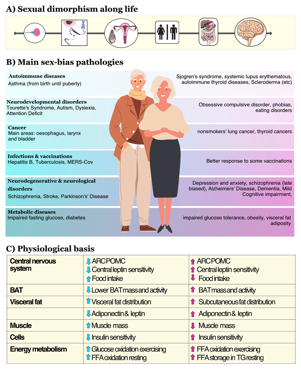
Figure 3. Overview of the main changes between men and women.
Panel A) Sexual dimorphism. Sexual dimorphism (SD) starts with the primary differences in gametes and gonads and continues through life, shaping differences in behaviour, morphology, and physiology between men and women. SD begins to manifest at the genetic level and must be influenced by the sex chromosomes, as these are the only genomic differences between the sexes (Mank, 2009). Panel B) Main sex-bias pathologies. Differential susceptibility of males and females to autoimmune diseases, neurodevelopmental disorders, some cancers, infectious & vaccination, neurodegenerative & neurological disorders, and metabolic and cardiovascular diseases has been reported (Mauvais-Jarvis, 2015; Migliore et al., 2021; Ullah et al., 2019). Panel C) Physiological bases. The physiological bases underlying these differences are reported in the table. Acronyms used: BAT, Brown Adipose Tissue; FFA, Free Fatty Acids; ARC POMC, Arcuate Nucleus Pro-opomelanocortin.
The expression of these sexually dimorphic traits is influenced by the sex-biased gene expression of a multitude of genes located on different chromosomes. However, the sex chromosomes (XX or XY) play a crucial role in this process as they are the only genomic difference between the sexes (Mank, 2009). Sexual dimorphism begins to manifest at the genetic level, where the pattern of growth hormone (GH) secretion is dependent on sex hormones (Birzniece & Ho, 2017). This, in turn, conditions the sexually dimorphic gene expression. Studies have shown that the regulation of growth hormone secretion and its downstream effects on gene expression are different between males and females and that these differences can contribute to the development of sexually dimorphic traits (Ahluwalia et al., 2004). While sexual dimorphism is the result of the interaction between many genes located on different chromosomes, the sex chromosomes must play a crucial role as they are the only genomic difference between the sexes, influencing the expression of sexually dimorphic traits through hormonal regulation of gene expression.
Sex differences in brain structure and function refer to the variations in the physical and functional properties of the brain between males and females. These differences have been observed throughout the lifespan and can have an impact on various aspects of cognitive and emotional processing. Research has shown that females typically have greater volume in certain brain regions compared to males. These regions include the prefrontal cortex, orbitofrontal cortex, superior temporal cortex, lateral parietal cortex, and insula. These regions are responsible for processing information related to decision-making, emotional regulation, social cognition, and sensory processing. On the other hand, males, on average, have greater volume in the ventral temporal and occipital regions. These regions are responsible for processing visual information and spatial perception (DeCasien et al., 2022; Liu et al., 2020b). These sex differences in brain structure have been recognized as a potential contributing factor in the development and progression of neurodegenerative diseases such as Alzheimer's and Parkinson's. These diseases can have different impacts on males and females, and understanding these sex differences can help in the development of more effective treatments and strategies for managing these conditions (Bruzelius et al., 2019; Meoni et al., 2020; Ullah et al., 2019).
Research has suggested that the GM of men and women may differ from each other, and this difference may contribute to the different sex-related predispositions to certain diseases (Santos-Marcos et al., 2019a). Studies have shown that the sex-specific microbiota, along with its interaction with sex hormones and the immune system, is an emerging field in the study of sex-biased disease susceptibility (Santos-Marcos et al., 2018; Vemuri et al., 2019). These findings suggest that GM may play a role in the sex-dependent incidence and prevalence of diseases such as dementia, MCI, AD, and PD (Figure 2). Therefore, it is important to consider gender differences when tailoring treatment, predicting outcomes, and meeting other individual and social needs in men and women.
Sex differences in dementia risk are not well understood, but some studies have found that women may have a greater risk of developing dementia compared to men (Levine et al., 2021). Several factors are thought to contribute to these differences, including biological factors such as hormonal differences, health factors such as cardiovascular disease and stroke, and social factors such as education and occupation (Carter et al., 2012; Rocca et al., 2014). Additionally, evidence suggests that the incidence of dementia has decreased over the past 25 years, but this decline has been more pronounced in men than in women (Wolters et al., 2020). This may be due to a combination of factors, including increased awareness and prevention efforts, better medical treatment, and improved living conditions. Further research is needed to understand the underlying causes of these sex differences and to develop effective strategies for reducing the risk of dementia in both men and women.
Research has suggested that the incidence of neurodegenerative diseases such as AD may vary between men and women due to the effects of sex hormones on oxidative stress (Tenkorang et al., 2018). However, it is also possible that sex hormones may affect the composition of gut microbiota (Cross et al., 2018), which can in turn influence the prevalence of AD. According to previous studies, the host organism may be affected differently by alterations in gut microbiota, which are frequently seen in situations where sex hormone levels are declining, such as aging in men and menopause in women. It is believed that the interaction between intestinal miRNA and gut microbiota mediates these effects (Barroso et al., 2020; Santos-Marcos et al., 2020). This demonstrates the intricate relationships that exist between gut microbiome, neurodegenerative illnesses, and sex hormones.
PD is associated with a depletion of SCFA-producing bacteria. Similarly, previous studies have demonstrated that menopause is also linked to a reduction in several SCFA-producing bacterial species (Mayneris-Perxachs et al., 2020; Santos-Marcos et al., 2018). However, these changes have been described to occur after menopause, and therefore premenopausal women might be potentially protected against PD. Indeed, PD is 1.5 times more common in males than in females (Elbaz et al., 2016). Moreover, females often develop a more benign PD phenotype (Cerri et al., 2019; Haaxma et al., 2007). Considering that PD means age is around 60 years, it is plausible to think that these variations could be due to lasting effects from the significant differences in sex steroid profiles between males and females in early life, which may have a persistent impact on gut microbiota. Alternatively, they could result from actual fluctuations in sex hormone levels in older men and women.
Overall, it is plausible the idea of a sex-dependent incidence and prevalence in brain-related disease is determined by sex differences in GM. Indeed, the composition of the intestinal microbiota depends on gender, besides other factors such as age, genetic makeup, and the nutritional habits of the host organism (Cӑtoi et al., 2020; Dominianni et al., 2015; Haro et al., 2016c; Tims et al., 2013). In line with this, accumulating evidence shows that sex hormones play an important role in shaping intestinal microbiota (Cross et al., 2018). These differences may contribute to sex-dependent differences in the incidence and prevalence of MCI, dementia, AD, or PD. This hypothesis is based on the potential contribution of GM alterations, and the gut-brain-axis influence in these pathologies.
Conclusions
Given the complexity and multifactorial nature of neurodegenerative diseases, continued research into the role of gut microbiota (GM) is crucial. Emerging evidence indicates that variations in GM, potentially influenced by sex hormones and other factors, may affect the development and progression of these conditions. However, the precise mechanisms driving these associations are still not fully understood. While animal studies have shown causal relationships, these findings require confirmation through human clinical trials.
As we move forward, it is vital that research continues to investigate microbial therapies for preventing and treating age-related pathologies. Future studies should be designed with a focus on sex-specific factors, taking into account the varying impacts of GM and sex hormones. The EU-funded project MEMOIR (Metagenomic Modelling of cOgnitive ImpaiRment) represents a step forward in this direction by examining how a healthy diet can influence gut microbiota and the progression of Mild Cognitive Impairment. Such projects, alongside others, will be instrumental in uncovering how GM affects neurodegenerative diseases and whether modulating GM can serve as a viable strategy for prevention and treatment.
List of abbreviations
AD, Alzheimer’s disease;
ANS, autonomic nervous system;
BB, blood-brain;
BMI, body mass index;
CNS, central nervous system;
CRF, corticotropin-releasing factor;
GB, gut-brain;
GI, gastrointestinal
GM, gut-microbiota;
HPA, hypothalamic pituitary adrenal;
LPS, lipopolysaccharide;
MCI, Mild Cognitive Impairment;
PD, Parkinson’s disease;
SCFA, short-change fatty acids;
WHO, Word Health Organization;