Introduction
Living cells proliferating in confined spaces eventually build up mechanical compressive stress exerted onto themselves and their surroundings. This growth-induced pressure (GIP) has the potential to decrease cell growth in all kingdoms of the living, including bacteria1,2, fungi3,4, plants5,6 or mammals7–9. It has recently been shown in the budding yeast Saccharomyces cerevisiae and in mammalian cells that GIP is accompanied by a decrease in the diffusion of genetically-encoded tracer nanoparticles10. This decreased diffusion has been attributed to an increase in intracellular density through a mechanism where the production of macromolecules with limited cell volume expansion leads to increased biomass within the cytoplasm. However, the accumulation of macromolecules has yet to be observed experimentally, as this decrease in diffusion could also be attributed to other effects, such as a decrease in biochemical activity which is known to fluidize the cytoplasm11,12.
The dry mass density of a living cell can be a direct proxy of macromolecular concentration. Numerous methods exist to measure the dry mass density of living cells, but their applications to confined cells developing GIP are inexistent. To develop GIP, cells are proliferating inside a confining microfluidic chamber and become densely packed3,7. The experimental conditions restrict access to several measurement techniques, such as suspended microchannel resonator (SMR) measuring single cells’ buoyancy suspended in media13,14, or cryoelectron tomography requiring cell fixation15,16. On the other hand, quantitative phase imaging (QPI) is an alternative technique that can non-invasively measure the dry mass density of optically transparent biological cells or tissues, which does not require sample preparation17,18.
QPI can quantify the refractive index (RI) distribution of a biological sample by detecting the phase difference of light passing through the sample with the surrounding media. This measured RI is directly proportional to the dry mass density of a biological sample19. Here, we investigated the changes in the dry mass density of the budding yeast Saccharomyces cerevisiae growing within a confined space using QPI. We observed that confined growth was associated with increased dry mass density. We measured the concentration of a fluorophore inside the cell and observed that the increase in RI was proportional to the increase in fluorophore concentration. A linear extrapolation between GIP and RI down to the refractive index of water predicted the nominal intracellular osmotic pressure of the cell.
Methods
Cell culture conditions
Saccharomyces cerevisiae strains were grown and maintained on a Synthetic Complete (DCS0019, Formedium) + 2% dextrose (SCD) media agar Petri dishes. Next, a single colony was inoculated in a fresh liquid SCD liquid media and incubated with orbital shaking at 200 rpm at 30°C overnight. Exponentially growing culture at OD = 0.3 was then loaded into the microfluidic chamber.
Microfabrication of PDMS devices
The mold with two layers of different heights is fabricated in a cleanroom at LAAS-CNRS using classical photolithography with negative photoresists. The first layer at a height of 0.8 μm defining the culture media channels was prepared with 2ml of negative photoresist (SU8 3000.5, Kayaku Advanced Materials), and the second layer at a height of 9.2 μm defining the cell growth chamber and the main cell-loading channel was prepared with 2ml of another negative photoresist (HARE-SQ10, Kemlab). After the lithography process, perfluorodecyltrichlorosilane was supplied in 100 sccm for 10 seconds and grafted with O2 plasma under 40 Torr of pressure onto the wafer surface using an SPD (Memsstar Technology) machine to improve the hydrophobicity and non-stiction. PDMS elastomer was prepared by mixing the base and curing agent in a 10:1 ratio, poured onto the mold, and cured overnight at 60°C. Both PDMS and a glass coverslip were activated with oxygen plasma treatment (Diener PICO; gas, oxygen; pressure, 0.3 mbar; power, 100%; activation, 20 s), and bonded to each other immediately after surface activation. The PDMS/glass chip was then baked for ≥5 h at 60°C.
Microfluidic device operation
The cell suspension was introduced to the chambers using a syringe. We next injected the SCD media through the main inlet channel using a Fluigent MFCS pressure control system. During the cell incubation and imaging, the pressure at the culture media inlet was maintained at ~1 bar. The chips were placed in a microscope environmental chamber at 30°C (TempControl-37, Leica Microsystems).
Refractive index measurement of bulk liquid/solid samples
Liquid and solid samples were measured using an Abbe refractometer (2WAJ, OPL) with white light at 30°C. Culture media (SCD) and distilled water samples were measured in spread, like a thin film, between the main and secondary prisms. A cured PDMS sample was prepared into a film of about 500 μm thickness and was carefully placed between the two prisms' surfaces without any air bubbles. The RI of each sample was consistent for three independent measurements, and errors were not stated because the variances were smaller than the refractometer's measurement resolution (10-4).
Bright field and fluorescent microscopy
All imaging acquisition in this study was conducted on a invertedmicroscope(DMi8, Leica Microsystems). The lateral deformation of PDMS chambers was measured to infer growth-induced pressure through bright-field microscopy simultaneously with the QPI. The relationship between pressure and chamber deformation was calibrated as done in previous work10, giving a value of 8.2 μm.MPa–1.
To measure GFP accumulation, cytosolic GFP expressed from HIS3 promoter were measured by fluorescent z-stacking (0.5 μm interval) with a spinning-disk confocal scanner unit (CSU-X1, Yokogawa). We acquired z-stack of the first cell layer of each chamber using the same way of measurement for the chamber height with ET525/50 nm red emission filter, a dichroic mirror (ZT405/488/561/638rpc, Chroma) and Hamamatsu scientific complementary metal–oxide–semiconductor camera (ORCA-Flash4.0 v3, Hamamatsu Photonics). In the same way, we imaged the cells without any GFP labeling to subtract the cellular autofluorescence which can be detected from GFP channels. The GFP expression of each cell was calculated by summing of pixel values across all z-stack slice of an chamber in ImageJ/Fiji20.
Refractive index and dry mass density measurement of cells with QPI
For transmission QPI, the microscope was equipped with conventional LED Köhler transillumination and a condenser of maximum numerical aperture NA = 0.55. To acquire QPI data, we imaged the samples with a 20x 0.8 NA objective (Leica) and a quadriwave lateral shearing interferometry (QWLSI) system (SID4 sc8, Phasics) mounted on the microscope’s lateral camera port. The QWLSI measures the local phase shift, also called optical path difference (OPD), introduced by a specimen placed under a microscope. Depending on the sample's thickness and the difference of sample's refractive index from the background material, the OPD was depicted in grayscale on the image, the so-called phase image.
To estimate RI of cells (ncell) on a culture plate, every cell was segmented from a phase image, which set the culture media as a background baseline, using Otsu algorithm method by CellProfiler20. The ncell is estimated by the relation, ncell = nbackground + OPD/dcell, where the OPD is difference between cell and background and dcell is the height of the cell. Since cells were assumed to be prolate ellipsoids, the dcell was taken as the length of the minor axis of the cell in the phase image. The corresponding OPD was taken as the brightness of the upper 5% of the intensity distribution within the cell from the phase image in which the background was subtracted.
To measure the average RI of cells within a microfluidic chamber, the averaged OPD value along the chamber area was measured, comparing to surrounding PDMS. To minimize the influence of local and variable OPD gradients occurring in phase images of cells within the PDMS microfluidics chip, we took the average value of the PDMS area in all directions surrounding the cell sample as the background baseline of the measurement. The chamber height under pressure was determined by the OPD of the calibrated culture medium of chambers expanded by predefined hydraulic pressure: dchamber = OPD/(nPDMS-nmedium). Measurement of chamber’s height at GIP = 0 MPa perfectly matched the measurement of the SU8 mold measured with a profilometer. For GIP > 0 MPa, the cellular RI was measured in comparison to PDMS: ncell(P) = nPDMS + OPD(P)/dchamber(P).
The dry mass density of a yeast cell is directly calculated from the mean RI value since the RI in biological samples is linearly proportional to the dry mass density inside cells as ncell = nmedia + αρ, where ncell is the average RI of each cell, nmedia is the RI of the surrounding media which was obtained with Abbe refractometer (nmedia = 1.336), α is an RI increment (α = 0.190 ml/g for proteins and nucleic acids19), and ρ is the dry mass density inside cells.
Data and statistical analysis
All image analysis was performed using ImageJ (National Institutes of Health, US). Statistical analysis was performed on Python with Statsmodels modules21.
Results
Refractive index and dry mass density of S. cerevisiae determined with QPI
We measured the RI and dry mass density of living yeast S. cerevisiae on a culture plate using QPI, which measures the phase difference between a sample and a reference in the image. Phase images displayed the optical path difference (OPD) between cells and the surrounding culture media in grayscale (Figure 1a). The OPD was the difference in RI of a sample with regard to a reference (here, the culture media), times the height of the sample. The pixel value along the cross section of a cell was related to the physical thickness and the local RI of different parts of living yeasts (Figure 1b). We measured the maximum OPD of each cell with respect to the culture media to be 180 ± 20 nm. We estimated the average cellular height to be 3.94 ± 0.50 μm (see Materials and Methods). We determined the mean RI and dry mass density of an asynchronous population of yeast S. cerevisiae, ncell = 1.384 ± 0.004 and ρcell = 271 ± 19 mg/ml (Figure 1c), which lied within the range of the RI measured for various yeast cells22–26.
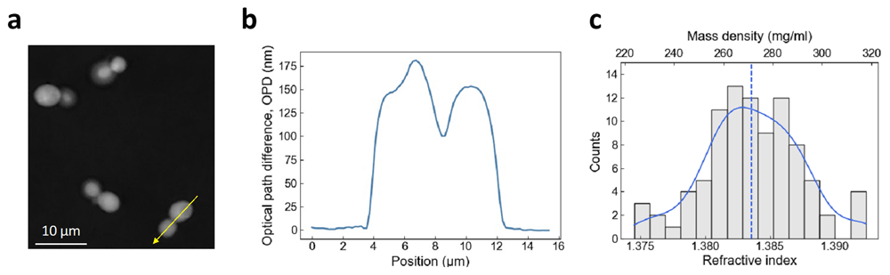
Figure 1. Measurement of cellular refractive index and dry mass density using QPI.
(a) Optical path difference (OPD) image of budding yeast cells in SCD medium. (b) Profile plot drawn from (a) following the yellow arrow line. (c) Histogram of Refractive index (RI) and dry mass density measurements of budding yeast cells grown at 30°C in SCD medium. A kernel density estimation was plotted (blue) with the median value (dashed line).
Increased intracellular density under GIP
We investigated the change in intracellular dry mass density as a function of growth-induced pressure (GIP). We used microfluidic elastic chambers to grow S. cerevisiae within a confined space to investigate the cellular RI under GIP (Figure 2a). To prevent nutrient depletion and exchange media, chambers were supplied with nutrients through microchannels on both sides. After the cells filled the space through proliferation, they developed GIP by pushing against their neighbors and onto their surroundings. GIP was measured through the deformation of the PDMS elastic wall.
We acquired phase images of cells in a chamber under GIP using QPI to examine cellular RI (Figure 2a). We measured that the average OPD of the cells with respect to the surrounding PDMS decreased linearly with pressure (Figure 2b). As PDMS has a higher RI than the cells on average, nPDMS = 1.405, the decrease in OPD implied that the RI of a cell approached the value of PDMS with increased GIP. We measured the effective height of the deformed confining chamber using OPD of the chamber filled with calibrated culture medium and pressurized by a measured hydraulic pressure (Figure 2c). We then extracted, assuming that the chamber was fully filled with cells, the mean cellular RI as a function of GIP (Figure 2d). We showed that RI increased roughly linearly with increased GIP under confinement.
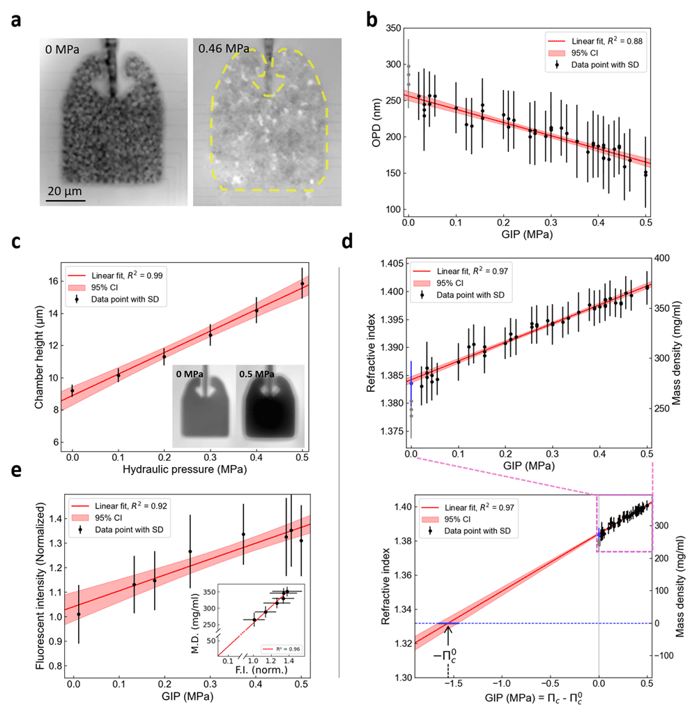
Figure 2. Intracellular dry mass density of budding yeast increases linearly with growth-induced pressure.
(a) OPD images of budding yeast cells growing within the confining space of a PDMS microfluidic chamber. Cells do not experience growth-inducing pressure until their growth fills the entire volume within the chamber (left, GIP = 0 MPa). After, confined growth leads to the build-up of growth-induced pressure (GIP), deforming the elastic chamber, where the expanded boundary is labeled with a yellow dashed line (right, GIP = 0.46 MPa). (b) Change in OPD of cells under GIP measured relative to surrounding PDMS. (c) The effective height of the PDMS chamber linearly increased with the GIP level, which is proportional to the OPD between the chamber and surrounding PDMS (inset). (d) The RI and dry mass density increase linearly as a function of GIP. The value estimated outside of the device previously is presented in a blue point. The measurements within the device at GIP = 0 are presented in grey, which were excluded from the regression as they are assumed to be underestimated. The nominal intracellular osmotic pressure () is estimated through linear extrapolation. An estimated value is indicated. (e) Fluorescence intensity (F.I.), corresponding to GFP expression level, linearly increases along GIP (n = 20). Dry mass (D.M.) density is proportional to fluorescence intensity (inset). The values in the inset graph is denoted by mean ± standard deviation in both the x and y-axis direction.
We noted that the RI of cells without pressure in the chambers was underestimated (gray points in Figure 1c, compared to the blue point measured outside of the device). We attributed this underestimation to the fact that cells were not tightly packed in the chamber, lowering the effective RI due to culture medium at a non-negligeable volume fraction in the chamber.
Proportional increase in GFP production with intracellular density under GIP
We measured the mean fluorescence intensity of a GFP expressed from the HIS3 promoter. We showed a linear increase of GFP concentration in the cell as a function of GIP (Figure 2e). Interestingly, we observed that the increase in mass density was proportional to the increase in GFP concentration (Figure 2e, inset), suggesting that the linearity between RI and protein concentration was kept in these conditions. Importantly, intracellular density extrapolated to 0 when the fluorescence intensity did. These results together demonstrated that the cellular biomass and GFP concentration increase roughly proportionally to GIP.
Estimation of the nominal intracellular osmotic pressure
The intracellular RI is proportional to the dry mass of the cell. Figure 2d showed that mass and intracellular osmotic pressure increased proportionally under confined growth. We performed a linear extrapolation of the pressure to a RI matching the one of water at 30°C (nwater = 1.332), without considering the points at GIP = 0 MPa measured in the microfluidic chip, but considering the RI of cells measured in culture medium (blue point). We assumed that when the cell would have the RI of water, it would be empty of its constituents, and its intracellular pressure would then be null, = 0 MPa. When the RI would match the RI of water, it would thus correspond to a point where the cell would be “empty” of macromolecules. The corresponding GIP, denoted , corresponding to the pressure difference between the intracellular osmotic pressure cell, , and the nominal intracellular osmotic pressure, , allowed us to estimate the nominal intracellular pressure of the cell: (Figure 2d). We found in this case a nominal pressure of 1.55 MPa and the 95% confidence interval ranges from 1.46 to 1.66 MPa, comparably larger than the 0.95 MPa estimated for the same S. cerevisiae in previous studies, with a different method10 (see Discussion).
Conclusion
In this study, the dry mass density under pressure was estimated by quantitative phase microscopy, and increased with GIP. The previous measurement of intracellular density under GIP has only been estimated indirectly by tracking fluorescently tagged tracer nanoparticles. It was assumed that the decrease in particle diffusion occurred due to the reduction of the free volume of the cytoplasm10. However, the macromolecular diffusion within the cytoplasm, a highly heterogeneous active media, can be limited not only by the accumulation of macromolecules but also by a change in macromolecule size distribution27, molecular electrostatic interactions28, or active random force29. Therefore, our results experimentally verify that the physical basis of the crowding-induced reduction in particle diffusion is compatible with an increase in dry mass density.
The previous theoretical model relating nanoparticle tracer diffusion with GIP assumed that the concentrations of osmolytes and macromolecules were produced proportionally10,33, but this was not confirmed experimentally. The linear relation between GIP and RI shown in our results indicated that the production ratio of macromolecules and osmolytes was kept constant under pressure in the yeast S. cerevisiae (Figure 2d). However, the nominal osmotic pressure of budding yeast estimated through linear extrapolation, 1.55 MPa, was slightly larger than the value obtained from the previous study's theoretical model, 0.95 MPa10, but remained in the same order of magnitude.
Ethics and consent
Ethical approval and consent were not required.
Data availability
Underlying data
Zenodo: Data of the subfigures of article "Intracellular dry mass density increases under growth-induced pressure", https://doi.org/10.5281/zenodo.1384264830
Data is available under the terms of the Creative Commons Attribution 4.0 International
Author contributions
HK, BA, NC, JR and MD did the experiments. HK and MD analyzed the data and wrote the manuscript. All authors proofread the manuscript.
Faculty Opinions recommendedReferences
- 1.
Stewart PS, Robertson CR:
Microbial growth in a fixed volume: studies with entrapped Escherichia coli.
Appl Microbiol Biotechnol.
1989; 30: 34–40. Publisher Full Text
- 2.
Chu EK, Kilic O, Cho H, et al.:
Self-induced mechanical stress can trigger biofilm formation in uropathogenic Escherichia coli.
Nat Commun.
2018; 9(1): 4087. PubMed Abstract
| Publisher Full Text
| Free Full Text
- 3.
Delarue M, Hartung J, Schreck C, et al.:
Self-driven jamming in growing microbial populations.
Nat Phys.
2016; 12(8): 762–766. PubMed Abstract
| Publisher Full Text
| Free Full Text
- 4.
Delarue M, Poterewicz G, Hoxha O, et al.:
SCWISh network is essential for survival under mechanical pressure.
Proc Natl Acad Sci U S A.
2017; 114(51): 13465–13470. PubMed Abstract
| Publisher Full Text
| Free Full Text
- 5.
Bengough AG, Croser C, Pritchard J:
A biophysical analysis of root growth under mechanical stress. In: Anderson HM, Barlow PW, Clarkson DT, Jackson MB, Shewry PR, eds. Plant Roots - From Cells to Systems. Springer Netherlands, 1997; 107–116. Publisher Full Text
- 6.
Breuil-Broyer S, Morel P, De Almeida-Engler J, et al.:
High-resolution boundary analysis during Arabidopsis thaliana flower development.
Plant J.
2004; 38(1): 182–192. PubMed Abstract
| Publisher Full Text
- 7.
Meriem ZB, Mateo T, Faccini J, et al.:
A microfluidic mechano-chemostat for tissues and organisms reveals that confined growth is accompanied with increased macromolecular crowding.
Lab Chip.
2023; 23(20): 4445–4455. PubMed Abstract
| Publisher Full Text
- 8.
Rizzuti IF, Mascheroni P, Arcucci S, et al.:
Mechanical control of cell proliferation increases resistance to chemotherapeutic Agents.
Phys Rev Lett.
2020; 125(12): 128103. PubMed Abstract
| Publisher Full Text
- 9.
Alessandri K, Sarangi BR, Gurchenkov VV, et al.:
Cellular capsules as a tool for multicellular spheroid production and for investigating the mechanics of tumor progression in vitro.
Proc Natl Acad Sci U S A.
2013; 110(37): 14843–14848. PubMed Abstract
| Publisher Full Text
| Free Full Text
- 10.
Alric B, Formosa-Dague C, Dague E, et al.:
Macromolecular crowding limits growth under pressure.
Nat Phys.
2022; 18(4): 411–416. PubMed Abstract
| Publisher Full Text
| Free Full Text
- 11.
Weber SC, Spakowitz AJ, Theriot JA:
Nonthermal ATP-dependent fluctuations contribute to the in vivo motion of chromosomal loci.
Proc Natl Acad Sci U S A.
2012; 109(19): 7338–7343. PubMed Abstract
| Publisher Full Text
| Free Full Text
- 12.
Sakaue T, Saito T:
Active diffusion of model chromosomal loci driven by athermal noise.
Soft Matter.
2016; 13(1): 81–87. PubMed Abstract
| Publisher Full Text
- 13.
Grover WH, Bryan AK, Diez-Silva M, et al.:
Measuring single-cell density.
Proc Natl Acad Sci U S A.
2011; 108(27): 10992–10996. PubMed Abstract
| Publisher Full Text
| Free Full Text
- 14.
Miettinen TP, Ly KS, Lam A, et al.:
Single-cell monitoring of dry mass and dry mass density reveals exocytosis of cellular dry contents in mitosis. Pines J, Cooper JA, Piel M, eds.
eLife.
2022; 11: e76664. PubMed Abstract
| Publisher Full Text
| Free Full Text
- 15.
Delarue M, Brittingham GP, Pfeffer S, et al.:
mTORC1 controls phase separation and the biophysical properties of the cytoplasm by tuning crowding.
Cell.
2018; 174(2): 338–349.e20. PubMed Abstract
| Publisher Full Text
| Free Full Text
- 16.
Asano S, Engel BD, Baumeister W:
In Situ cryo-electron tomography: a post-reductionist approach to structural biology.
J Mol Biol.
2016; 428(2 Part A): 332–343. PubMed Abstract
| Publisher Full Text
- 17.
Park Y, Depeursinge C, Popescu G:
Quantitative phase imaging in biomedicine.
Nature Photon.
2018; 12(10): 578–589. Publisher Full Text
- 18.
Nguyen TL, Pradeep S, Judson-Torres RL, et al.:
Quantitative Phase Imaging: recent advances and expanding potential in biomedicine.
ACS Nano.
2022; 16(8): 11516–11544. PubMed Abstract
| Publisher Full Text
| Free Full Text
- 19.
Zhao H, Brown PH, Schuck P:
On the distribution of protein refractive index increments.
Biophys J.
2011; 100(9): 2309–2317. PubMed Abstract
| Publisher Full Text
| Free Full Text
- 20.
Stirling DR, Swain-Bowden MJ, Lucas AM, et al.:
CellProfiler 4: improvements in speed, utility and usability.
BMC Bioinformatics.
2021; 22(1): 433. PubMed Abstract
| Publisher Full Text
| Free Full Text
- 21.
Seabold S, Perktold J:
Statsmodels: econometric and statistical modeling with python. 2010; 92–96. Publisher Full Text
- 22.
Abuhattum S, Kim K, Franzmann TM, et al.:
Intracellular mass density increase is accompanying but not sufficient for stiffening and growth arrest of yeast cells.
Front Phys.
2018; 6. Publisher Full Text
- 23.
Odermatt PD, Miettinen TP, Lemière J, et al.:
Variations of intracellular density during the cell cycle arise from tip-growth regulation in fission yeast. Balasubramanian MK, Barkai N, Piel M, eds,
eLife.
2021; 10: e64901. PubMed Abstract
| Publisher Full Text
| Free Full Text
- 24.
Bianco V, D’Agostino M, Pirone D, et al.:
Label-free intracellular multi-specificity in yeast cells by phase-contrast tomographic flow cytometry.
Small Methods.
2023; 7(11): e2300447. PubMed Abstract
| Publisher Full Text
- 25.
Boltyanskiy R, Odete MA, Cheong FC, et al.:
Label-free viability assay using in-line holographic video microscopy.
Sci Rep.
2022; 12(1): 12746. PubMed Abstract
| Publisher Full Text
| Free Full Text
- 26.
Midtvedt D, Olsén E, Höök F, et al.:
Label-free spatio-temporal monitoring of cytosolic mass, osmolarity, and volume in living cells.
Nat Commun.
2019; 10(1): 340. PubMed Abstract
| Publisher Full Text
| Free Full Text
- 27.
Kalwarczyk T, Tabaka M, Holyst R:
Biologistics—Diffusion Coefficients for complete proteome of Escherichia coli.
Bioinformatics.
2012; 28(22): 2971–2978. PubMed Abstract
| Publisher Full Text
| Free Full Text
- 28.
Zhou HX, Pang X:
Electrostatic interactions in protein structure, folding, binding, and condensation.
Chem Rev.
2018; 118(4): 1691–1741. Publisher Full Text
- 29.
Brangwynne CP, Koenderink GH, MacKintosh FC, et al.:
Cytoplasmic diffusion: molecular motors mix it up.
J Cell Biol.
2008; 183(4): 583–587. PubMed Abstract
| Publisher Full Text
| Free Full Text
- 30.
Kim H, Delarue M:
Data of the subfigures of article "Intracellular dry mass density increases under growth-induced pressure".Zenodo.2024. http://www.doi.org/10.5281/zenodo.13842648