Introduction
Aquaculture has become a type of animal food production with the fastest growth in recent decades1. Fish are among the most important groups of allergenic foods worldwide2,3. Allergic reactions result in sensitized consumers from an excessive IgE-mediated response of the immune system against specific proteins called allergens. The major allergen responsible for fish allergies in humans is parvalbumin4. Parvalbumins are heat-stable intracellular EF-hand proteins involved in muscle relaxation via calcium buffering5. Unfortunately, the development of specific immunotherapeutics for preventing fish allergies is limited. As a result, prophylactic management of this allergy relies only on the elimination of fish products from the diet of sensitized patients4. In salmonids, a single parvalbumin-beta1 protein was identified as an allergen in some patients4. Conceptually, a simple way to suppress trout allergy is to inactivate the gene encoding parvb1. Nevertheless, one cannot rule out the possibility that the invalidation of parvb1 might prevent fish development and/or affect muscle physiology. Consistent with this remark, we observed that crossbreeding of two distinct trout (F1) lines carrying one null parvb1 allele did not lead to the generation of homozygous F2 mutants (Lebret et al., unpublished data). Given that parvb1 exon 2 encodes an allergenic peptide4, we aimed to generate Parvb1 alleles that specifically lack this exon. To this end, we attempted to perform exon 2 skipping and swapping by applying microhomology-mediated end-joining (MMEJ) knock-in, a process that facilitates the insertion of donor DNA flanked by a small region (approximately 25nt) homologous to the Crispr target site6,7.
Methods
Animals
All experiments reported in this study were performed on rainbow trout Oncorhynchys mykiss (walbaum, 1792).
Guide RNA (gRNA) design and synthesis
The guide RNA design was performed using the Crispor tool (http://crispor.tefor.net/), which identifies gRNAs, evaluates potential off-targets, and predicts on-target activity, and was based on scanning of the trout genome which has been sequenced, assembled and annotated8. gRNAs and cas9 mRNA were prepared as previously described9. Briefly, the DR274 vector (Addgene #42250) containing the universal guide RNA sequence was linearized with Bsa1 and purified. PCR amplifications were performed using linearized DR274 as a template and specific primers for each sgRNA. Forward primers containing sgRNA target sequences (bold and underlined) between the T7 promoter sequence at the 5' end and the conserved tracrRNA domain sequence were as follows: forward primer (#site 1), 5' GAAATTAATACGACTCACTATA GGAGGGGGAAGTAAACGAACT GTTTTAGAGCTAGAAATAGCAAG-3'. Forward primer (#site 2): 5'-GAAATTAATACGACTCAC TATA GGTACGAAACATAGCATACAGTTTTAGAGCTAGAAATAGCAAG-3'; universal Reverse primer: 5'-AAAAGCACCGACTCGGTGCCACT-3'. Subsequently, the residual plasmid was digested using Dpn1. The final product was purified and used as the DNA template for transcription. sgRNAs were transcribed using the MegaShortScript T7 Transcription Kit (Ambion (cat. AM1354)) according to the manufacturer’s instructions. The double-stranded templates (Figure 1B and Figure 4B) added to the injection mix for MMEJ Knock-In experiments were provided by GeneStrand Synthesis, Eurofin Genomics.
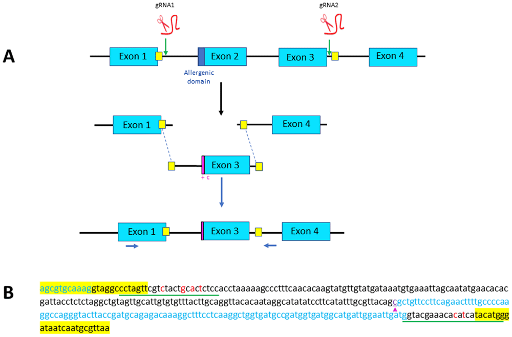
Figure 1. Strategy designed for Exon 2 skipping through microhomology-mediated end joining (MMEJ).
(A) Exons 2 and 3 of parvb1 locus are excised using two gRNAs, one (gRNA1) located in the intron between exons 1 and 2, and one (gRNA2) in the intron between exons 3 and 4. The two cutting-sites targeted by the gRNAs are flanked by short sequence (yellow squares) that are added as homology arm at the 5’ and 3’ ends of the donor DNA (containing only parvb1 exon 3) to be inserted. Position of primers used for PCR screening are indicated (blue arrows). (B) Nucleotide sequence of the donor DNA used for exon skipping. The short homology arms are highlighted in yellow. The gRNA sites of the donor DNA (underlined in green) have been slightly modified (red letters) to prevent cas9-mediated re-cutting of the donor DNA after its integration into parvb1 locus. A cytosine (purple arrowhead) has been added at the 5’ extremity of exon 3 to keep the reading frame of exon 1.
Preparation of embryos and microinjection
Trout eggs were collected by gentle manual stripping under anesthesia (2-phenoxyethanol, 0.05%). The eggs were then fertilized by careful mixing with the sperm solution. For microinjection, a mixture containing gRNA (100ng/μl), cas9 mRNA (200ng/μl) and double-stranded donor DNA (10 ng/μl) was prepared and microinjected using a FemtoJet (Eppendorf) microinjector during a period of 2 to 6 h after egg fertilization (one cell stage prior to the first cleavage). Once injected, embryos were transferred to hatchery trays and incubated in running water (10°C) until first feeding (65 dpf). Subsequently, the fingerlings were transferred into indoor tanks.
Screening for mutations
Genotyping was performed using genomic DNA extracted from the fin tissue of 70 dpf fingerlings. Specifically, fingerlings were anesthetized in 2-phenoxyethanol (0.5 ml/L) and a small part (2mm2) of the caudal fin was clipped. Fin-clip samples were then incubated in 100 μl of 5% Chelex 100 Molecular Biology Grade resin (BioRad (cat.1421253)) and 50 μl of proteinase K solution (20mg/ml, Ambion (cat. AM2546)) for 2 h at 55°C and then for 10 min at 99°C with continuous stirring. After centrifugation at 5000 g for 10 min, the supernatant was collected and subsequently used for PCR. PCR reactions were performed taking one μl (~ 100 ng) of extracted genomic DNA and using the jumpstart Taq DNA polymerase (Sigma (cat. D9307)), the PCR nucleotide mix from Promega (cat. C1145) and Parvb1 gene specific primers (Eurofins genomics) at 10μM each. For the exon skipping experiment, PCR was performed using the forward primer 5’-CAATGCTCAATACCGAACGTTGA-3’ and the reverse primer 5’-AGAGGCAGTAGTGATGACCAG-3’ present in parvb1 exon1 and intron3 (Figure 1A), respectively. For the exon swapping experiment, PCR was performed using the forward primer 5’-CAATGCTCAATACCGAACGTTGA-3’ present in parvb1 exon1, the reverse primer 5’-AGAGGCAGTAGTGATGACCAG-3’ present in parvb1 intron3 and the reverse primer 5’-TCCTCAATGAAGCCACTCTTG-3’ specifically present in parvb2 exon 2 (Figure 4A). The PCR cycling conditions were as follows: 95°C for 5 min and 35 cycles of 95°C for 15 s, 60°C for 30s, and 72°C for 30 s. The reaction products were separated and visualized using agarose gel electrophoresis. Of note, gel electrophoresis of PCR products was appropriate to uncover alleles resulting from exon2 skipping or swapping but did not reveal unwanted allele displaying small indel-associated mutations. PCR products of interest were further sequenced by the Eurofins Genomics Sequencing Service (https://www.eurofinsgenomics.eu).
Results
Exon 2 skipping
Parvb1 is composed of four exons (Figure 1A). The 5’ subdomain of exon 2 encodes the allergenic peptide, whereas the 3’ subdomain of exon 2, along with the 5’ subdomain of exon 3, encode one of the two EF-hand motifs involved in calcium binding. The 3’ subdomain of exon 3 along with exon 4 encode the second EF-hand motif4. Exons 1 and 3 of parvb1 are not in frame, so a single excision of exon 2 followed by NHEJ would not result in appropriate splicing of the remaining exons. Therefore, to perform correct exon 2 skipping, we sought to delete both exon 2 and exon 3 using gRNA1 and gRNA2 (Figure 1A) which displayed reliable activity when injected independently into zygotes, and to elicit, at the cutting sites, the integration of an injected donor DNA containing parvb1 intron 1, exon 3 (with an additional nucleotide immediately following the splice acceptor site to maintain the reading frame of exon 1), and the 5’ domain of intron 3 (Figure 1 A and B). The donor DNA was flanked by short homology arms for proper insertion through microhomology-mediated end joining (MMEJ). Out of approximately 600 fertilized eggs injected to achieve exon2 skipping, only 191 developed up to the fingerling stage (approximately 70 d post-fertilization). The fingerlings were genotyped using appropriate primers (Figure 1A). In most cases, only one PCR band corresponding to the wild-type allele was found in fingerlings. Truncated alleles of various sizes were sometimes observed (10 fingerlings out of 191) in addition to the wild-type allele (Figure 2). Only one individual exhibited, in addition to the wild type allele, a truncated allele which size was compatible with a correct insertion of donor DNA (Figure 2). Sequencing of this allele confirmed the correct insertion of donor DNA but also revealed a tandem duplication of the 3’ microhomology arm in intron 3 (Figure 3). Given its position within the gene, it is unlikely that this tandem duplication would affect the processing of the parvb1 primary transcript. Unfortunately, the premature death of fingerlings carrying this mutation divested the F1 generation for further phenotyping. Overall, this study shows that microhomology-mediated end joining can be used to delete exon 2 of parvb1, although the success rate was very low.
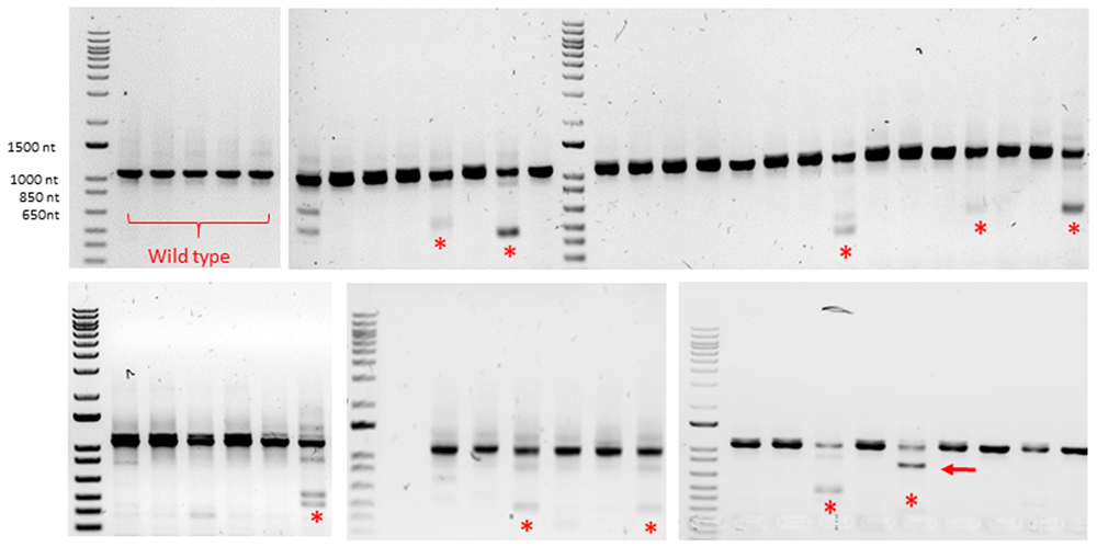
Figure 2. Exon skipping experiment: detection of Crispr/Cas9-introduced mutations by genotyping.
Gel electrophoresis of several PCR products. Top left corner (lanes 1-5): wild type trout DNA. Other lanes: DNA from F0 trout originating from eggs injected with donor DNA designed for exon 2 skipping: truncated alleles of various sizes (red stars) were sometimes found in addition to the wild type allele; only one individual displayed an allele carrying a deletion of the expected size (red arrow).
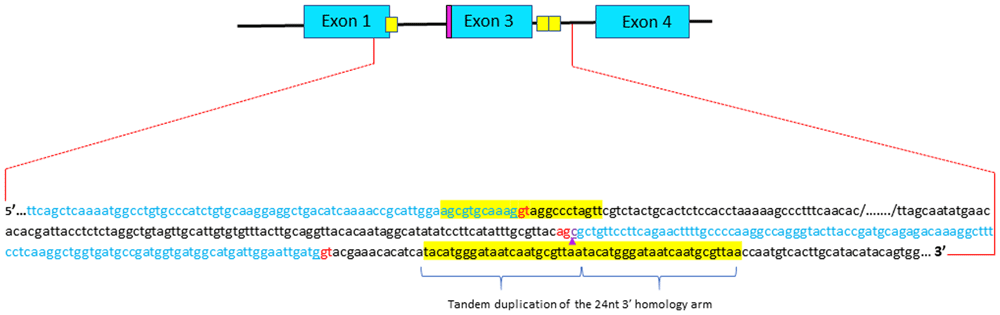
Figure 3. Sequencing analysis of integration junctions of the donor DNA into parvalbumin beta 1 locus in the animal displaying exon2 skipping.
Short homology arms are highlighted in yellow. Although the donor DNA is correctly inserted, one can observed a tandem duplication of the 3’ short homology arm. The additional cytosine that kept the reading frame of exon 1 is outlined (purple arrow). Donor and acceptor splice sites are in red.
Exon 2 swapping
To replace parvb1 exon2 with parvb2 exon2, which does not encode the allergenic peptide, we excised exons 2 and 3 using the same gRNAs employed for parvb1 Exon2 skipping (Figure 1A). Knock- In was carried out using donor DNA containing parvb1 intron 1, parvb2 exon 2, parvb1 intron 2, parvb1 exon 3, and the 5’ domain of parvb1 intron 3 (Figure 4 A and B). Short homology arms were added at the extremities of the donor DNA to achieve proper insertion through microhomology-mediated end joining (MMEJ) (Figure 4B). Approximately 500 fertilized eggs were injected, 157 of which developed up to the fingerling stage. These fingerlings were genotyped, but none of them were found to display the correct donor DNA insertion into the parvb1 locus, as shown after genotyping using an internal parvb2 specific primer (Figure 4A). Interestingly, in contrast to our initial exon-skipping experiment, no truncated junk alleles were observed. This second part of our study confirms an apparently intrinsically low incidence for the targeted integration of DNA templates through microhomology-mediated end joining in trout.
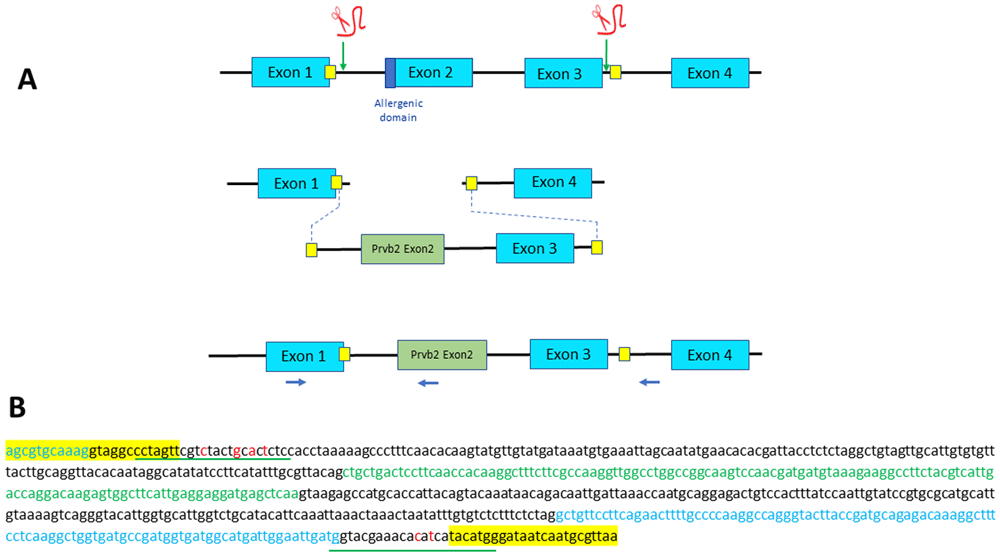
Figure 4. Strategy designed for Exon 2 swapping through microhomology-mediated end joining (MMEJ).
(A) Exons 2 and 3 of parvb1 locus are excised using two gRNAs, one located in the intron 5’ to exon 2 and one in the intron between exons 3 and 4. The two cutting-sites targeted by the gRNAs are flanked by a short sequence (yellow squares) that are added as homology arm at the 5’ and 3’ ends of the donor DNA (containing parvb2 exon 2 and parvb1 exon 3) to be inserted. Position of primers used for PCR screening are indicated (blue arrows). (B) Nucleotide sequence of the donor DNA used for exon 2 swapping. Parvb2 Exon 2 and parvb1 exon 3 are in green and blue letters, respectively. Short homology arms are highlighted in yellow. The gRNA sites (underlined in red) have been slightly modified (red letters) to prevent cas9-mediated re-cutting of the donor DNA after its integration into parvalbumin beta 1 locus.
Discussion
Parvalbumin beta-1 is a major allergen in the trout. Therefore, the invalidation of parvb1 encoding gene could prevent allergic reactions in sensitive persons. However, one cannot rule out the possibility that knockout of the gene encoding parvb1 could have deleterious developmental and/or physiological effects on the whole animal. In keeping with this point, we were unable to generate homozygous mutants after crossing two distinct F1 lines carrying one null allele (Lebret, unpublished work). Hence, precise engineering that allows the production of active parvb1 specifically deprived of the allergenic domain remains the only possible alternative strategy to suppress fish allergenicity. Given that the allergenic domain of parvb1 is encoded by exon2, our editing strategy was to use the Crispr/cas9 system to generate F0 trout carrying either an allele lacking exon 2 (exon skipping) or displaying the replacement of parvb1 exon 2 with that of parvalbumin beta-2 (parvb2), which does not encode the allergenic peptide (exon swapping). For this purpose, we tested the possibility of inserting specific donor DNA through the microhomology-mediated end-joining pathway (MMEJ). (MMEJ)-based methods are designed to integrate donor DNA into a targeted cutting site of the genome, provided that the donor DNA contains, at its 5’ and 3’ ends, short arms that are homologous to the sequences flanking the targeted cutting site6,7. Building on the work by Paquet et al.10 and feedback from preliminary experiments on medaka (Herpin, unpublished results), we introduced silent CRISPR/cas9 blocking mutations in the guide RNA sequence adjacent to the microhomology arms to prevent re-cutting of the target sequence once the donor DNA has been integrated into the genome. In our study, only one individual (out of 191) displayed exon 2 exon skipping through microhomology-mediated end-joining Knock-In, and not a single animal (out of 157) exhibited exon swapping. The efficiency of the procedure was disappointingly low and clearly below our expectations when compared with the success rate obtained in medaka. Some success has been reported regarding Crispr/cas9-mediated knock-in in farmed fish, such as carp (Laboeo rohita)11 and salmon (Salmo salar L.)12. Nevertheless, in the first report11, the two homology arms of the donor DNA to be inserted were considerably longer (800 nt) than those used for MMEJ (20-30nt) and in the second report12, the sequence to be inserted through microhomology-mediated end-joining Knock-In was very short (<100 nucleotides). Therefore, gene editing to insert any desired sequence in farmed fish genomes needs to be carefully considered. Not only is knock-in efficiency obviously uncertain, but germinal mosaicism in the F0 generation is also important. Being often long, the generation time of farmed fish species (two years for trout) might be an obstacle for producing heterozygous F1 and homozygous F2 lines. Regarding knock-in efficiency, improvements can be made by testing templates of different lengths. The length of the homologous arms, as well as the use of asymmetric donor DNA containing homology arms of different sizes at the 5’ and 3’ ends, are also parameters that might impact KI efficiency13. Chemical intervention, blocking the NHEJ pathway, and promoting the MMEJ pathway, such as NU744114, might also be a suitable approach. Beyond the fact that effective harnessing of MMEJ remains challenging15, it is important to point out that genome editing using donor DNA is likely to lead, at some point, to uncontrolled and random insertion of the donor DNA into the host genome. To illustrate this aspect, it is worth noting that 60% of trout embryos injected with a donor DNA containing a myosin promoter coupled with GFP cDNA gave rise to F0 individuals displaying mosaic GFP fluorescence in skeletal muscle16. Therefore, in addition to potential off-target mutations, animals edited by knock-in are likely to display high rates of unintentional transgene insertions. This highlights the necessity of conducting careful whole-genome sequencing of knock-In farmed animals to limit introgression of the template inserted in non-targeted loci during successive crossings. This implies that gene editing must be performed solely in animals in which the genome has been fully sequenced and annotated. Phenotyping gene-edited animals is an essential step that follows gene modification. If parvb1 knock-in finally became successful to a certain extent, then the next objective would be to prepare muscle extracts from knock-in trout and to test them against sera of specific patients. In keeping with this point, collaborations with allergology laboratories would be particularly valuable for future studies.
In July 2018, the Court of Justice of the European Union delivered a judgement in which it held that organisms modified by genome-editing techniques are genetically modified organisms (GMOs) and fall within the scope of EU legislation on GMOs (see footnote: infocuria Jurisprudence). This judgement, which makes no difference between a gene-edited animal displaying a single base pair substitution and a transgenic animal carrying exotic DNA, is highly disputed, as recently reported by a European commission study concluding that the present legislation is not suitable for new genomic techniques (NGT) and needs some adaptations to follow rapid scientific and technological progress (see footnote: New genomic techniques, European commission study and first reactions). In any event, we believe that revision of the law determining the legal status of gene-edited animals would gain much from a basic distinction between those edited using donor DNA (HDR, MMEJ, etc.) and those edited without donor DNA (NHEJ). The former is potentially transgenic if one posits that the insertion(s) of any donor DNA outside of its natural locus falls under transgenesis. Whether the Crispr/cas9-mediated KI gene should be developed in aquaculture breeding programs depends on consumer acceptance as well as government regulation. There have been lively debates on this point, notably in the European parliament. We believe that our data can contribute to feed debates regarding the feasibility and limitations of CRISPR-mediated knock-Ins in farmed fish, as well as future regulatory actions for their generation and use.
Ethical statement
Fish care and sampling were conducted at the INRAE UR1037 fish facilities (DDSP approval D35-238-6) in strict accordance with the European policies and the guidelines of the National Legislation on Animal Care and Use Ethical Committee (Decree No.2013–118, February 1st, 2013; European Directive 2010–63, September 22, 2010). Experimental protocols involving animals were approved by the “Comité Rennais d’éthique pour l’expérimentation animale’ (CREEA, ref. APAFIS #31048-2021041509574912V3).
Animal ethics
All efforts were made to prevent fish suffering. In particular, trout eggs used for microinjection were collected by gentle manual stripping of mature anesthetised females. Also, regarding fingerling genotyping, fin tissue sampling was carried out on anesthetised animals according to current standards.
Availability of data and materials
Reporting guidelines
This study is reported in accordance with ARRIVE guidelines:
DOI: http://dx.doi.org/10.5281/zenodo.1380370718
Data are available under the terms of the Creative Commons Attribution 4.0 International license (CC-BY 4.0)
Author contributions
Lebret V: Investigation, Methodology, Visualization; Duret C: Investigation, Methodology; Herpin A: Conceptualization, Investigation, Methodology, Writing-Review and Editing; Rescan PY: Conceptualization, Funding acquisition, Investigation, Methodology, Project administration, Supervision, Visualization, Writing-Original draft, Writing-Review and Editing.
Acknowledgement
Not applicable.
Faculty Opinions recommendedReferences
- 1.
FAO:
The state of world fisheries and aquaculture-sustainability in action. 2020. Publisher Full Text
- 2.
Moonesinghe H, Mackenzie H, Venter C, et al.:
Prevalence of fish and shellfish allergy: a systematic review.
Ann Allergy Asthma Immunol.
2016; 117(3): 264–272.e4. PubMed Abstract
| Publisher Full Text
- 3.
Sharp MF, Lopata AL:
Fish allergy: in review.
Clin Rev Allergy Immunol.
2014; 46(3): 258–71. PubMed Abstract
| Publisher Full Text
- 4.
Kuehn A, Swoboda I, Arumugam K, et al.:
Fish allergens at a glance: variable allergenicity of parvalbumins, the major fish allergens.
Front Immunol.
2014; 5: 179. PubMed Abstract
| Publisher Full Text
| Free Full Text
- 5.
Arif SH:
A Ca2+-binding protein with numerous roles and uses: parvalbumin in molecular biology and physiology.
Bioessays.
2009; 31(4): 410–21. PubMed Abstract
| Publisher Full Text
- 6.
McVey M, Lee SE:
MMEJ repair of double-strand breaks (director's cut): deleted sequences and alternative endings.
Trends Genet.
2008; 24(11): 529–38. PubMed Abstract
| Publisher Full Text
| Free Full Text
- 7.
Wierson WA, Welker JM, Almeida MP, et al.:
Efficient targeted integration directed by short homology in zebrafish and mammalian cells.
eLife.
2020; 9: e53968. PubMed Abstract
| Publisher Full Text
| Free Full Text
- 8.
Berthelot C, Brunet F, Chalopin D, et al.:
The rainbow trout genome provides novel insights into evolution after whole-genome duplication in vertebrates.
Nat Commun.
2014; 5: 3657. PubMed Abstract
| Publisher Full Text
| Free Full Text
- 9.
Imarazene B, Du K, Beille S, et al.:
A supernumerary "B-sex" chromosome drives male sex determination in the Pachón cavefish, Astyanax mexicanus.
Curr Biol.
2021; 31(21): 4800–4809.e9. PubMed Abstract
| Publisher Full Text
| Free Full Text
- 10.
Paquet D, Kwart D, Chen A, et al.:
Efficient introduction of specific homozygous and heterozygous mutations using CRISPR/Cas9.
Nature.
2016; 533(7601): 125–9. PubMed Abstract
| Publisher Full Text
- 11.
Chakrapani V, Patra SK, Panda RP, et al.:
Establishing targeted carp TLR22 gene disruption via homologous recombination using CRISPR/Cas9.
Dev Comp Immunol.
2016; 61: 242–7. PubMed Abstract
| Publisher Full Text
- 12.
Straume AH, Kjærner-Semb E, Ove Skaftnesmo K, et al.:
Indel locations are determined by template polarity in highly efficient in vivo CRISPR/Cas9-mediated HDR in Atlantic salmon.
Sci Rep.
2020; 10(1): 409. PubMed Abstract
| Publisher Full Text
| Free Full Text
- 13.
Richardson CD, Ray GJ, DeWitt MA, et al.:
Enhancing homology-directed genome editing by catalytically active and inactive CRISPR-Cas9 using asymmetric donor DNA.
Nat Biotechnol.
2016; 34(3): 339–44. PubMed Abstract
| Publisher Full Text
- 14.
Aksoy YA, Nguyen DT, Chow S, et al.:
Chemical reprogramming enhances homology-directed genome editing in zebrafish embryos.
Commun Biol.
2019; 2: 198. PubMed Abstract
| Publisher Full Text
| Free Full Text
- 15.
Seol JH, Shim EY, Lee SE:
Microhomology-mediated end joining: good, bad and ugly.
Mutat Res.
2018; 809: 81–87. PubMed Abstract
| Publisher Full Text
| Free Full Text
- 16.
Gabillard JC, Rallière C, Sabin N, et al.:
The production of fluorescent transgenic trout to study in vitro myogenic cell differentiation.
BMC Biotechnol.
2010; 10: 39. PubMed Abstract
| Publisher Full Text
| Free Full Text
- 17.
Lebret V, Duret C, Herpin A, et al.:
Microhomology-mediated end-joining Knock-In approaches to delete the allergenic domain of trout parvalbumin beta-1. Preliminary results in F0 animals and feedback. Underlying data [Data set].
Zenodo.
2024. http://www.doi.org/10.5281/zenodo.13804556
- 18.
Lebret V, Duret C, Herpin A, et al.:
Microhomology-mediated end-joining Knock-In approaches to delete the allergenic domain of trout parvalbumin beta-1. Preliminary results in F0 animals and feedback. Reporting guidelines.
Zenodo.
2024. http://www.doi.org/10.5281/zenodo.13803707








