Introduction
Dictyostelia evolved multicellularity independently from animals, plants and fungi within the single-celled Amoebozoa. They feed on bacteria but unlike their amoebozoan relatives, which survive starvation and other stressors as individual dormant cysts, dictyostelid amoebas aggregate instead to form fruiting bodies with dormant spores and up to three supporting cell types. Molecular phylogenetics subdivides Dictyostelia in four major and some minor groups (Schaap et al., 2006; Schilde et al., 2019). Group 4 contains the model organism Dictyostelium discoideum (Ddis) and displays several traits that set it apart from the other groups. Group 4 species uniquely use cyclic AMP (cAMP), secreted in pulses, as chemoattractant for aggregation. They mostly form large solitary fruiting bodies with spores supported by stalk, basal disc and upper and lower cup cells and have an intermediate migrating slug stage in which future (pre) spore and supporting cells initiate differentiation in appropriate proportions required for fruiting bodies construction. Species in groups 1–3 generally form smaller clustered and/or branched structures with at most two cell types, spores and stalk cells. Here most cells differentiate as prespore cells and transdifferentiate into stalk cells at the fruiting body tip. Species in these groups often use the dipeptide glorin as chemoattractant. Many group 1, 2 and 3 species can also still encyst individually when stressed, but this alternative life cycle is lost in group 4 (Romeralo et al., 2013; Schilde et al., 2014).
In addition to its role as chemoattractant, secreted cAMP acting on cAMP receptors (cARs) also induces the differentiation of prespore cells in group 4 (Schaap & Van Driel, 1985; Wang et al., 1988), while intracellular cAMP acting on cAMP dependent protein kinase (PKA) induces the maturation of spores and stalk cells (Harwood et al., 1992; Hopper et al., 1993; Mann et al., 1994). Evolutionary comparative studies showed that the roles of intracellular cAMP and PKA are conserved throughout Dictyostelia and are ancestrally derived from roles for cAMP and PKA as intermediates for stress-induced encystation in the solitary Amoebozoa (Du et al., 2014; Funamoto et al., 2003; Kawabe et al., 2015; Ritchie et al., 2008). While secreted cAMP pulses did not mediate aggregation in groups 1–3, they did organise fruiting body morphogenesis in the group 3 species D. minutum and the group 2 species Polysphondylium pallidum (Ppal) (Alvarez-Curto et al., 2005; Schaap et al., 1984). Secreted cAMP acting on cARs was also essential for induction of prespore differentiation in P. pallidum (Kawabe et al., 2009).
These observations suggested an evolutionary scenario in which the roles of cAMP in spore and stalk maturation, induction of prespore differentiation, organisation of morphogenesis and finally in group 4 aggregation were sequentially derived from the ancestral role of cAMP as intermediate for stress-induced encystation (Schaap, 2016). Recent studies in Polysphondylium violaceum (Pvio), which resides in a small sister clade to group 4, question the universality of this scenario. Unlike Ppal, where deletion of car genes disorganizes post-aggregative morphogenesis and prevents prespore and spore differentiation, deletion of the single Pvio carA gene leaves both its morphogenesis and spore differentiation intact (Kawabe & Schaap, 2023).
While it remains possible that a second car remained unsequenced in its draft genome (Narita et al., 2020), lack of prespore markers made direct assessment of prespore gene induction by cAMP impossible. In this study, I identified relatives of three Ddis prespore genes in Pvio. Pvio cells transformed with gene fusions of the promoters of either gene and the LacZ reporter gene were used to study activation of the promoters by cAMP, glorin and signals known to promote or inhibit prespore differentiation in Ddis. The study indicated that the Pvio lineage invented a novel mechanism for prespore induction.
Methods
Cell culture
Polysphondylium violaceum strain QSvi11 (Kalla et al., 2011) was grown in association with Klebsiella aerogenes on 1/3rd SM agar (13.9 g SM agar (catalogue number SMA0102 Formedium), 11 g agar (AGA03, Formedium), 1.27 g KH2PO4 (P5379, Sigma-Aldrich), 0.87 g K2HPO4.3H2O (P3786, Sigma-Aldrich) and 0.67 g MgSO4.7H2O (230391, Sigma-Aldrich) per litre H2O). Cells were harvested in PB (5 mM Na2HPO4 (71643, Sigma-Aldrich), 5 mM KH2PO4, pH 6.5), when about 1/3rd of the bacterial lawn was cleared and developed on NN agar (1.5% agar in 8.8 mM KH2PO4 and 3.4 mM Na2HPO4) at 1.7 × 10⁶ cells/cm2 until the desired developmental stage was reached.
Promoter-lacZ constructs
To prepare promoter-lacZ fusions of the putative Pvio prespore genes g4562 (psp1), g2696 (psp2) and g2380 (psp3), their respectively 2.1, 1.2 and 2.2 kb 5′intergenic regions were amplified from Pvio QSvi11 genomic DNA using Phusion high-fidelity DNA polymerase (F530S, ThermoFisher Scientific) and the oligonucleotide primer pairs g4562pF/g4562pR, g2696pF/g2696pR or g2380pF/g2380pR (Table 1) which harbour XbaI and BglII sites in the forward (F) and reverse (R) primers respectively. After XbaI/BglII digestion, the amplified products were cloned into XbaI/BglII digested vector pDdgal-17 (Harwood & Drury, 1990), which fuses the start codon and usually the first few amino-acids of the inserted gene in frame with LacZ. The constructs were validated by DNA sequencing using primers Gal17F and Gal17R (Table 1) and transformed into Pvio QSvi11 as described by (Narita et al., 2020), except that initial overnight starvation was at 5×10⁶ cells/ml instead of 5×10⁵.
Table 1. Oligonucleotide primers.
| Primer ID | Sequence | gene
name |
|---|
| g4562pF | GGGTCTAGAGGGTAGGAGAGGTAACAAATG | psp1 |
| g4562pR | GGGAGATCTTGACTTCATTTTATTCTTATTAATTTAATTC | |
| g2696pF | CCCTCTAGACATATGATAGGTGTATGCAGATTTAAG | psp2 |
| g2696pR | CCCAGATCTCATTGTACTAGTTATATTTTAAATTATTTTAGTAATGTTAC | |
| g2380pF | GTGTCAGACATTTTATGAATATATATAGTTATCTAGAAAG | psp3 |
| g2380pR | CCAGATCTTGGCTTCATTTATTTATGTATATGTG | |
| Gal17F | TATGGTAAAACTTGAATTGATCCTCTAG | |
| Gal17R | CATCCTGCAGTAAGCTTGGTACCGAGATC | |
Transformants were initially selected at 50 μg/ml G418 (G4181, Formedium) on 1/3rd SM agar with G418 resistant Escherichia coli (Narita et al., 2020), which was raised to 100 μg/ml G418 for psp1-lacZ and psp2-lacZ transformed cells and to 200 μg G418/ml for psp3-lacZ, which was expressed relatively weakly. The generated plasmids are deposited in the Dictyostelium Stock Center http://dictybase.org/StockCenter/StockCenter.html.
β-galactosidase histochemistry
Cells transformed with lacZ constructs were washed free from bacteria and 20 μl aliquots of 108 cells/ml were plated on 2×2 cm squares of nitrocellulose filters (GE10600002, Merck) supported by NN agar. Filters with developing structures were transferred to squares of Whatman 3MM paper (WHA3030931, Merck) soaked in 0.5% glutaraldehyde (354400, Sigma-Aldrich) in Z buffer (60 mM Na2HPO4, 40 mM NaH2PO4 (71469, Sigma-Aldrich), 10 mM KCl (P3911, Sigma-Aldrich) and 1 mM MgSO4, pH 7.0) and incubated for 5 min. Structures were next fully submersed in 0.25% glutaraldehyde and 1% Tween-20 (P2287, Sigma-Aldrich) in Z buffer for 10 min and then washed and stained with X-gal (XGAL001, Formedium) as described previously (Dingermann et al., 1989).
Prespore gene induction and β-galactosidase assay
For time courses, Pvio cells transformed with psp1-LacZ or psp2-LacZ were developed on nitrocellulose filters as above, harvested from filters by mixing on a Vortex Genie 2 (Z258423, Sigma-Aldrich) in 0.5 ml 1x Z-buffer, frozen at -20°C and lysed by freeze-thawing three times, with thawing under vigorous shaking. β-galactosidase activity was assayed in 100 μl lysate and protein in 20 μl lysate in triplicate.
For gene induction cells were starved for 4 h on NN agar, aggregates were gently dissociated and cells resuspended to 2×10⁶ cells/ml. 50 μl of cell suspension was incubated in triplicate with 50 μl of variables or water in microtiter plates for 6 h at 22°C, while shaking on an IKA MTS4 microplate shaker (0003208002 IKA, Oxford, UK). Cells were frozen at -20°C and lysed as above.
To assay β-galactosidase activity, lysed cells were supplemented with 10 μl 40 mM chlorophenolred-β-D-galactopyranoside (CPRG)(59767 Sigma-Aldrich) and for the gene induction experiments with 30 μl 2.5x Z-buffer (1% mercapto-ethanol (M3148, Sigma-Aldrich) in 150 mM Na2HPO4, 100 mM NaH2PO4, 25 mM KCl, 7.5 mM MgSO4, pH 7.0). The optical density at 574 nm was measured at regular intervals using a Multiskan SkyHigh Microplate Spectrophotometer (A51119500C, Thermo Fisher Scientific). The β-galactosidase activity (ΔOD574/min) was calculated from one or more intervals where OD574 increased linearly and standardized on the protein content of the lysates as assayed with Bradford reagent (5000205 Bio-rad).
Results
Identification of Pvio prespore genes
D. discoideum prespore genes were originally identified as genes that were highly enriched in the prespore fraction of dissociated slugs, separated on density gradients (Mehdy et al., 1983; Ratner & Borth, 1983). Many of these genes were found to encode spore coat proteins, which accumulate as an inner lining of Golgi-derived prespore vesicles in the posterior of early slugs (Fosnaugh & Loomis, 1989). In the final phase of fruiting body formation, the vesicles fuse with the plasma membrane and their inner lining forms the first layer of the spore wall. Another prespore gene, pspA, encodes a cell surface protein, recognized by monoclonal antibody MUD1 (Early et al., 1988). The expression of all these genes starts shortly after aggregation and is induced and maintained by micromolar concentrations of cAMP (Mehdy & Firtel, 1985; Schaap & Van Driel, 1985). The genes encoding the vesicle-associated proteins also require intracellular cAMP acting on PKA for expression (Hopper et al., 1993).
The spore coat proteins are members of a large family that share FOLN (Follistatin-N-terminal domain-like, EGF-like) domains and often Dicty_spore_N domains. The family shows gene gain and loss in the different dictyostelid taxon groups, precluding unequivocal assessment of orthology. While many Ddis members have known functions in spore coat assembly (Srinivasan et al., 1999; West, 2003), functions in the stalk or cyst wall for others cannot be excluded. To identify Pvio prespore genes, I therefore sought for the closest relatives of the well-characterized Ddis spore coat genes cotA, cotB, cotC, cotD, cotE, pspA, pspB and pspD (Fosnaugh & Loomis, 1989; Grant et al., 1985; Metcalf et al., 2007; Smith et al., 1989; Takemoto et al., 1990; Yoder et al., 1994) and SP45, a prespore gene of the group 2 species Ppal (Gregg & Cox, 2000). To assist subdivision in orthologous groups, a second group 4 species, D. purpureum (Dpur), and the group 3 species D. lacteum (Dlac) were also included in the search. A phylogeny was inferred from the closest BLASTp hits to the 7 Ddis genes, which was annotated with protein functional domains and developmental and cell-type specific expression profiles of the genes (Figure 1). PspA was not detected in the Pvio genome. The relatives of cotB, cotA, cotD and pspB separate into clades with single members in each investigated species. Across species, these genes and most of the others increase expression in aggregates, but down-regulate expression in mature fruiting bodies. While in Ddis slugs, their expression is strongly prespore-enriched, later in mature fruiting bodies, expression is higher in cup and stalk cells than in spores. Note that the developmental- and cell-type specificity profiles are obtained from separate RNAseq experiments, which means that expression data between profiles cannot be compared in an absolute sense and expression in all mature cell types may be relatively low. cotE is expressed late in development and is directly transported to the spore coat (Metcalf et al., 2007). A previously identified Pvio spore gene Pvio_g1612 (Narita et al., 2020) belongs to a similar class of late spore genes. From the six detected Pvio prespore gene candidates, I selected three genes Pvio_g4562 (psp1), Pvio_g2696 (psp2) and Pvio_g2380 (psp3) with relatively high expression levels and for which the full 5’intergenic sequence was present in the (draft) genome.
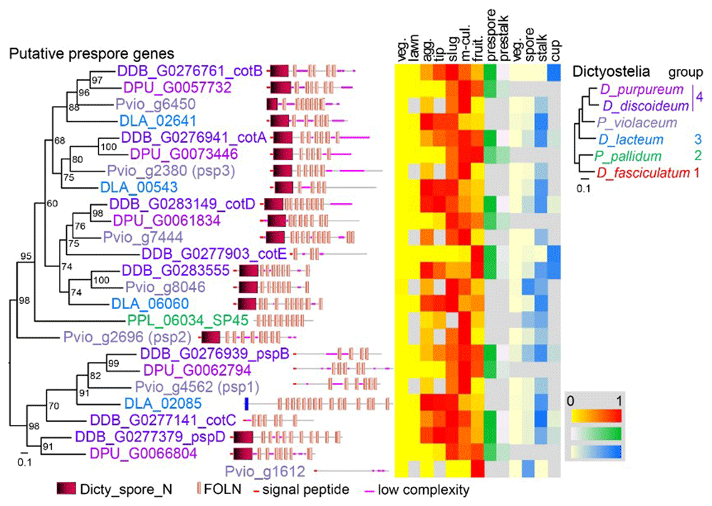
Figure 1. Search for Pvio prespore genes.
The Pvio (Narita et al., 2020), D. lacteum (Glöckner et al., 2016) and D.purpureum (Sucgang et al., 2011) proteomes were queried for relatives of 7 well-documented D. discoideum and one P. pallidum prespore genes using BLASTp. The top hits were aligned using ClustalOmega (Sievers & Higgins, 2014) and a phylogenetic tree was inferred with IQtree (Nguyen et al., 2015). The tree is annotated with the functional domains of the proteins, as analysed by SMART (Schultz et al., 1998), and with relative expression levels of the genes during the Ddis developmental cycle and in its purified cell types, as retrieved from published RNAseq experiments (Glöckner et al., 2016; Kawabe et al., 2023; Kin et al., 2018; Narita et al., 2020; Parikh et al., 2010). A multigene phylogeny of taxon-group representative Dictyostelia (Schilde et al., 2019) is shown in top right corner.
Expression pattern of putative Pvio prespore genes
The 5’intergenic regions of the three candidate prespore genes were fused to the lacZ reporter gene and Pvio cells, transformed with either construct, were developed on non-nutrient agar and stained with X-gal (Figure 2A). The promoter activity patterns of the three genes were very similar. β-galactosidase activity was first observed inside newly formed aggregates with little to no activity in the inflowing streams and then partitioned to the rear 4/5th of the emerging sorogen (es). The tip region, which was initially free of β-galactosidase activity, became proportionally smaller as the sorogen matured and the newly formed stalks contained few or no stained cells. While in freshly formed fruiting bodies the spore heads were deeply stained, β-galactosidase activity disappeared as the spores matured further.
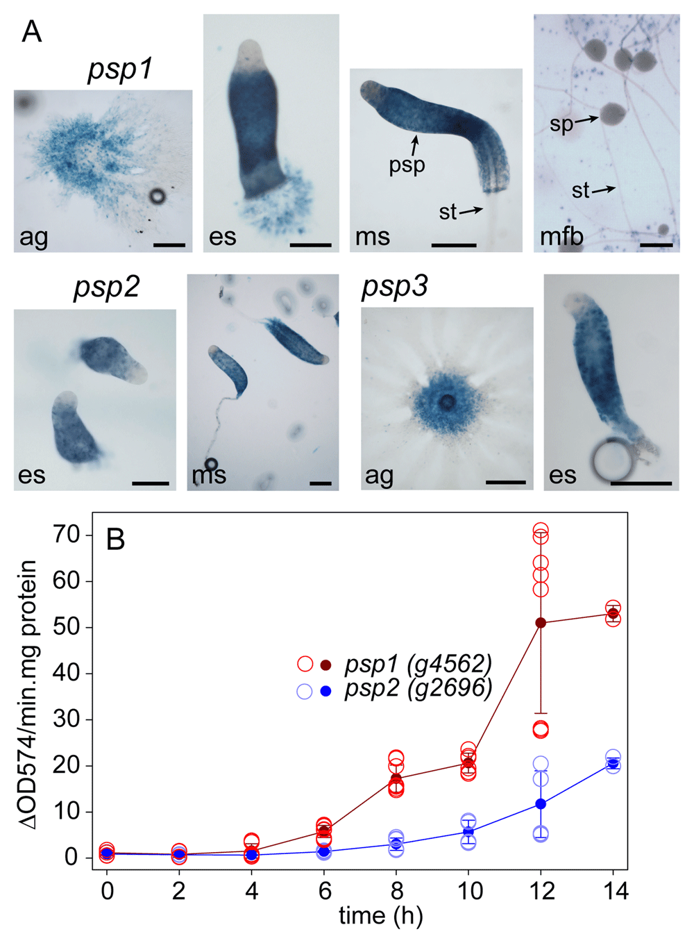
Figure 2. Pattern and timing of Pvio prespore gene expression.
A. Pattern. Pvio cells transformed with lacZ fusions with the 5’intergenic regions of the Pvio psp1 (g4562), psp2 (g2696) and psp3 (g2380) genes were developed on nitrocellulose filters supported by NN agar. Developing structures were fixed and stained with X-gal from 4 h and imaged under transmitted light. Bars: 200 μm. ag: aggregates, es and ms: early and mid sorogens, mfb: mature fruiting bodies, psp, sp and st: prespore, spore and stalk cells. B. Developmental timing. 4x10⁶ cells developed on 2x2 cm2 filters were harvested and frozen at the indicated time periods of starvation, and then lysed and assayed spectrophotometrically for β-galactosidase activity as described in Methods. Two time courses with triplicate filters were examined for the psp1-LacZ cells and one time course for the psp2-LacZ cells. The open circles represent the activity standardized on protein of the individual filters and the closed circles and error bars the mean and SD of all measurements. The underlying data are archived at https://osf.io/bawv6/ PvioPresporeGeneInduction.xlsx, sheet Fig.2BData. (Schaap, 2024).
Overall the expression pattern of the Pvio prespore genes was similar to that of the Ddis prespore genes pspA and cotA,B and C, except the anterior prespore free region remains considerably larger in Ddis throughout development. Compared to Ddis sorogens, the signature prespore vesicles are also present much closer to the tip in Pvio sorogens, where they undergo degradation as the cells are differentiating into stalk cells (Schaap et al., 1985).
Genes psp1, psp2 and psp3 are therefore reliable markers of prespore differentiation.
To estimate the developmental stage when cells were competent for gene induction by signal molecules, psp1-lacZ and psp2-lacZ cells, developed on filters, were also assayed for β-galactosidase activity by a quantitative spectrophotometric assay. Figure 2B showed that activity started to increase at 4 h of starvation (when cells had formed streaming aggregates) up to the last time point at 14 h (when cells were in the mid-sorogen stage). Psp1-lacZ cells showed about 2.5 fold higher activity as psp2-lacZ cells.
Effects of Ddis and Pvio signal molecules on Pvio prespore gene expression
Prespore gene expression in Ddis is induced by cAMP acting on cARs and further promoted by GABA (gamma-aminobutyric acid), an unknown steroid and the peptides SDF1 and SDF2. The latter four signals eventually activate PKA, which is also directly activatable by the membrane-permeant cAMP analogue 8Br-cAMP (Anjard et al., 1998; Anjard & Loomis, 2006; Anjard & Loomis, 2008; Anjard et al., 2009; Wang et al., 1999). c-di-GMP activates PKA in prestalk cells to induce stalk cell maturation (Chen & Schaap, 2012; Chen et al., 2017), while DIF-1 induces basal disc differentiation in Ddis (Saito et al., 2008). cAMP and glorin are the chemoattractants that mediate Ddis and Pvio aggregation respectively, while folate mediates attraction to the bacterial food source (Konijn et al., 1967; Pan et al., 1972; Shimomura et al., 1982). cGMP is a second messenger mediating chemotaxis (Mato et al., 1977). Except for SDF1 and SDF2 which are not commercially available, I compared the effect of all these compounds on the induction of Pvio psp1-lacZ and psp2-lacZ expression in 4 h starved cells shaken in buffer for 6 h with cells developing on solid substratum for the same period. The compounds were added in the concentration range where they are active in Ddis. These are 10-5 to 10-3 M for cAMP (Schaap & Van Driel, 1985), >10-10 M for DIF-1 (Masento et al., 1988), 10-7 to 10-5 M for c-di-GMP (Chen & Schaap, 2012), 10-9 to 10-4 M for GABA (Anjard & Loomis, 2006), 10 to 15 mM for 8Br-cAMP (Yamada et al., 2018). Glorin, folate and cGMP were used in the range where cAMP induces prespore gene expression.
Figure 3 shows that without added compounds psp1-lacZ activity increased about 4-fold over 6 h incubation, while psp2-lacZ activity increased about 6-fold (compare black diamonds to coloured circles at 0 M). None of the added compounds had any significant effect on this increase over their entire concentration range. Note that the control (water) values fluctuate a bit around 1 for the individual concentration series, which likely reflects accumulated pipetting errors, since this value was set at 1 for the averaged control values of all series. The fluctuations from 1 when variables were added is mostly not greater than that at the control, indicating that they represent the pipetting error. On the other hand, the cells developed for 6 h on solid substratum (to mid-sorogens) showed 5 to 60-fold higher activity for psp1 and psp2, respectively, than those incubated in dilute suspensions.
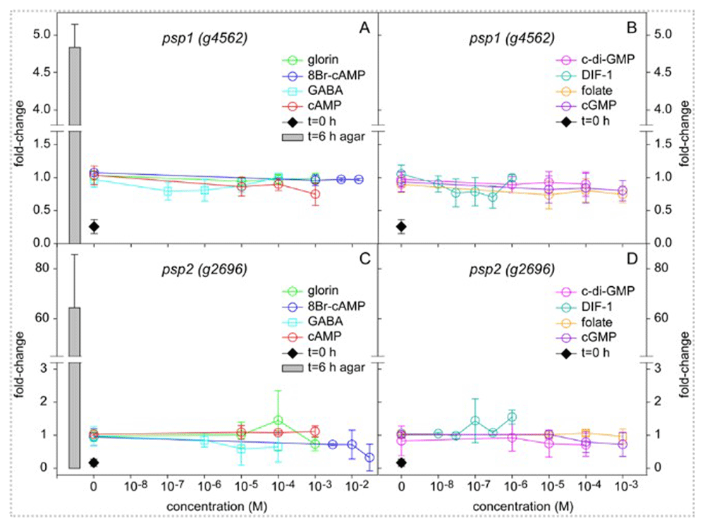
Figure 3. Induction of prespore gene expression by signal molecules.
Pvio psp1-lacZ and psp2-lacZ cells were starved for 4 h on NN agar, harvested and incubated at 106 cells/ml in PB for 6 h with the indicated concentrations of signal molecules or developed on as 106 cells per 1 cm2 filter on NN agar. At 0 and 6 h of incubation cells were frozen, lysed and assayed for β-galactosidase activity and protein content. The results are expressed as fold-change of the ΔOD574/min.mg protein values over the averaged control (water) values for all concentration series at t=6 h. Data represent means and SD of 2 or 3 independent experiments performed in triplicate for each variable. The underlying data are archived at https://osf.io/bawv6/ PvioPresporeGeneInduction.xlsx, sheet Fig.3Data (Schaap, 2024).
Evidently, the cells received some signal during normal development, that is different from any of the signals that were added during incubation in suspension, although a small amount of an endogenous inducer was also produced by the suspended cells.
Discussion
Three marker genes for prespore differentiation were identified in Pvio. Their promoters were fused to LacZ and a convenient bioassay for detection of prespore-inducing signals was developed from Pvio cells expressing these constructs. Unlike Ddis in group 4 (Schaap & Van Driel, 1985) and Ppal in group 2 (Kawabe et al., 2009) who use secreted cAMP acting on cARs to induce prespore genes, Pvio, a sister species to group 4 did not require its supposedly single cAR for (pre)spore differentiation (Kawabe & Schaap, 2023). Using the novel constructs, it was evident that Pvio prespore genes are not inducible by cAMP (Figure 3A,C), which indicates that normal development of the Pvio cAR knock-out was not due to an undiscovered cAR taking over its function.
The Pvio chemoattractant glorin could also not induce expression of prespore genes and neither did folate, a chemoattractant used for food seeking in group 4 (Pan et al., 1972) and for aggregation in the group 3 species D. minutum (De Wit & Konijn, 1983). Dissimilar to Ddis, the Pvio prespore-inducing signal is unlikely to be a chemoattractant. cGMP, an intermediate of cAMP- and folate-induced chemotaxis (Mato et al., 1977) had also no effect on prespore gene induction. Competent Pvio cells showed a low level of prespore gene induction in suspension, but this endogenous induction was neither stimulated nor inhibited by the Ddis signals c-di-GMP and DIF-1 (Figure 3B,D) which induce stalk and basal disc expression, respectively (Chen & Schaap, 2012; Saito et al., 2008), and in case of DIF-1 inhibit prespore differentiation (Wang et al., 1987).
Many signals act late in Ddis development to regulate the timely maturation of spore and stalk cells by controlling the levels of intracellular cAMP and thereby the activation of PKA. These signals such as GABA, SDF1 and SDF2 and an unidentified steroid act in synergy with cAR activation by extracellular cAMP to induce maximal expression of prespore genes and exocytosis of prespore vesicles, which are lined with the first layer of the spore coat. The effects of these late signals can be mimicked by the membrane-permeant PKA agonist 8Br-cAMP (Anjard et al., 1998; Anjard & Loomis, 2006; Anjard & Loomis, 2008; Anjard et al., 2009; Wang et al., 1999). A secreted glycoprotein, PsiA, promotes prespore differentiation in the presence of cAMP in an unknown manner (Kawata et al., 2004). psiA is member of a clade of 15 PA14 genes, which are amplified only in group 4, and it therefore has no ortholog in Pvio.
Neither GABA nor up to 30 mM of 8Br-cAMP stimulated Pvio prespore gene expression. Pvio cells can take up to 8Br-cAMP, since earlier experiments showed that 10 mM 8Br-cAMP could restore aggregation and spore differentiation in Pvio lacking the two adenylate cyclases acaA and acrA. In Ddis, AcrA has a specific role in synthesizing cAMP for PKA activation in spore maturation and the late functions of AcrA and PKA were apparently conserved in Pvio (Kawabe & Schaap, 2023). Only the early role of secreted cAMP in prespore induction is not conserved and the question arises what the missing signal might be.
Dictyostelia contain many species- or group-specific genes involved secondary metabolism such as polyketide synthases and terpene synthases (Chen et al., 2018; Heidel et al., 2011; Zucko et al., 2007). Several secondary metabolites were previously identified that act on cell differentiation, such as DIF-1 (Morris et al., 1987), MPBD (Narita et al., 2011) and dictyodene D (Günther et al., 2022). In addition, dictyostelid genomes contain large and highly variable families of cell surface or secreted glycoproteins, such as the IPT/TIG and PA40 domain families with known members like tgrB1/C1 and psiA being involved in developmental transitions (Hirose et al., 2015; Kawata et al., 2004). In the assay used here cells still clumped together during incubation, making it unlikely that direct cell-cell contact was the missing signal. While it will be a daunting task to identify any candidate as the Pvio prespore inducer, the availibitily of a sensitive bioassay based on the psp1_LacZ transformed Pvio cells is a step towards its completion.
The loss of secreted cAMP function in Pvio multicellular development does not only concern prespore induction but also the regulation of multicellular morphogenesis, which is in groups 4 (Singer et al., 2019), 3 (Alvarez-Curto et al., 2005; Schaap et al., 1984) and 2 (Kawabe et al., 2009; Kawabe et al., 2012) mediated by cAMP as chemoattractant. With the associated proteins involved in the timely synthesis and processing of the novel signal(s), this is from an evolutionary perspective an immense lineage-specific innovation in a pathway essential for species survival. It emphasizes that evolution does not always involve small stepwise improvements, but can proceed in remarkably large spurts.
Ethics and consent
Ethical approval and consent were not required.
Author contributions
P.S. designed the study, performed the experiments and wrote the manuscript.
Faculty Opinions recommendedReferences
- Alvarez-Curto E, Rozen DE, Ritchie AV, et al.:
Evolutionary origin of cAMP-based chemoattraction in the social amoebae.
Proc Natl Acad Sci U S A.
2005; 102(18): 6385–6390. PubMed Abstract
| Publisher Full Text
| Free Full Text
- Anjard C, Chang WT, Gross J, et al.:
Production and activity of Spore Differentiation Factors (SDFs) in Dictyostelium.
Development.
1998; 125(20): 4067–4075. PubMed Abstract
| Publisher Full Text
- Anjard C, Loomis WF:
GABA induces terminal differentiation of Dictyostelium through a GABAB receptor.
Development.
2006; 133(11): 2253–2261. PubMed Abstract
| Publisher Full Text
- Anjard C, Loomis WF:
Cytokinins induce sporulation in Dictyostelium.
Development.
2008; 135(5): 819–827. PubMed Abstract
| Publisher Full Text
- Anjard C, Su Y, Loomis WF:
Steroids initiate a signaling cascade that triggers rapid sporulation in Dictyostelium.
Development.
2009; 136(5): 803–812. PubMed Abstract
| Publisher Full Text
| Free Full Text
- Chen X, Köllner TG, Shaulsky G, et al.:
Diversity and functional evolution of terpene synthases in dictyostelid social amoebae.
Sci Rep.
2018; 8(1): 14361. PubMed Abstract
| Publisher Full Text
| Free Full Text
- Chen ZH, Schaap P:
The prokaryote messenger c-di-GMP triggers stalk cell differentiation in Dictyostelium.
Nature.
2012; 488(7413): 680–683. PubMed Abstract
| Publisher Full Text
| Free Full Text
- Chen ZH, Singh R, Cole C, et al.:
Adenylate Cyclase A acting on PKA mediates induction of stalk formation by Cyclic diguanylate at the Dictyostelium organizer.
Proc Natl Acad Sci U S A.
2017; 114(3): 516–521. PubMed Abstract
| Publisher Full Text
| Free Full Text
- de Wit RJ, Konijn TM:
Identification of the acrasin of Dictyostelium minutum as a derivative of folic acid.
Cell Differ.
1983; 12(4): 205–210. PubMed Abstract
| Publisher Full Text
- Dingermann T, Reindl N, Werner H, et al.:
Optimization and in situ detection of Escherichia coli β-galactosidase gene expression in Dictyostelium Discoideum.
Gene.
1989; 85(2): 353–362. PubMed Abstract
| Publisher Full Text
- Du Q, Schilde C, Birgersson E, et al.:
The cyclic AMP phosphodiesterase RegA critically regulates encystation in social and pathogenic amoebas.
Cell Signal.
2014; 26(2): 453–459. PubMed Abstract
| Publisher Full Text
| Free Full Text
- Early AE, Williams JG, Meyer HE, et al.:
Structural characterization of Dictyostelium Discoideum prespore-specific gene D19 and of its product, cell surface glycoprotein PsA.
Mol Cell Biol.
1988; 8(8): 3458–3466. PubMed Abstract
| Publisher Full Text
| Free Full Text
- Fosnaugh KL, Loomis WF:
Spore coat genes SP60 and SP70 of Dictyostelium Discoideum.
Mol Cell Biol.
1989; 9(11): 5215–5218. PubMed Abstract
| Publisher Full Text
| Free Full Text
- Funamoto S, Anjard C, Nellen W, et al.:
cAMP-dependent Protein Kinase regulates Polysphondylium pallidum development.
Differentiation.
2003; 71(1): 51–61. PubMed Abstract
| Publisher Full Text
- Glöckner G, Lawal HM, Felder M, et al.:
The multicellularity genes of dictyostelid social amoebas.
Nat Communs.
2016; 7: 12085. PubMed Abstract
| Publisher Full Text
| Free Full Text
- Grant WN, Welker DL, Williams KL:
A polymorphic, prespore-specific cell surface glycoprotein is present in the extracellular matrix of Dictyostelium Discoideum.
Mol Cell Biol.
1985; 5(10): 2559–2566. PubMed Abstract
| Publisher Full Text
| Free Full Text
- Gregg KY, Cox EC:
Spatial and temporal expression of a Polysphondylium spore-specific gene.
Dev Biol.
2000; 224(1): 81–95. PubMed Abstract
| Publisher Full Text
- Günther M, Reimer C, Herbst R, et al.:
Yellow polyketide pigment suppresses premature hatching in social amoeba.
Proc Natl Acad Sci U S A.
2022; 119(43): e2116122119. PubMed Abstract
| Publisher Full Text
| Free Full Text
- Harwood AJ, Drury L:
New vectors for expression of the E.coli lacZ gene in Dictyostelium.
Nucleic Acids Res.
1990; 18(14): 4292. PubMed Abstract
| Publisher Full Text
| Free Full Text
- Harwood AJ, Hopper NA, Simon MN, et al.:
Culmination in Dictyostelium is regulated by the cAMP-dependent Protein Kinase.
Cell.
1992; 69(4): 615–624. PubMed Abstract
| Publisher Full Text
- Heidel AJ, Lawal HM, Felder M, et al.:
Phylogeny-wide analysis of social amoeba genomes highlights ancient origins for complex intercellular communication.
Genome Res.
2011; 21(11): 1882–1891. PubMed Abstract
| Publisher Full Text
| Free Full Text
- Hirose S, Santhanam B, Katoh-Kurosawa M, et al.:
Allorecognition, via TgrB1 and TgrC1, mediates the transition from unicellularity to multicellularity in the social amoeba Dictyostelium Discoideum.
Development.
2015; 142(20): 3561–3570. PubMed Abstract
| Publisher Full Text
| Free Full Text
- Hopper NA, Harwood AJ, Bouzid S, et al.:
Activation of the prespore and spore cell pathway of Dictyostelium differentiation by cAMP-dependent protein kinase and evidence for its upstream regulation by ammonia.
EMBO J.
1993; 12(6): 2459–2466. PubMed Abstract
| Publisher Full Text
| Free Full Text
- Kalla SE, Queller DC, Lasagni A, et al.:
Kin discrimination and possible cryptic species in the social amoeba Polysphondylium violaceum.
BMC Evol Biol.
2011; 11: 31. PubMed Abstract
| Publisher Full Text
| Free Full Text
- Kawabe Y, Du Q, Narita TB, et al.:
Emerging roles for diguanylate cyclase during the evolution of soma in dictyostelia.
BMC Ecol Evol.
2023; 23(1): 60. PubMed Abstract
| Publisher Full Text
| Free Full Text
- Kawabe Y, Morio T, James JL, et al.:
Activated cAMP receptors switch encystation into sporulation.
Proc Natl Acad Sci U S A.
2009; 106(17): 7089–7094. PubMed Abstract
| Publisher Full Text
| Free Full Text
- Kawabe Y, Schaap P:
Development of the dictyostelid Polysphondylium violaceum does not require secreted cAMP.
Biol Open.
2023; 12(2): bio059728. PubMed Abstract
| Publisher Full Text
| Free Full Text
- Kawabe Y, Schilde C, Du Q, et al.:
A conserved signalling pathway for amoebozoan encystation that was co-opted for multicellular development.
Sci Rep.
2015; 5: 9644. PubMed Abstract
| Publisher Full Text
| Free Full Text
- Kawabe Y, Weening KE, Marquay-Markiewicz J, et al.:
Evolution of self-organisation in Dictyostelia by adaptation of a non-selective phosphodiesterase and a matrix component for regulated cAMP degradation.
Development.
2012; 139(7): 1336–1345. PubMed Abstract
| Publisher Full Text
| Free Full Text
- Kawata T, Nakagawa M, Shimada N, et al.:
A gene encoding, prespore-cell-inducing factor in Dictyostelium Discoideum.
Dev Growth Differ.
2004; 46(4): 383–392. PubMed Abstract
| Publisher Full Text
- Kin K, Forbes G, Cassidy A, et al.:
Cell-type specific RNA-seq reveals novel roles and regulatory programs for terminally differentiated Dictyostelium cells.
BMC Genomics.
2018; 19(1): 764. PubMed Abstract
| Publisher Full Text
| Free Full Text
- Konijn TM, Van De Meene JG, Bonner JT, et al.:
The acrasin activity of adenosine-3',5'-cyclic phosphate.
Proc Natl Acad Sci U S A.
1967; 58(3): 1152–1154. PubMed Abstract
| Publisher Full Text
| Free Full Text
- Mann SKO, Richardson DL, Lee S, et al.:
Expression of cAMP-dependent protein kinase in prespore cells is sufficient to induce spore cell differentiation in Dictyostelium.
Proc Natl Acad Sci U S A.
1994; 91(22): 10561–10565. PubMed Abstract
| Publisher Full Text
| Free Full Text
- Masento MS, Morris HR, Taylor GW, et al.:
Differentiation-inducing factor from the slime mould Dictyostelium discoideum and its analogues. Synthesis, structure and biological activity.
Biochem J.
1988; 256(1): 23–28. PubMed Abstract
| Publisher Full Text
| Free Full Text
- Mato JM, Krens FA, Van Haastert JPM, et al.:
3': 5'-cyclic AMP dependent 3': 5'-cyclic GMP accumulation in Dictyostelium discoideum.
Proc Natl Acad Sci U S A.
1977; 74(6): 2348–2351. PubMed Abstract
| Publisher Full Text
| Free Full Text
- Mehdy MC, Firtel RA:
A secreted factor and cyclic AMP jointly regulate cell-type- specific gene expression in Dictyostelium discoideum.
Mol Cell Biol.
1985; 5(4): 705–713. PubMed Abstract
| Publisher Full Text
| Free Full Text
- Mehdy MC, Ratner D, Firtel RA:
Induction and modulation of cell-type-specific gene expression in Dictyostelium.
Cell.
1983; 32(3): 763–771. PubMed Abstract
| Publisher Full Text
- Metcalf T, van der Wel H, Escalante R, et al.:
Role of SP65 in assembly of the Dictyostelium discoideum spore coat.
Eukaryot Cell.
2007; 6(7): 1137–1149. PubMed Abstract
| Publisher Full Text
| Free Full Text
- Morris HR, Taylor GW, Masento MS, et al.:
Chemical structure of the morphogen differentiation inducing factor from Dictyostelium discoideum.
Nature.
1987; 328(6133): 811–814. PubMed Abstract
| Publisher Full Text
- Narita TB, Kawabe Y, Kin K, et al.:
Loss of the polyketide synthase StlB results in stalk cell overproduction in Polysphondylium violaceum.
Genome Biol Evol.
2020; 12(5): 674–683. PubMed Abstract
| Publisher Full Text
| Free Full Text
- Narita TB, Koide K, Morita N, et al.:
Dictyostelium hybrid polyketide synthase, SteelyA, produces 4-methyl-5-pentylbenzene-1,3-diol and induces spore maturation.
FEMS Microbiol Lett.
2011; 319(1): 82–87. PubMed Abstract
| Publisher Full Text
- Nguyen LT, Schmidt HA, von Haeseler A, et al.:
IQ-TREE: a fast and effective stochastic algorithm for estimating maximum-likelihood phylogenies.
Mol Biol Evol.
2015; 32(1): 268–274. PubMed Abstract
| Publisher Full Text
| Free Full Text
- Pan P, Hall EM, Bonner JT:
Folic acid as second chemotactic substance in the cellular slime moulds.
Nat New Biol.
1972; 237(75): 181–182. PubMed Abstract
| Publisher Full Text
- Parikh A, Miranda ER, Katoh-Kurasawa M, et al.:
Conserved developmental transcriptomes in evolutionarily divergent species.
Genome Biol.
2010; 11(3): R35. PubMed Abstract
| Publisher Full Text
| Free Full Text
- Ratner D, Borth W:
Comparison of diffentiating Dictyostelium discoideum cell types separated by an improved method of density gradient centrifugation.
Exp Cell Res.
1983; 143(1): 1–13. PubMed Abstract
| Publisher Full Text
- Ritchie AV, van Es S, Fouquet C, et al.:
From drought sensing to developmental control: evolution of cyclic AMP signaling in social amoebas.
Mol Biol Evol.
2008; 25(10): 2109–2118. PubMed Abstract
| Publisher Full Text
| Free Full Text
- Romeralo M, Skiba A, Gonzalez-Voyer A, et al.:
Analysis of phenotypic evolution in Dictyostelia highlights developmental plasticity as a likely consequence of colonial multicellularity.
Proc Biol Sci.
2013; 280(1764): 20130976. PubMed Abstract
| Publisher Full Text
| Free Full Text
- Saito T, Kato A, Kay RR:
DIF-1 induces the basal disc of the Dictyostelium fruiting body.
Dev Biol.
2008; 317(2): 444–453. PubMed Abstract
| Publisher Full Text
| Free Full Text
- Schaap P:
Evolution of developmental signalling in Dictyostelid social amoebas.
Curr Opin Genet Dev.
2016; 39: 29–34. PubMed Abstract
| Publisher Full Text
| Free Full Text
- Schaap P:
PvioPresporeGeneInduction.xlsx.
Open Science Framework.
2024. http://www.doi.org/10.17605/OSF.IO/BAWV6
- Schaap P, Konijn TM, Van Haastert PJ:
cAMP pulses coordinate morphogenetic movement during fruiting body formation of Dictyostelium minutum.
Proc Natl Acad Sci U S A.
1984; 81(7): 2122–2126. PubMed Abstract
| Publisher Full Text
| Free Full Text
- Schaap P, Pinas JE, Wang M: Patterns of cell differentiation in several cellular slime mold species.
Dev Biol.
1985; 111(1): 51–61. Publisher Full Text
- Schaap P, Van Driel R: Induction of post-aggregative differentiation in Dictyostelium discoideum by cAMP. Evidence of involvement of the cell surface cAMP receptor.
Exp Cell Res.
1985; 159(2): 388–398. PubMed Abstract
| Publisher Full Text
- Schaap P, Winckler T, Nelson M, et al.: Molecular phylogeny and evolution of morphology in the social amoebas.
Science.
2006; 314(5799): 661–663. PubMed Abstract
| Publisher Full Text
| Free Full Text
- Schilde C, Lawal HM, Kin K, et al.: A well supported multi gene phylogeny of 52 dictyostelia.
Mol Phylogenet Evol.
2019; 134: 66–73. PubMed Abstract
| Publisher Full Text
| Free Full Text
- Schilde C, Skiba A, Schaap P: Evolutionary reconstruction of pattern formation in 98 Dictyostelium species reveals that cell-type specialization by lateral inhibition is a derived trait.
EvoDevo.
2014; 5: 34. PubMed Abstract
| Publisher Full Text
| Free Full Text
- Schultz J, Milpetz F, Bork P, et al.: SMART, a simple modular architecture research tool: identification of signaling domains.
Proc Natl Acad Sci U S A.
1998; 95(11): 5857–5864. PubMed Abstract
| Publisher Full Text
| Free Full Text
- Shimomura O, Suthers HLB, Bonner JT: Chemical identity of the acrasin of the cellular slime mold Polysphondylium violaceum.
Proc Natl Acad Sci U S A.
1982; 79(23): 7376–7379. PubMed Abstract
| Publisher Full Text
| Free Full Text
- Sievers F, Higgins DG: Clustal omega, accurate alignment of very large numbers of sequences.
Methods Mol Biol.
2014; 1079: 105–116. PubMed Abstract
| Publisher Full Text
- Singer G, Araki T, Weijer CJ: Oscillatory cAMP cell-cell signalling persists during multicellular Dictyostelium development.
Commun Biol.
2019; 2: 139. PubMed Abstract
| Publisher Full Text
| Free Full Text
- Smith E, Gooley AA, Hudson GC, et al.: Glycoproteins that exhibit extensive size polymorphisms in Dictyostelium discoideum.
Genetics.
1989; 122(1): 59–64. PubMed Abstract
| Publisher Full Text
| Free Full Text
- Srinivasan S, Alexander H, Alexander S: The prespore vesicles of Dictyostelium discoideum. Purification, characterization, and developmental regulation.
J Biol Chem.
1999; 274(50): 35823–35831. PubMed Abstract
| Publisher Full Text
- Sucgang R, Kuo A, Tian X, et al.: Comparative genomics of the social amoebae Dictyostelium discoideum and Dictyostelium purpureum.
Genome Biol.
2011; 12(2): R20. PubMed Abstract
| Publisher Full Text
| Free Full Text
- Takemoto K, Takeuchi I, Tasaka M: cAMP regulation of the expression of prespore-specific genes, SP96 and Dp87, in disaggregated slug cells of Dictyostelium discoideum.
Cell Differ Dev.
1990; 31(2): 89–96. PubMed Abstract
| Publisher Full Text
- Wang N, Soderbom F, Anjard C, et al.: SDF-2 induction of terminal differentiation in Dictyostelium discoideum is mediated by the membrane-spanning sensor kinase DhkA.
Mol Cell Biol.
1999; 19(7): 4750–4756. PubMed Abstract
| Publisher Full Text
| Free Full Text
- Wang M, Van Driel R, Schaap P: Cyclic AMP-phosphodiesterase induces dedifferentiation of prespore cells in Dictyostelium discoideum slugs: evidence that cyclic AMP is the morphogenetic signal for prespore differentiation.
Development.
1988; 103(3): 611–618. Publisher Full Text
- Wang M, Van Haastert PJM, Schaap P: Multiple effects of differentiation-inducing factor on prespore differentiation and cyclic-AMP signal transduction in Dictyostelium.
Differentiation.
1987; 33(3): 24–28. Publisher Full Text
- West CM: Comparative analysis of spore coat formation, structure, and function in Dictyostelium.
Int Rev Cytol.
2003; 222: 237–293. PubMed Abstract
| Publisher Full Text
- Yamada Y, Cassidy A, Schaap P: The transcription factor Spores Absent A is a PKA dependent inducer of Dictyostelium sporulation.
Sci Rep.
2018; 8(1): 6643. PubMed Abstract
| Publisher Full Text
| Free Full Text
- Yoder BK, Mao J, Erdos GW, et al.: Identification of a new spore coat protein gene in the cellular slime mold Dictyostelium discoideum.
Dev Biol.
1994; 163(1): 49–65. PubMed Abstract
| Publisher Full Text
- Zucko J, Skunca N, Curk T, et al.: Polyketide synthase genes and the natural products potential of Dictyostelium discoideum.
Bioinformatics.
2007; 23(19): 2543–2549. PubMed Abstract
| Publisher Full Text