Introduction
Q fever is a widespread zoonotic disease caused by the bacterium, Coxiella burnetii. Infections caused by C. burnetii are referred to as coxiellosis in animals and Q(uery) fever in humans (Plummer et al., 2018; WOAH, 2018). However, since the publication of case definition guidelines in the WOAH Terrestrial Code in 2024, the preferred terminology promoted by WOAH is 'Infection by Coxiella burnetii' (WOAH, 2024). Clinical symptoms in humans range from flu-like symptoms to persistent and potentially fatal infections, although asymptomatic infections and flu-like symptoms are more common than the latter (Angelakis & Raoult, 2010; Eldin et al., 2017; Maurin & Raoult, 1999). Transmission is usually airborne and results from inhalation of infectious dust particles or aerosols (Angelakis & Raoult, 2010; Clark & Soares Magalhães, 2018). Ruminant livestock, particularly sheep and goats, are the primary sources of human infections, although C. burnetii can infect a wide range of other animals, including wildlife, domestic birds, and pets (Angelakis & Raoult, 2010; Celina & Cerny, 2022; EFSA, 2010; Marrie et al., 1988; Maurin & Raoult, 1999; WOAH, 2018). It can also infect ticks, which may act as vectors and reservoirs, although their role in the epidemiology of the disease has likely been overestimated in some studies owing to diagnostic difficulties in distinguishing C. burnetii from Coxiella-like endosymbionts within tick populations (Duron et al., 2015; Jourdain et al., 2015; Körner et al., 2021; Yessinou et al., 2022).
In ruminants, C. burnetii can cause abortion, stillbirth, premature birth, and weak offspring (the ASPW complex). Coxiellosis may therefore lead to significant economic losses, particularly in small ruminants (goats and, to a lesser degree, sheep), in which abortion waves affecting all breeding females of a herd may occur (EFSA, 2010). In cattle, most infections are asymptomatic and abortions are sporadic. Disorders such as subfertility, endometritis/metritis, and retained fetal membranes (RFMs) have been associated with C. burnetii infection status in cattle, although direct evidence linking C. burnetii infection with these disorders is currently lacking (Agerholm, 2013). Furthermore, a recent systematic review concluded that although associations between C. burnetii infection status and RFMs and infertility/subfertility have been suggested, the association between endometritis/metritis in cattle and C. burnetii infection is far from clear (Gisbert et al., 2024).
Overall, the host range and infection outcome are highly variable. Although host factors may contribute to this outcome, our understanding of how C. burnetii genotype contributes to this variation is limited. Currently, the main genotyping methods used for C. burnetii generate limited genomic information and are difficult to standardize between laboratories. Whole genome sequencing (WGS) provides comprehensive genetic information and is easily standardized between laboratories and consequently, WGS has revolutionized the molecular epidemiology and surveillance of many zoonotic pathogens. However, due to difficulties in isolating the bacteria from field samples, few C. burnetii strains are currently available for WGS.
In 2023, a consortium consisting of partners from Belgium, France, Germany, Spain, the Netherlands, and the United Kingdom began with the EU-funded project Q-Net-Assess as part of the ICRAD (International Coordination of Research on Infectious Animal Diseases) initiative. The project aims to generate the most comprehensive understanding of C. burnetii genetic variation and to determine the implications of this genetic variation for zoonotic risk, livestock pathogenicity, and control of C. burnetii infection.
Work package (WP) 1 is aimed at collecting C. burnetii positive field samples from a range of hosts and environmental sources to obtain strains for isolation methods optimisation (WP2), WGS (WP3), as well as phenotypic and genotypic analyses (WP4). The collection included both animal and human samples and was built from archived and prospectively collected samples. Archived samples were obtained from the biobanks of human and animal origin samples from different consortium partners. Prospective animal sample collection mainly includes samples from cases of abortion in ruminants, as well as other types of samples from ruminant farms, such as bulk tank milk (BTM), environmental samples, and wildlife whenever available. Prospective sampling of C. burnetii associated with abortions in ruminants is being performed using coxiellosis surveillance systems already in place and under local mandatory monitoring programs in some of the participating countries. Laboratory procedures used at different laboratories for routine testing for concurrent abortifacients were shared and compared to ensure that they all comply with the minimum requirements for the investigation of C. burnetii and share equivalent criteria for case definition.
This manuscript compiled the information gathered from a review of the protocols used by different project partners and/or countries. It identifies the essential and complementary samples needed for an accurate diagnosis, describes the interpretation and validity of each type of sample and analytical techniques to detect C. burnetii as the causative agent of the abortion, and establishes the minimum requirements for C. burnetii investigation and the criteria for case definition.
Methods
At the commencement of the project, all partners involved were requested to submit the protocols applied in their institute/country to investigate abortions in ruminants. Partners included national reference laboratories for C. burnetii in animals (ANSES, APHA, FLI) and both animals and humans (Sciensano), as well as research organizations (APHA, INRAE, MRI, NEIKER, and Royal GD), all specializing in animal health, including coxiellosis. Some of these institutes perform (NEIKER, APHA, MRI) or even centralize the diagnostic process in their country (Royal GD), while others (ANSES, FLI, INRAE, Sciensano) do not routinely investigate abortions in ruminants and have reported the methods collected from networks of veterinary diagnostic laboratories in their respective countries. During the in-person kick-off meeting of the project (31 May 2023), concept protocols for abortion diagnostics were discussed. Different types of samples, diagnostic tools used, and agents investigated were inventoried using standardized templates, which were the drafts of the Table 2–Table 3 that are finally incorporated within this manuscript. Partners gathered this information during discussions with national institutes involved in abortion diagnostics. These protocols were completed by partners and discussed in four online project working group meetings (7 July 2023, 13 October 2023, 12 January 2024, 12 April 2024). After these meetings, a preliminary draft report was written and shared with partners, which was discussed during the annual internal in-person project meeting (30 May 2024). Based on the input from all partners, the final version of the report was written, subsequently discussed with all partners during two virtual working group meetings (12 July 2024 and 11 October 2024) and then approved by all partners before submission (December 2024). The protocols presented here are those in use as of February 2025.
Results
3.1 Investigation of ruminant abortion submissions
The analysis of the ruminant abortion protocols shared by the consortium partners showed that the pathogens investigated as the possible cause of abortion in ruminants, and the techniques used varied among different institutes and/or countries. The agents that were investigated included viruses (Schmallenberg virus, pestiviruses, bovine herpes virus), bacteria (C. burnetii, Chlamydia abortus, Listeria spp., Campylobacter spp., Salmonella spp., Leptospira spp., Mycoplasma spp. and other abortifacient bacteria which can be detected through aerobic and anaerobic culture), parasites (Neospora caninum and Toxoplasma gondii), and mycotic agents (Table 1). Testing for certain agents is restricted to specific ruminant species (e.g., T. gondii in sheep and goats; bovine herpesvirus only in cattle) or specific epidemiological contexts (i.e., presence of agents in a certain region) and therefore differs among countries, while other pathogens are the subject of EU and/or country-specific legislation (i.e., Brucella). The pathogens investigated and the diagnostic techniques used by each institute/country are presented in Table 2a–f. Protocols usually include a panel of pathogens that are routinely investigated, which vary according to the specific epidemiological context of each region, plus additional agents that are only investigated when specific clinical signs or pathological findings are observed or a diagnosis is not achieved using the standard protocol. Some countries follow standardized protocols that include the animals to be sampled, the type of sample(s), the tests to be performed, and the interpretation of the results for each disease (e.g. the Observatory of Causes of Abortions in Ruminants (OSCAR) protocol in France); others define (mandatory) tests supported by the government (e.g. the Belgian “Abortion Protocol” funded by the Federal Agency for the Safety of the Food Chain - FASFC). In other countries (e.g., Germany and Spain), there are no general guidelines for the diagnosis of abortions in ruminants. A combination of histopathology, bacterial culture, serology, and/or real-time PCR is used by all partners, using specific approaches that vary depending on the targeted etiological agent.
Table 1. Pathogens investigated as possible cause of abortion in ruminants in the different institutes and/or countries.
| Reporting partner | ANSES | APHA | FLI | NEIKER | Royal GD | Sciensano |
|---|
| Scope | France1 | UK | Germany2 | Spain3 | The
Netherlands | Belgium5 |
|---|
| Pathogens | Coxiella burnetii | ✓ | ✓ | ✓ | ✓ | ✓ | ✓ |
| Anaplasma sp. | ✓ | (✓) | NR | (✓) | NR | (✓) |
| Brucella sp. | ✓ | ✓ | ✓ | ✓ | NR4 | ✓ |
| Campylobacter sp. | ✓ | ✓ | ✓ | (✓) | ✓ | ✓ |
Campylobacter fetus
subsp. venerealis | ✓ | (✓) | ✓ | (✓) | NR4 | NR |
| Chlamydia abortus | ✓ | ✓ | ✓ | ✓ | ✓ | ✓ |
| Leptospira sp. | ✓ | ✓ | ✓ | ✓ | ✓ | (✓) |
| Mycoplasma sp. | NR | (✓) | (✓) | (✓) | ✓ | NR |
| Tritrichomonas foetus | NR | (✓) | (✓) | (✓) | NR | NR |
| Ureaplasma sp. | NR | (✓) | NR | (✓) | NR | (✓) |
Other bacteria (e.g., Salmonella sp.,
Listeria sp., Yersinia sp., Trueperella
pyogenes) | ✓ | ✓ | ✓ | ✓ | ✓ | ✓ |
| Neospora caninum | ✓ | ✓ | ✓ | ✓ | ✓ | ✓ |
| Toxoplasma gondii | ✓ | ✓ | (✓) | ✓ | ✓ | ✓ |
| Bluetongue virus | (✓) | (✓) | (✓) | NR | (✓) | (✓) |
| Bovine herpesvirus | NR | (✓) | ✓ | ✓ | ✓ | (✓) |
Pestivirus (Border disease virus-BD;
Bovine viral diarrhoea virus-BVD) | ✓ | ✓ | ✓ | ✓ | ✓ | ✓ |
| Schmallenberg virus | (✓) | (✓) | ✓ | (✓) | (✓) | (✓) |
| Mycoses (particularly Aspergillus sp.) | ✓ | ✓ | (✓) | (✓) | ✓ | ✓ |
Table 2a. Abortion diagnosis in France (reporting partners: ANSES and INRAE).
| Pathogen / Test1 | Histopathology | Bacteriology (culture) | Serology | PCR or RT-PCR |
|---|
| Coxiella burnetii | | | S | VS; P; F; FSC |
| Anaplasma sp. | | | S | - A. marginale: B
- A phagocytophilum: B; P; VS |
| Brucella sp. | | VS (if serology positive) | S (mandatory) | VS (if serology positive) |
Campylobacter fetus (subsp.
fetus and venerealis - cattle) | | | | ES |
| Chlamydia abortus | | | S | VS; P; F; FSC |
| Leptospira sp. | | | S | P; FSC; VS |
Other bacteria (e.g.
Salmonella sp., Listeria sp.,
Yersinia sp., Trueperella sp.,
Bacillus sp.) | | Salmonella sp. & Listeria
sp.: FSC; F (spleen, liver,
brain); VS; P | Salmonella sp.:
S (SAT) | Salmonella sp.: FSC (primarily);
F (spleen, liver); VS |
| Neospora caninum | F (brain) | | S | F (brain) |
| Toxoplasma gondii | | | S | F (brain, primarily; umbilical
cord, possible); P) |
| Bluetongue virus* | | | | B |
| Pestivirus (BD / BVD) | | | S | - BD: B (stunted or sick
newborns); F (spleen
primarily, brain, liver);
P - BVD: F (spleen); P |
| Schmallenberg virus* | Congenital
malformations | | | F (brain primarily, blood, spleen) |
Mycoses (particularly
Aspergillus) | If culture positive | P, FSC | | |
Table 2b. Abortion diagnosis in Great Britain, UK (reporting partner: APHA).
| Pathogen / Test | Histopathology | Bacteriology | Serology
(ancillary test) | PCR or RT-PCR |
|---|
| Culture | MZN |
|---|
| Coxiella burnetii | P | | P; FSC | B (ELISA) | P; FF |
| Anaplasma sp.* | F | | | | B |
| Brucella sp. | | FSC; P | P; FSC | B (RBT, ELISA) | |
| Campylobacter sp. | | FSC | | | |
Campylobacter fetus subsp.
venerealis* (cattle) | | FSC | | | |
| Chlamydia abortus | P | | P; FSC | B (ELISA) | P |
| Leptospira sp. | F | | | B (MAT, ELISA) | F (kidney) |
| Mycoplasma sp.* | F | See PCR1 | | B (M. bovis ELISA) | FSC (culture / DGGE
/ PCR) |
| Tritrichomonas foetus* | | VF, SW2 | | | |
| Ureaplasma sp.* | F | See PCR1 | | | FSC (culture / DGGE / PCR) |
Other bacteria (e.g.
Salmonella sp., Listeria sp.,
Yersinia sp., Trueperella sp.,
Bacillus sp.) | P, F | FSC | | | |
| Neospora caninum | F | | | B (ELISA) | F (brain) |
| Toxoplasma gondii | P, F | | | B (LAT) | P (cotyledon) |
| Bluetongue virus* | F | | | B (ELISA) | B, F (spleen, brain) |
| Bovine herpesvirus (BHV-1)* | F | | | B (ELISA) | F (liver, lung) |
| Pestivirus (BD / BVD) | P, F | | | B (Antibody
ELISA; antigen
ELISA for BVD) | F (spleen or thymus) |
| Schmallenberg virus* | F | | | B (ELISA) | B; F (brain, spinal
cord); FF |
Mycoses (particularly
Aspergillus sp.) | P, F | FSC (P) | | | |
Table 2c. Abortion diagnosis in Germany1 (reporting partner: FLI).
| Pathogen / Test | Histopathology1 | Bacteriology
(culture) | Serology | PCR or RT-PCR |
|---|
| Coxiella burnetii | F, P | | S, Pm, M (ELISA) | F, FSC, LF, M, P, VS |
| Brucella spp. | F, P | | S (RBT, CFT, ELISA) | F, P, VS |
| Campylobacter spp. (small ruminants) | | F, LF, P, VS | | |
| Campylobacter fetus subsp. venerealis (cattle) | | F, LF, P, PS, VS | | |
| Chlamydia abortus | F, P | | S (ELISA) | F, LF, VS, P |
| Leptospira sp. | P | | S (MAT) | F, VS |
| Mycoplasma sp.* | | M (M. bovis) | S, M (ELISA, M. bovis) | |
| Tritrichomonas foetus* | | VS, PS | | VS, PS |
Other bacteria (e.g. Salmonella sp., Listeria
sp., Yersinia sp.,, Trueperella sp., Bacillus sp.) | | F, FO, P, VS | | |
| Neospora caninum | F, P | | S (ELISA) | F, LF |
| Toxoplasma gondii* | | | | F (brain), P |
| Bluetongue virus* | | | S (ELISA) | B, S, F |
| BHV-1 (cattle) | F, P | | S (ELISA) | LF, VS |
| Pestivirus (BD / BVD) | F, P | | S (ELISA Ab+Ag) | B, M, NS (BVD), EP
(BVD) |
| Schmallenberg virus | F, P | | S (ELISA) | B, F (brain), LF, VS |
| Mycoses* (particularly Aspergillus sp.) | | P, F | | |
Table 2d. Abortion diagnosis in the Basque Country, Spain (reporting partner: NEIKER).
| Pathogen / Test | Histopathology1 | Bacteriology | Serology | PCR or RT-PCR |
|---|
| Culture2 | MZN3 |
|---|
| Coxiella burnetii | F; P | | P (F; VS) | S (ELISA) | P; F; VS |
| Anaplasma phagocytophylum* | | | | | B |
| Brucella sp. | F; P | P; F; FSC; VS | P (F; VS) | S (RBT; CFT) | |
| Campylobacter sp. (small ruminants)* | F; P | P; F; FSC; VS | | | |
| Campylobacter fetus subsp. v enerealis* | F; P | P; F; FSC; VS; PS | | | VS; PS |
| Chlamydia abortus | F; P | | P (F; VS) | S (ELISA) | P; VS; F |
| Leptospira | F; P | | | S; FF4 (MAT) | F (lung; kidney); U; M |
| Mycoplasma* | F; P | P; VS | | | |
| Tritrichomonas foetus* | F; P | P; F; VS; PS | | | VS; PS |
| Ureaplasma* | F; P | P; F; VS | | | |
Other bacteria (e.g. Salmonella sp.,
Listeria sp., Yersinia sp., Trueperella sp.,
Bacillus sp.) | F; P | P; F; FSC; VS | | | |
| Neospora caninum | F; P | | | S (ELISA) | P, F |
| Toxoplasma gondii | F; P | | | S (ELISA) | P, F |
| Bovine herpesvirus (BHV-1) | F; P | | | S (ELISA) | P, F |
| Pestivirus (BD / BVD) | F; P | | | S; FF4
(ELISA) | P; F; B |
| Schmallenberg virus* | F; P | | | S (ELISA) | P; F; Me; B |
| Mycoses (Aspergillus)* | F; P | P | | | |
Table 2e. Abortion diagnosis in The Netherlands (reporting partner: Royal GD).
| Pathogen / Test | Histopathology1 | Immuno-
histochemistry
(IHC)1 | Bacteriology
(culture) | Serology | PCR or RT-PCR |
|---|
| Coxiella burnetii2 | F; P | F; P | | S (ELISA) | F; P |
| Brucella sp.3 | | | | Mandatory sampling
of maternal sera
from aborted cattle
(RBT) | |
| Campylobacter sp. | F; P | | FSC | | |
| Chlamydia abortus | F; P | F; P | | S (ELISA) | F; P |
| Leptospira sp. | F; P | F; P | | S (cattle) | |
| Mycoplasma | | | | S (M. bovis in cattle) | |
Other bacteria (e.g. Salmonella
sp., Listeria sp., Yersinia sp.,
Trueperella sp., Bacillus sp.) | F; P | | FSC | Salmonella in cattle:
S (ELISA) | |
| Neospora caninum | F; P | | | S (cattle) | |
| Toxoplasma gondii | F; P | F; P | | | F; P |
| Bluetongue (BTV)* | F | | | Fetal serum (ELISA) | F (if the fetus shows
typical abnormalities) |
| Bovine herpesvirus (BHV-1) | F; P | F | | S (IgE ELISA) | F |
Pestivirus (BD/BVD-small
ruminants*; BVD-cattle) | F | | | S (ELISA) | F (antigen ELISA) |
| Schmallenbergvirus* | F | | | Fetal serum (ELISA) | F (if the fetus shows
typical abnormalities) |
Table 2f. Abortion diagnosis in Belgium (reporting partner: Sciensano-DGZ).
| Pathogen / Test | Bacteriology | Serology | PCR | Other |
|---|
| Culture | MZN |
|---|
| Coxiella burnetii | | P; F
(SR1) | S (ELISA – C2, SR2) | P; F (C2, SR1) | |
| Anaplasma phagocytophilum* | | | | P; F (C2) | |
| Brucella spp. | P; F (C1, SR1
if MZN is
positive) | P; F
(SR1) | S (SAW, MAT, ELISA
– C1, SR1) | | |
| Campylobacter spp. | F (C2, SR1) | | | | |
Chlamydia abortus, Chlamydia
spp. | | P; F
(SR1) | S (ELISA C. abortus
- SR1) | Chlamydia spp.: P; F (C2,
SR1 if MZN-positive) | |
| Leptospira sp.* | | | S (L. Hardjo ELISA - C2) | P; F (C2) | |
| Ureaplasma sp.* | | | | U. diversum: F (C2) | |
Other bacteria (e.g.
Salmonella sp., Listeria sp.,
Yersinia sp., Trueperella sp.,
Bacillus sp.) | F (C2, SR1) | | | | |
| Neospora caninum | | | S (ELISA – C1, SR2);
F-blotting paper
(ELISA – C2, SR2) | F; P (C2) | histopathology on F (heart
and brain) – C2 |
| Toxoplasma gondii | | | | F; P (SR1) | |
| Bluetongue virus* | | | S (C2, SR2) | F – C2, SR2 | |
| Bovine herpesvirus (BHV-4)* | | | | F (C2) | |
| Pestivirus (BD / BVD) | | | | | F (ear notch) antigen ELISA
– C1; S (if F tests positive)
antigen ELISA – C1 |
| Schmallenberg virus* | | | S (C2, SR2) | F-C2SR2 | |
Methods for the diagnosis of C. burnetii infection are presented in Table 3. C. burnetii PCR is conducted by all (6/6) partners in cases of abortion in ruminants based on several matrices, mainly the placenta, fetal tissues, and vaginal fluids from aborted females. Histopathology of the fetus and placenta is performed by four out of six (4/6) partners, although only Royal GD combines this with immunohistochemistry (IHC) in cases of placentitis as a diagnostic tool in (small) ruminants. Bacteriology for C. burnetii is mainly based on microscopic examination of smears (placenta, fetal stomach content, and/or vaginal discharge) stained using modified Ziehl-Nielsen, and it is applied by four out of six (4/6) partners. The presence of antibodies against C. burnetii in maternal serum is available by serology in six out of six (6/6) partners, although different commercial ELISA kits were used.
Table 3. Coxiella burnetii investigation in abortion submissions by the different partners.
Reporting
Partner | Histopathology | Bacteriology1 | Serology | Real-time PCR |
|---|
| ANSES | | | S (ELISA) | VS; P; F; FSC2 |
| APHA | P | P; FSC (MZN) | S3 (ELISA) | P; FF4 |
| FLI | | P; F; FSC; VS; M
(culture) | S; Pm; M (ELISA) | P; F; FSC; VS; M |
| NEIKER | P; F5 | P; F; VS (MZN)6 | S (ELISA) | P; F; VS |
| Royal GD | P; F (including IHC7) | | S (ELISA) | VS8; F; P (cattle) |
| Sciensano | F | P; F (MZN) | S (ELISA) | F9; P; VS; B |
3.2 Interpretation of results of abortion submissions in relation to Coxiella burnetii
The interpretation of C. burnetii results by different methods and in different sample matrices in relation to abortion in ruminants is presented in Table 4 and Figure 1. Samples that are mainly investigated by the consortium partners are placenta (including cotyledons), fetal samples (including fetal fluid), vaginal swabs, and maternal blood samples. The interpretation of results can be challenging and difficult to harmonize. Histopathology combined with IHC on placental membranes and cotyledons is considered the most reliable diagnostic technique for abortion diagnostics, but it is not as sensitive as real-time PCR.
Table 4. Interpretation of Coxiella burnetii results by different test methods and in different matrices/samples in relation to abortion in ruminants.
| Technique | Sample | Prove of
causality1
(-, +/-, +, ++,
+++) | Interpretation and conclusion |
|---|
| MZN Smears | Placenta/fetus | + | Unspecific result on its own; needs
confirmation of presence of Cb by PCR. |
Histopathology (HP)
Typical pathological
lesions and IHC positive | Placenta (fetus)2 | +++ | Lesions compatible with Cb are not
pathognomonic; needs positive IHC or IHC
positivity to prove Cb causality of the abortion. |
Real-time PCR
In general, Cb PCR on its own does not prove a relationship between the detection of the pathogen and the
abortion, although both the type of sample investigated and the level of positivity influence the likelihood of
causality. The higher the level of positivity, the more likely causality. Causality can be proven by HP in combination
with immunohistochemistry. |
| Fetus | ++ | Can be used to confirm the detection of Cb, in
association with compatible histopathological
findings. In the absence of compatible lesions,
PCR positivity makes the relationship between
pathogen and abortion more likely if other
abortifacients have been ruled out. |
| Placenta | ++ | Same interpretation as fetus but only
if Cb PCR positivity level is high; low PCR
positivity levels can be the result of sample
contamination. |
| Vaginal discharge | + | Indication for a putative relationship
between abortion and Cb that needs further
confirmation. Shedding of Cb can also occur
after normal parturition. |
| Bulk tank milk or individual
milk sample
(independently of Ct-value) | - | Detects shedding of Cb in a herd/flock or
at the individual level. Does not prove a
relationship between abortion and pathogen. |
| Environmental sample
dust/air/…)
(independently of Ct-value) | - | Detects environmental contamination with Cb
which may result in human exposure. Does
not prove a relationship between abortion
and pathogen. Tracing the most likely source
of shedding may be advised depending on
the context of the result. |
| Serology3 | Maternal serum | - | Seropositivity shows previous contact with
Cb, but no causality with abortion. The
relationship between shedding/abortion and
serology is poor. |
| Milk | - |
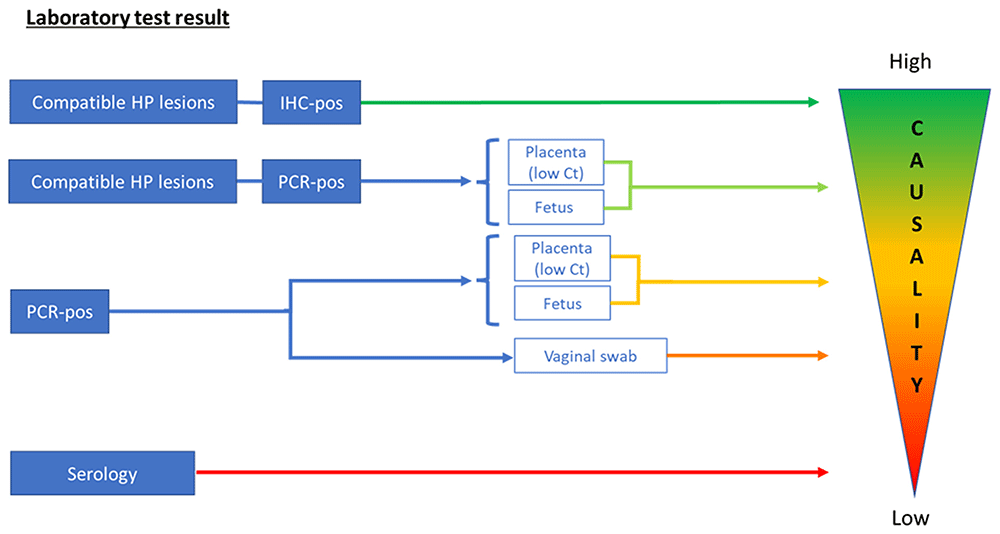
Figure 1. Interpretation of laboratory test results for Coxiella burnetii in relation to causality of abortion.
Scheme of proposed interpretation of laboratory test results in relation to their value for the diagnosis of Coxiella burnetii as the cause of the abortion (causality). For immunohistochemistry (IHC) and histopathology (HP), both the fetus and placenta can be used, and their diagnostic values are comparable. Ct, Cycle threshold; Pos, positive.
The most common PCR target used was the multicopy IS1111 element (used by 6/6 partners), as targeting a multicopy gene is anticipated to increase PCR sensitivity over a single-copy PCR target. PCR of the placenta, abomasal content of the fetus, and vaginal swabs was considered the most sensitive approach, particularly when IS1111 PCR was used. However, only when PCR is positive for placenta with relevant histopathological findings and/or fetal tissues, fetal fluid, or stomach contents from aborted fetuses test positive by PCR can C. burnetii be considered a potential cause of abortion. In other circumstances, causality can be suspected but not confirmed following a PCR-positive test. In these instances, other abortifacient agents need to be ruled out, and herd background and other laboratory results should be considered before reaching a diagnosis.
Serological results from maternal blood samples are even more difficult to relate to the cause of abortion, as not all infected animals seroconvert. In addition, positive serology (in non-vaccinated dams) provides information on previous exposure to C. burnetii, but not on the time of exposure; thus, causality cannot be established. Nevertheless, in some countries, serological sampling recommendations are used to investigate animals within a certain time period after they show reproductive disorders. Furthermore, it is currently not possible to differentiate between animals exposed to C. burnetii and those vaccinated against coxiellosis. In cases where C. burnetii is suspected to be the cause of abortion, additional sampling and/or testing should be conducted.
Discussion
Diagnosis of the cause of abortion in ruminants is complex, and identification of the specific etiology is not always achieved. Success depends on the samples available for laboratory analyses, the techniques used, and the additional clinical and epidemiological information available (e.g., serial abortions, history of affected animals, stressful conditions, possible co-infections, and other investigations that point towards an abortive infection). Here, a comparison of ruminant abortion protocols submitted by all participants showed some variation in the way abortion diagnostics were performed. This enabled the discussion of the minimum and best practice requirements for the detection of abortifacient pathogens, including C. burnetii.
To establish the best standardized protocol for ruminant abortion diagnosis, it is essential to consider that these protocols may need to evolve as scientific knowledge and available tools advance. In addition, cost-benefit considerations play a crucial role. Each additional test will improve the likelihood of detecting an agent but will also increase costs. The pathogens to be investigated should be adjusted according to the current epidemiological situation of animal diseases and the evidence available in each region. Diagnosis requires detection of the pathogen, usually by isolation and/or PCR, with histopathology aiding diagnosis by identifying compatible lesions, which, in some cases, may be pathognomonic (e.g., neosporosis). Currently, the protocols for abortion investigation in ruminants contain a tailored combination of histopathology, bacteriology, serology, and/or real-time PCR for abortifacient agents known to be present in a certain region. If routine laboratory techniques fail to reach a diagnosis when an infectious cause is probable, further efforts should concentrate on detecting less likely or new pathogens. An advantage of post-mortem examination is that it allows the detection of new pathogens and novel histopathological changes (Borel et al., 2014; Van den Brom et al., 2012; Van den Brom et al., 2021). In the future, molecular techniques capable of identifying multiple agents simultaneously would improve the broadness of abortion diagnostics in ruminants, although interpretation of the findings would need to be done with caution.
For the diagnosis of C. burnetii abortion, the most suitable samples are the placenta, fetus, and vaginal swabs from aborted animals, of which the latter have biosecurity advantages regarding transport and laboratory handling. The highest bacterial loads are found in placental membranes, with up to 109 bacteria per gram (Babudieri, 1959; Fournier et al., 1998). When placental samples are submitted, both intercotyledonary tissue and cotyledons should be included (Roest et al., 2012). C. burnetii replicates intensively within trophoblast cells of the placenta; these cells are present in the villi of cotyledons and in smaller numbers in the intercotyledonary tissue. It is important that the veterinarian collects appropriate samples from a fresh placenta and submits them according to laboratory requirements. The fetal samples of choice were the spleen, lung, liver, and abomasal contents (fetal stomach contents). Of these, the stomach content is the most practical sample to be collected. Furthermore, C. burnetii infection can progress in stages within fetal organs and may colonize the spleen before other organs. This suggests that multiple organs may need to be tested for fetal diagnosis, as non-detection of C. burnetii in one organ does not rule out its presence in other organs at any given time.
Histopathology of the fetus and placenta can identify lesions compatible with C. burnetii infection (Agerholm, 2013), but the placenta and many fetuses will have some degree of autolysis, which will compromise histopathological examination. As histopathological changes alone are not pathognomonic for C. burnetii, suspected lesions need to be further corroborated by additional staining of histological samples (e.g., IHC). Thus, histopathology combined with IHC of the placenta and fetus is a useful reference for establishing the role of C. burnetii as the cause of abortion in ruminants, although C. burnetii specific antibodies for IHC are not easily available in all routine laboratories.
PCR is a sensitive and specific method for detecting C. burnetii. PCR can also be used to confirm the presence of C. burnetii in samples with positive MZN smears (an unspecific result on its own), and when compatible histopathological lesions are observed in placenta/fetal samples, and IHC is not performed. Real-time PCR analysis of the placenta, fetal tissues, and vaginal swabs (from aborted animals within eight days post-abortion) was used by all partners to test for C. burnetii. However, causality of abortion cannot be proven based solely on the detection of C. burnetii DNA by PCR, although this is more likely when the estimated bacterial loads are high. The observation of low cycle threshold (Ct) values from real-time PCR is indicative of high bacterial loads, but caution must be taken since an “Inter-laboratory proficiency testing programme report” revealed significant differences in Ct values obtained by different laboratories (Rousset, 2021). The inclusion of a standard of known bacterial gene copy numbers in each PCR reaction could help control intra- and inter-laboratory variations in qPCR.
PCR tests targeting IS1111 are more sensitive than PCR tests designed to detect single-copy genes (EFSA, 2010; Jones et al., 2011). Because of this high sensitivity, PCR results with low bacterial load should be interpreted with care, as they can be the result of environmental contamination rather than shedding by the animal. In addition, because the IS1111 element is present in variable numbers in different C. burnetii strains, from seven to 110 copies according to Klee et al. (2006), the sensitivity of this method is biased towards samples that contain Coxiella isolates with higher IS1111 copy numbers. A French expert group has proposed a quantitative PCR threshold above which C. burnetii can be considered the likely cause of abortion, a guideline adopted at the European level (EFSA, 2010) that French partners are currently applying. Under this proposal, a causative relationship between abortion in ruminants and C. burnetii is considered highly probable when at least 104 bacteria per gram of placenta or vaginal swab is detected. However, this threshold may be more appropriate for vaginal swabs than for placental tissue, where bacterial loads are often much higher because of the intense replication of C. burnetii in trophoblasts. Therefore, a higher threshold for placental samples might be considered to rule out contamination. The C. burnetii strain Nine Mile RSA493, which displays 20 copies of the IS1111 element, is considered as a reference for copy number estimation (EFSA, 2010). However, the use of a quantitative PCR threshold based on IS1111 amplification is debatable because the copy numbers of the target vary among C. burnetii strains. Accurate quantification of the bacterial load would require the use of a single-copy gene, such as com1 or icd, as the target; however, its use in routine analyses is hampered by a loss in sensitivity.
Serology detects antibodies against a specific agent following previous exposure of the sampled animal, but cannot discriminate between vaccinated and naturally exposed individuals. However, the presence of antibodies in individual maternal serum does not prove a relationship between abortion and a particular agent. In the case of C. burnetii, the relationship between positive serology and shedding of C. burnetii is poor at the individual level (Hogerwerf et al., 2014; Rousset et al., 2009). Several factors have contributed to this complexity. First, some animals may not generate an antibody response that is sufficiently strong to be detected by serological tests, falling below the detection threshold. In addition, some animals may exhibit delayed seroconversion, with antibody levels rising slowly and crossing the detection threshold one to two weeks after abortion. This delayed seroconversion can lead to false-negative results if samples are collected too early (Arricau-Bouvery et al., 2005; de Cremoux et al., 2012; Rousset et al., 2009). Consequently, the usefulness of serology for the diagnosis of abortions in individual animals is limited. However, at the herd level, seroprevalence levels of approximately 50% of the sampled animals may be indicative of active C. burnetii infection, requiring further testing for confirmation (EFSA, 2010). Thus, serology performed at the herd level provides valuable complementary information to the results of direct diagnostic methods (IHC and real-time PCR) performed on aborted individuals, but is insufficient to diagnose C. burnetii as the cause of abortion in individual cases.
In several countries, vaccination against Q fever with a non-DIVA (Differentiating Infected from Vaccinated Animals) compatible vaccine is applied, which should be considered when serology results are interpreted. In addition, the sensitivity and specificity of ready-to-use commercial kits should be considered because recent studies have revealed that significant variations exist between available ELISAs depending on the ruminant species (Lurier et al., 2021).
Although coxiellosis frequently results in clinical signs (the ASPW complex), particularly in small ruminants, shedding of C. burnetii also occurs in the absence of clinical signs after normal parturition (Álvarez-Alonso et al., 2020). Therefore, notification and investigation of ruminant abortions will not detect all possible sources of C. burnetii. Additional surveillance systems are required to detect shedding, subsequent environmental contamination, and human exposure. In the case of asymptomatic infection, detection of shedding in dairy flocks/herds is possible by the collection and testing of bulk tank milk (BTM) (Boarbi et al., 2014; Jansen et al., 2022; Van den Brom et al., 2015), a method routinely used in the laboratories of two out of six (2/6) partners. Environmental samples (dust and aerosols) can also be tested for C. burnetii by PCR, but positive results only provide information on the presence of the bacterium in the tested sample/material, and not on the source of environmental contamination with C. burnetii (De Bruin et al., 2013; Hurtado et al., 2017). In addition, PCR detection in the environment provides no information on viability or infectivity, which requires in vitro and/or in vivo infection models (Mori et al., 2013). However, it may prove to be a useful indicator, as it suggests that subsequent human and animal exposures are likely. Depending on the epidemiological context, such environmental indicators may highlight the importance of source tracing and additional measures to prevent further shedding (Hurtado et al., 2023). Additionally, human syndromic surveillance (where clusters of suspected Q fever cases are identified) in combination with geographic information system analyses can also help detect possible sources of C. burnetii shedding animals (van den Wijngaard et al., 2011).
Conclusion
In conclusion, it appears that abortion diagnosis protocols for ruminants are tailored by each of the partners, based on the infectious agents present in each region. Histopathology, including IHC, on fetal and placental membranes supplemented with bacteriology, serology, and real-time PCR, is currently used for the detection of infectious causes of abortion in ruminants. This is the broadest diagnostic approach to relate a detected agent to the pathological changes in the fetus and/or placenta. For the identification of C. burnetii as the causative agent of abortion, based on the consortium expert opinion, we provide some guidelines for the interpretation of laboratory test results in relation to their diagnostic value. PCR is the preferred diagnostic tool for the detection of C. burnetii shedding because it is very sensitive. However, because of this high sensitivity, real-time PCR results with low bacterial load should be interpreted with caution because they can be the result of environmental contamination rather than shedding by the animal.
Despite advances in diagnostic techniques, establishing a standardized protocol that balances sensitivity, specificity, practicality, and cost remains challenging. In the case of C. burnetii, future efforts should focus on harmonizing diagnostic protocols across regions and improving our understanding of its role in ruminant abortion to improve control and prevention strategies.
Ethics statement
Ethical approval and consent were not required.