Introduction
In the late 1990s, the first evidence of voice-selective cortical areas in humans was obtained using functional magnetic resonance imaging (fMRI) (for review see Belin (2018)). This series of studies provided significant evidence of specialized brain regions for processing vocal sounds in humans, named temporal voice areas (TVAs) or “voice patches”. Voice patches are not exclusive to humans and have also been observed in other species. Specifically, they have been observed in macaques, suggesting that they already existed in some rudimentary form in the last common ancestor of macaques and humans, indicating a long evolutionary history. Studies using fMRI and positron emission tomography (PET) have identified macaques’ TVAs as those that respond preferentially to conspecific vocalizations in the superior temporal gyrus (STG) (Bodin et al., 2021; Gil-da-Costa et al., 2004; Gil-Da-Costa et al., 2006; Joly et al., 2012; Ortiz-Rios et al., 2015; Petkov et al., 2006; Petkov et al., 2008; Poremba et al., 2004).
In recent years, marmosets have gained popularity in neuroimaging studies due to their small size (Belin et al., 2023; Miller et al., 2016), which enables the use of high-field small animal MRI with much higher signal and spatial resolution. In 2015, a study was conducted using fMRI at 7T on six anesthetized marmosets to investigate the existence of conspecific voice sensitive areas (Sadagopan et al., 2015). The results showed that regions along the lateral sulcus, near the temporal pole, exhibited a preference for conspecific vocalizations (CVs), with the most rostro-lateral region showing the highest preference. These findings are consistent with similar cortical specializations and CV sensitivity as observed in macaques, suggesting a homologous structure-function relationship in the processing of conspecific vocalizations in primates. This suggests the evolutionary conservation of cortical specialization for vocalization processing, with indications pointing to a shared ancestor approximately 40 million years ago. Additional detail was provided by a recent study (Jafari et al., 2023) that used ultra-high field fMRI (9.4T) in five awake marmosets. The results reveal an extensive voice-selective network, including symmetrical regions in the temporal cortex and subcortical areas, exhibiting heightened activity during conspecific vocalizations compared to either nonvocal sounds or scrambled vocalizations. Additionally, by utilizing open-access databases, the authors uncovered robust connections between temporal and cingulate voice-selective regions. This study suggests that the marmoset's vocal brain offers a more straightforward model for understanding human cerebral voice processing, particularly with more invasive techniques like fMRI-guided electrophysiology and reinforce the notion of evolutionarily conserved cortical specialization for vocalization processing. Such findings underscore the potential of marmosets in advancing our understanding of the neural mechanisms underlying vocal processing (Belin et al., 2023).
Although clinical MRI scanners are more accessible than preclinical high-field MRI scanners, facilitating their use for a variety of studies, research conducted at 3T on such MRI scanners is limited to anatomical imaging. Building on this knowledge, the aim of this study is to replicate previous results (Jafari et al., 2023; Sadagopan et al., 2015) in a more traditional 3T scanner with a large magnetic field. To compensate for the low magnetic field intensity, this research opts for a larger number of animals (n=15), a marmoset-specific 16-channel head antenna, a "clustered sparse" design optimized for auditory stimulation, and the use of a contrast agent (MION). This study is part of a larger project called Covoprim, which aims to compare fMRI brain responses in three distinct species: humans, macaques and marmosets (Belin et al., 2018; Bodin et al., 2021). Using the same scanner and the same sounds, this comparative method involving three species is necessary to obtain general conclusions about neural bases of voice perception and its evolutionary origin.
Methods
Animals
This study involved 15 adult offspring of common marmosets (Callithrix jacchus) (Table 1). These marmosets belonged to a colony consisting of three families and were housed in cages measuring 1.05 m × 0.85 m × 2 m in the same room. The marmosets were kept on a 12-hour light/dark cycle that began at 8 a.m., and the room was soundproofed to prevent exposure to human voices. The animals involved in the study were not reproductive parents or young marmosets, but rather adult offspring. Both the research team and the veterinary staff monitored animals daily. Health was monitored by a general assessment of animal activity, and fur condition. All efforts were made to enhance the welfare of animals (i.e. social, structural and food enrichments). The ARRIVE reporting guidelines (Percie du Sert et al., 2020) was used.
Table 1. Characteristics of the 15 monkey subjects.
The rightmost column indicates whether the monkeys received a contrast agent injection during the scanning process for the localizer protocol or not.
| ID | Sex | Date of birth | MION |
|---|
| M1 | F | 2018/08/08 | YES |
| M2 | F | 2019/01/11 | YES |
| M3 | M | 2019/11/21 | YES |
| M4 | F | 2021/02/13 | YES |
| M5 | F | 2021/02/13 | YES |
| M6 | M | 2021/07/21 | YES |
| M7 | F | 2022/01/26 | YES |
| M8 | M | 2022/05/02 | YES |
| M9 | F | 2019/01/11 | NO |
| M10 | F | 2019/11/21 | NO |
| M11 | M | 2020/12/07 | NO |
| M12 | F | 2021/07/21 | NO |
| M13 | M | 2021/10/04 | NO |
| M14 | F | 2021/10/04 | NO |
| M15 | M | 2022/05/02 | NO |
Auditory stimuli
A total of 48 stimuli were used, which were divided into two categories: marmoset vocalizations (n=24), and temporally matched scrambled versions of these vocalizations (n=24).
The marmoset vocalizations were unfamiliar to the monkeys and were kindly provided by Asif Ghazanfar (Ghazanfar & Liao, 2018). Within this category, the vocalizations consisted of affiliative (trill 25%, n = 6), neutral (phee 25%, n = 6, twitter 25%, n = 6), and agonistic (tsik 25%, n = 6) calls.
To generate the scrambled versions of the vocalizations their frequency distribution was randomly rearranged while maintaining the amplitude temporal envelope minimally changed (Belin et al., 2002). Short-term Fourier transforms were performed with windows of 512 points (equivalent to 11.6 ms at a sampling rate of 44.1 kHz); amplitude components were scrambled within each window, only with other amplitude components, and the phase components were scrambled with other phase components; then an inverse Fourier transform was applied to reconstruct the scrambled signal (e.g., Figure 1B). The key characteristic of the scrambled sounds was that the amplitude values remained unchanged, preserving the temporal energy of the original signal. However, the spectral distribution of these sounds was altered, rendering them unrecognizable as vocalizations.
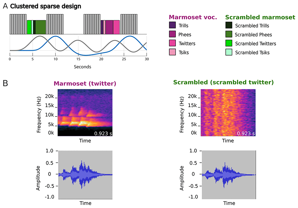
Figure 1. Marmoset Voice Localizer protocol.
(A) Design of the acquisition protocol. Each acquisition block (in gray) was composed of seven volumes and lasted 3.92 s. Colored blocks correspond to the two main sound categories: marmoset vocalizations (in pink shade) and scrambled versions of vocalizations (in green shade). The lines represent the theoretical hemodynamic response to stimuli (in blue) or to the noise produced by the scanner (in gray). The sound categories are composed of 4 subcategories. (B) Two examples of sound stimuli, a twitter for the marmoset vocalization category and the scrambled version of the same twitter for the scrambled category.
A 10-ms cosine ramp was applied to the onset and offset of all stimuli and they were normalized by their root mean square amplitude. Stimuli were processed digitally at a 48828 Hz sampling rate using TDT System 3 hardware (Tucker Davis Technologies, Alachua, FL, USA) and were delivered by an MRI-compatible loudspeaker (SPS-53-T00 Piezoelectric Miniature Speaker, Sonitron, Belgium) drived by a specific amplifier (PAA-LT3469-01, Sonitron, Belgium) at a sound pressure level of approximately 90 dB.
Scanning protocol
Functional neuroimaging studies of auditory processing face challenges, such as intense masking noise from the MR scanner (Hall et al., 1999; Peelle, 2014; Petkov et al., 2009). The continuous quasi-tonal stimulation created by this noise elevates the baseline activation level, hindering the detection of auditory activation in response to stimuli (Bandettini et al., 1998; Shah et al., 1997).
To address these challenges, we used a ‘clustered-sparse’ protocol aiming for an optimal compromise between number of brain volumes acquired (directly related to statistical power) and presentation of the stimuli on a silent background (to optimize voice/nonvoice perception). After pilot trials in human participants (Trapeau et al., 2024), we converged to series of 7 successive brain volumes acquired in succession in 3.92s, followed by a silent interval of 4.1s during which a block of either 4 vocalizations, 4 scrambled vocalizations or silence was played. Seven volumes were acquired between blocks of stimuli for a duration of 3.92 s. (Figure 1A).
Monkeys M1 to M8 were scanned with ferrous oxide contrast agent (monocrystalline iron oxide nanoparticle, MION, 10 mg/kg), on the other hand, monkeys M9 to M15 were scanned without contrast agent and the acquisition was reduced to six volumes for a duration of 5.88 s. MION contrast agents enhance the signal-to-noise ratio (SNR) and augment the distinction between various tissue types (Gautama et al., 2003). This results in a reduction of the hemodynamic response window, allowing for the acquisition of more volumes within the same session duration compared to sessions without MION.
All scans were performed under anesthesia with a protocol inspired by a previous study (Sadagopan et al., 2015). Anesthesia was induced by injecting alfaxalone (7–10 mg/kg, Alfaxan, Jurox, Ireland), followed by 2% sevoflurane gas (Baxter, France) administration through a mask. During anatomical scanning, the concentration of sevoflurane gas was lowered to 1–1.5%, and during functional scanning, it was reduced to 0.5–1% by adding nitrous oxide (Air Liquide, France) at a 70/30 ratio to oxygen (Air Liquide, France) at a flow rate of 1.8 L/min. The animal's physiological characteristics, which includes heart and respiratory rate, capnometry, and oximetry, were monitored during the operation with sensors, and a warming blanket was employed to prevent hypothermia.
fMRI acquisition
Monkeys were scanned using a 3-Tesla scanner (Prisma, Siemens Healthcare, Erlangen, Germany) and a 16-channels marmoset head coil (Rogue Research, Canada). A high-resolution T1-weighted anatomical volume was acquired (MP2RAGE sequence, TE = 2.58 ms, TR = 2200 ms, flip angle = 8°, matrix size = 384 × 324 × 256, resolution 0.18 × 0.18 × 0.18 mm3) as well as a T2-weighted anatomical volume (SPACE sequence, TE = 55.8 ms, TR = 3 s, matrix size = 256 × 324 × 384, acquisition resolution 0.36 × 0.36 × 0.36 mm3, interpolated to 0.18×0.18×0.18 mm3).
Functional volumes were then acquired using EPI sequences (multiband acceleration factor: 2, TR = 588 ms, TE = 30 ms, flip angle = 48°, matrix size = 40 × 40, 18 slices, no gap, resolution 1.25 × 1.25 × 1.25 mm3). The functional volumes collected on monkeys using MION agent contrast used different EPI sequences (multiband acceleration factor: 2, GRAPPA acceleration = 2, TR = 392 ms, TE = 16.6 ms, flip angle = 42°, matrix size = 40 × 40, 18 slices, no gap, resolution 1.25 × 1.25 × 1.25 mm3).
Quantification and statistical analysis
fMRI data preprocessing. To address minor muscle spasms during anesthesia, inter-run registration, and spatial smoothing, a preprocessing of the functional data was performed. To mitigate potential motion artifacts, each functional volume was realigned and unwrapped using a reference volume. This reference volume was chosen from a stable period in the session and was spatially closest to the average across all sessions. Spatial smoothing was applied using a three-dimensional Gaussian kernel with a full width at half-maximum that was twice the size of the functional voxels (2.5 mm). Tissue segmentation and brain extraction were performed on the structural scans using a custom segmentation pipeline. This pipeline consisted of several steps: volume cropping (FSL), bias field correction (T1w × T2w debiasing; Rilling et al., 2012), denoising (spatially adaptive nonlocal means; SPM), an initial brain extraction (FSL), and registration of the marmoset brain mapping template (Liu et al., 2021) to the anatomical scan to provide priors for SPM’s old_segment algorithm. A second brain extraction was then performed on the segmented tissues.
Transformation matrices between anatomical and functional data were generated using non-linear registration (ANTS). These matrices were used to align the tissue segmentation with the functional data and to register the functional results to the high-resolution anatomical scan.
fMRI data analysis. The fMRISTAT software (Worsley et al., 2002) was used to calculate the general linear model estimates of the responses to two contrasts. First all sounds relative to silence (i.e., all > silence then the responses to conspecific versus scrambled vocalizations (i.e., CV > scrambled).
The GLM design matrix included several principal components of principal component analyses (PCAs) that served as covariates of no interest. Two PCAs were performed on eroded masks of the functional voxels identified as containing white matter and cerebrospinal fluid tissues separately. The number of principal components used as covariates of no interest was optimized to enhance cortical auditory activation (all > silence contrast). The first two components typically captured the MRI signal decay to steady-state equilibrium, resulting from stopping the scanner between clusters of volume acquisitions, while the remaining components captured respiratory, cardiac or motion artifacts.
For BOLD data (M9 to M15), a hemodynamic response function (HRF) with a peak at 5 s and an undershoot at 10,8 s was used during analysis (Baumann et al., 2010). To analyze the MION data, a response function was designed to have a reversed sign, an earlier peak (4 s), a prolonged tail, and no undershoot, which was comparable to descriptions provided in prior literature (Leite et al., 2002).
Voxel significance was determined by applying a threshold to T-maps at *p* < 0.05, with corrections for multiple comparisons based on Gaussian Random Field Theory (Worsley et al., 2002). The quality of functional data in monkeys is influenced by various factors that cannot be quantitatively measured, such as anesthesia level and posture during MRI scans. Under these conditions, the overall fMRI response to sound serves as a quality assessment metric for each run, allowing for the identification and exclusion of low-quality runs (Petkov et al., 2008). A jackknife procedure was used to assess the contribution of each individual run to the overall sound response. This involved systematically removing each run from the dataset and measuring both the spatial extent of significant voxels and the maximum t-value in the all > silence contrast. This approach enabled the evaluation of each run’s impact on the global sound response. Only runs that contributed positively to the sound response were conserved. The study did not control for potential confounders. Additionally, there was no randomization or blinding in the experimental design.
Results
Contrast of all sounds vs. silence
We first performed individual-level analyses that revealed statistically significant (p<0.05 corrected) auditory activation maps (contrast: all sounds vs silence) in cortical areas for 7 out of the 15 individuals. Individual activation maps of these seven monkeys are available in the Extended data section (Obliger-Debouche et al., 2025). The other 8 monkeys showed few activations, and only in subcortical areas such as the inferior colliculus (IC).
The individual statistical maps for all monkeys were then combined using a fixed-effects model, as commonly used in non-human primate studies. Figure 2 shows the group-level activation map obtained in the 15 individuals. The activation included all auditory cortex subdivisions (AuA1, AuAL, AuCL, AuCM, AuCPB, AuML, AuR, AuRM, AuRT, AuRTL and AuRPB) but also subcortical areas (IC and Thal). We also noted an unexpected activation of visual area (V1).
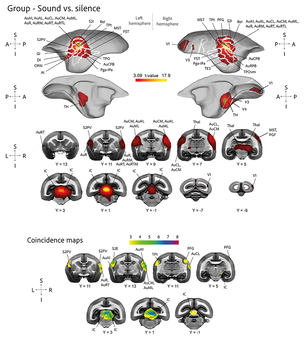
Figure 2. Fixed effect group analysis (n = 15) for the contrast sound versus silence.
(A) T-maps displayed on both the left and right fiducial brain surface template (Liu et al., 2021), with the lateral view presented at the top and the medial view at the bottom. The delineation of regions based on an atlas (Paxinos et al., 2012) is depicted by white lines. (B) Volumetric activations are superimposed onto coronal slices of template anatomical brain (Liu et al., 2021). C. Coincidence maps show the regions of the brain where the activation patterns of multiple individuals overlap.
The coincidence map (indicating the number of participants out of 15 with significant activation in each voxel, Figure 2 C.), shows that cortical auditory activations (all > silence) were only observed in a minority of our 15 participants. This may be due to deeper levels anesthesia (activations only observed subcortically), or to non-ideal auditory stimulation in those monkeys (no activations at all). Only data from monkeys that yielded significant and substantial cortical auditory activations were selected to compute our main contrast of interest (CV > scrambled). Seven monkeys (M1, M3, M4, M5, M8, M9, and M10) were kept because of this selection.
Figure 3 shows the fixed effect contrast between vocalizations and scrambled vocalizations in these seven auditory-responsive individuals. The results demonstrate a bilateral activation of the anterior and temporal cortex. Cortical activity in the left hemisphere includes the insular cortex's granular (GI) and dysgranular (DI) areas, as well as the visual area V1. Subcortically, activity is seen in the nucleus accumbens (Acb), caudate nucleus (Cd), and hippocampal formation (HipF). Conversely, within the right hemisphere, cortical activation manifests in the orbital periallocortex (OPAl), rostrotemporal medial area of the auditory cortex (AuRTM), ventral area of the secondary somatosensory cortex (S2PV), the V1 visual area, and the gustatory cortex (Gu). Subcortically, the hippocampal formation (HipF) shows an activation.
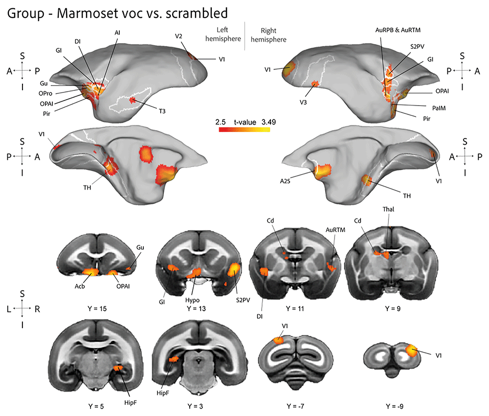
Figure 3. Fixed effect group analysis (n = 7) for the contrast marmoset vocalization versus scrambled.
(A) T-maps displayed on both the left and right fiducial brain surface template (Liu et al., 2021), with the lateral view presented at the top and the medial view at the bottom. The delineation of regions based on an atlas (Paxinos et al., 2012) is depicted by white lines. (B) Volumetric activations are superimposed onto coronal slices of template anatomical brain (Liu et al., 2021).
Discussion
To engage in social interactions and communicate, primates need to perceive and process vocalizations. Studies have revealed unique brain areas, referred to as "voice patches" (Belin, 2018; Pernet et al., 2015), which are specialized to processing voices, especially sounds that are conspecific to their species (Fecteau et al., 2004). While human voice areas have received much attention, research into similar regions in non-human primates are currently the focus of intense investigation and may reveal important insights into the brain mechanisms and evolutionary history of voice processing. In this study, marmosets (Callithrix jacchus), a species known for its complex vocal repertoire and social communication, are studied to better understand the brain mechanisms of conspecific voice perception (Agamaite et al., 2015; Bezerra & Souto, 2008). Due to its small size and advanced cognitive abilities, the marmoset is ideal for functional MRI (fMRI) research.
The goal of this work was to test whether voice patches could be identified in anesthetized animals using conventional 3T fMRI instead of high-field small-animal MRI. We optimized several aspects of the scanning protocol, the implementation of a specific clustered sparse design, the administration of the MION agent to half of the subjects who exhibited suboptimal resolution without it, the utilization of a 16-channel head coil to improve signal detection, and the maintenance of a low anesthesia level by employing a reduced concentration of sevoflurane in conjunction with nitrous oxide. This was the first time that this specially designed 3T marmoset head antenna has been used in a functional study. Previous studies using this antenna focused on resting-state imaging (Hayashi et al., 2021).
Despite these attempts, the observed auditory activations are not satisfying. Only half of the participants showed observable activations in auditory areas. This result contrasts with that of Jafari et al., whose investigation showed a stronger and more consistent auditory activation pattern in all participants. The disparities in auditory activation patterns between our study and the high-field fMRI study by Jafari et al. emphasize the inherent limitations of 3T large bore fMRI and of anesthesia.
One major disadvantage of 3T fMRI is a lower signal-to-noise ratio (SNR) than high-field devices. Despite efforts to increase signal detection with customized settings such as clustered sparse design and a 16-channel head coil, SNR at 3T remains dramatically lower than at higher field strengths. This decreased SNR may make it more difficult to distinguish tiny neural responses (voxel size in Jafari et al. (2023): 0.5mm isotropic; current study: 1.25mm), particularly in regions associated with complex cognitive activities such as auditory perception. Furthermore, the spatial resolution of functional images provided by 3T fMRI may be less precise than that achieved with higher field strengths. This limitation may impair the proper and exact localization of neuronal activations in cortical areas, thus lowering the sensitivity of activation pattern recognition.
Despite the difficulties in obtaining robust auditory activation using conventional anesthesia protocols, our fixed-effect and coincidence maps yielded valuable insights. In particular, the coincidence maps revealed common activation patterns in our cohort of subjects. Specifically, analysis of these maps showed significant activation in common subcortical brain areas, notably the inferior colliculus (IC), as well as activation in cortical auditory regions. This illustrates that low-field fMRI may reveal fundamental auditory activation patterns. Future research might focus on refining anesthetic procedures to boost the sensitivity of detecting auditory activations. The use of dexmedetomidine, a selective α2-adrenergic agonist with sedative and analgesic properties, may provide benefits compared to common anesthetics like sevoflurane used here. Dexmedetomidine-induced sedation has little impact on respiratory function and cerebral blood flow regulation, making it an attractive option for neuroimaging studies. Dexmedetomidine may improve the reliability of auditory activation signals acquired during 3T fMRI studies by lowering potential adverse effects such vasodilation and neuronal activity suppression. Using a combination of medetomidine and isoflurane proves to be more effective in eliciting visual BOLD responses compared to using isoflurane alone (Ortiz-Rios et al., 2023). Moreover, the resting-state networks under medetomidine-isoflurane anesthesia closely resembled those observed in awake states.
Seven animals showed brain activity in both contrasts (all > silent and CV > scrambled), despite the unsatisfactory sedation procedure. According to previous research, voice patches were found for the CV > scrambled contrast in the anterior temporal area (Jafari et al., 2023; Sadagopan et al., 2015). In marmosets, conspecific vocalizations activate a frontotemporal voice perception network, as shown by Sadagopan et al. (2015). Like the human activations observed, this network includes both cortical and subcortical locations. Our study's activation of subcortical areas such the caudate nucleus, nucleus accumbens, and hippocampal formation is consistent with subcortical involvement according to Sadagopan et al.’s research. These findings imply that both cortical and subcortical brain areas are involved in the preservation of the brain architecture for vocalization processing. The activation of the nucleus accumbens and the creation of the hippocampus highlights their roles in the processing of vocalizations associated with emotions and memories, which is crucial for social interaction and communication.
In the common marmoset's auditory cortex, Jafari et al. (2023) found a caudal-rostral gradient, with the anterior temporal lobe showing the greatest preference for conspecific vocalizations. This study identifies a brain specialty for vocalization processing, indicating that primates may share this specialized prior to the split of their lineages. Their results are consistent with ours, showing a comparable gradient and specialization in the bilateral activity of the anterior and temporal cortex. An equivalent functional architecture for vocalization processing is suggested by the participation of areas such as the insular cortex in the left hemisphere and the rostrotemporal medial area of the auditory cortex (AuRTM) in the right hemisphere. This analogy give credits to the idea that specific cortical regions involved in vocalization may have deep evolutionary roots, serving essential roles in social communication and recognition of vocalization types and contexts.
The merging of our results with those of Sadagopan et al. and Jafari et al. highlights the significance of vocalization processing networks in evolution. The cortical and subcortical activations that we identified are consistent with the hypothesis that humans and other primates share specialized brain areas for vocalization processing, pointing to a common evolutionary path that preceded the split of New and Old World monkeys.
To conclude, our findings strongly suggest that specialized, evolutionarily conserved brain networks exist for the processing of vocalizations. These networks include subcortical structures, which are essential for affective and memory-related elements of vocalization, as well as cortical areas, which oversee intricate auditory and multisensory processing. The intricate vocal communication systems found in primates, such as marmosets, are actively supported by this coherent and consistent brain architecture.
Conclusion
Despite the challenges of using a low-field scanner and administering anesthesia, we were able to identify the same brain regions as those highlighted by the literature. This correspondence is a valuable replication of these previous studies, especially as such studies are still rare for the marmoset model. Our results thus reinforce the hypothesis that humans and other primates share brain areas specialized in conspecific voice processing. This finding is crucial as it provides further evidence of functional parallelism between the neural systems of non-human primates and those of humans, suggesting a common evolutionary basis for the processing of vocal communication. Consequently, our study makes a significant contribution to our understanding of the neurobiological mechanisms underlying vocalization in primates, while highlighting the importance of replicability of results in scientific research.
Declarations
Ethics approval
All animal experimental protocols were conducted in accordance with the European directive (2010/63/UE) guidelines and approved under protocol 2019010911313842 by the Ethics Board of Institut de Neurosciences de la Timone, on 24/03/2019. Efforts were made to ameliorate the suffering of animals using endpoints, environmental enrichment, and careful monitoring of health and well-being.
Data and software availability
Reporting guidelines
ARRIVE Checklist For "Investigating the cortical mechanisms of conspecific voice perception in anesthetized marmosets using 3T fMRI", https://doi.org/10.5281/zenodo.14983863
Data are available under the terms of the Creative Commons Attribution 4.0 International.
Authors' contributions
Conceptualization, M.O.D., R.T., and P.B.; Methodology, M.O.D., R.T., J.S., B.N., J.L.A., and P.B.; Data acquisition, M.O.D., R.T., L.R., and J.S.; Analyses, M.O.D., and R.T.; Funding, P.B.; Writing, M.O.D., R.T., S.R., and P.B; Reviewing, R.T., J.S., B.N., and P.B.; Supervising of the study, S.R., and P.B.
Acknowledgments
We thank Lina Boudin and Shabnam Bahramiasl for their contributions during their internships. We also thank Dr. David Meunier, Dr. Mélina Cordeau, Dr. Charly Lamothe and Dr. Margherita Giamundo for useful discussions. The authors of this paper have no competing interests to declare.
Faculty Opinions recommendedReferences
- Agamaite JA, Chang CJ, Osmanski MS, et al.:
A quantitative acoustic analysis of the vocal repertoire of the common marmoset (Callithrix jacchus).
J Acoust Soc Am.
2015; 138(5): 2906–2928. PubMed Abstract
| Publisher Full Text
| Free Full Text
- Bandettini PA, Jesmanowicz A, Van Kylen J, et al.:
Functional MRI of brain activation induced by scanner acoustic noise.
Magn Reson Med.
1998; 39(3): 410–416. PubMed Abstract
| Publisher Full Text
- Baumann S, Griffiths TD, Rees A, et al.:
Characterisation of the BOLD response time course at different levels of the auditory pathway in non-human primates.
NeuroImage.
2010; 50(3): 1099–1108. PubMed Abstract
| Publisher Full Text
| Free Full Text
- Belin P:
The ‘Vocal Brain’: core and extended cerebral networks for voice processing. In: S. Frühholz & P. Belin (Eds.), The oxford handbook of voice perception. Oxford University Press, 2018; 37–60. Publisher Full Text
- Belin P, Bodin C, Aglieri V:
A “voice patch” system in the primate brain for processing vocal information?
Hear Res.
2018; 366: 65–74. PubMed Abstract
| Publisher Full Text
- Belin P, Trapeau R, Obliger-Debouche M:
A small, but vocal, brain.
Cell Rep.
2023; 42(6): 112651. PubMed Abstract
| Publisher Full Text
- Belin P, Zatorre RJ, Ahad P:
Human temporal-lobe response to vocal sounds.
Brain Res Cogn Brain Res.
2002; 13(1): 17–26. PubMed Abstract
| Publisher Full Text
- Bezerra BM, Souto A:
Structure and usage of the vocal repertoire of Callithrix jacchus.
Int J Primatol.
2008; 29(3): 671–701. Publisher Full Text
- Bodin C, Trapeau R, Nazarian B, et al.:
Functionally homologous representation of vocalizations in the auditory cortex of humans and macaques.
Curr Biol.
2021; 31(21): 4839–4844.e4. PubMed Abstract
| Publisher Full Text
| Free Full Text
- Fecteau S, Armony JL, Joanette Y, et al.:
Is voice processing species-specific in human auditory cortex? An fMRI study.
NeuroImage.
2004; 23(3): 840–848. PubMed Abstract
| Publisher Full Text
- Gautama T, Mandic DP, Van Hulle MM:
Signal nonlinearity in fMRI: a comparison between BOLD and MION.
IEEE Trans Med Imaging.
2003; 22(5): 636–644. PubMed Abstract
| Publisher Full Text
- Ghazanfar AA, Liao DA:
Constraints and flexibility during vocal development: insights from marmoset monkeys.
Curr Opin Behav Sci.
2018; 21: 27–32. PubMed Abstract
| Publisher Full Text
| Free Full Text
- Gil-da-Costa R, Braun A, Lopes M, et al.:
Toward an evolutionary perspective on conceptual representation: species-specific calls activate visual and affective processing systems in the macaque.
Proc Natl Acad Sci U S A.
2004; 101(50): 17516–21. PubMed Abstract
| Publisher Full Text
| Free Full Text
- Gil-da-Costa R, Martin A, Lopes MA, et al.:
Species-specific calls activate homologs of Broca’s and Wernicke’s areas in the macaque.
Nat Neurosci.
2006; 9(8): 1064–1070. PubMed Abstract
| Publisher Full Text
- Hall DA, Haggard MP, Akeroyd MA, et al.:
“Sparse” temporal sampling in auditory fMRI.
Hum Brain Mapp.
1999; 7(3): 213–223. PubMed Abstract
| Publisher Full Text
| Free Full Text
- Hayashi T, Hou Y, Glasser MF, et al.:
The nonhuman primate neuroimaging and neuroanatomy project.
NeuroImage.
2021; 229: 117726. PubMed Abstract
| Publisher Full Text
| Free Full Text
- Jafari A, Dureux A, Zanini A, et al.:
A vocalization-processing network in marmosets.
Cell Rep.
2023; 42(5): 112526. PubMed Abstract
| Publisher Full Text
- Joly O, Pallier C, Ramus F, et al.:
Processing of vocalizations in humans and monkeys: a comparative fMRI study.
NeuroImage.
2012; 62(3): 1376–1389. PubMed Abstract
| Publisher Full Text
- Leite FP, Tsao D, Vanduffel W, et al.:
Repeated fMRI using iron oxide contrast agent in awake, Behaving macaques at 3 Tesla.
NeuroImage.
2002; 16(2): 283–294. PubMed Abstract
| Publisher Full Text
- Liu C, Yen CCC, Szczupak D, et al.:
Marmoset brain mapping V3: population multi-modal standard volumetric and surface-based templates.
NeuroImage.
2021; 226: 117620. PubMed Abstract
| Publisher Full Text
| Free Full Text
- Miller CT, Freiwald WA, Leopold DA, et al.:
Marmosets: a neuroscientific model of human social behavior.
Neuron.
2016; 90(2): 219–233. PubMed Abstract
| Publisher Full Text
| Free Full Text
- Obliger-Debouche M, Trapeau R, Robinet L, et al.:
Extended data - marmoset fMRI voice localizer [Dataset].
Zenodo.
2025. http://www.doi.org/10.5281/ZENODO.14983863
- Obliger-Debouche M, Trapeau R, Sein J, et al.:
Marmoset fMRI data - voice localizer.
Zenodo.
2024. http://www.doi.org/10.5281/ZENODO.14499745
- Ortiz-Rios M, Kuśmierek P, DeWitt I, et al.:
Functional MRI of the vocalization-processing network in the macaque brain.
Front Neurosci.
2015; 9: 113. PubMed Abstract
| Publisher Full Text
| Free Full Text
- Ortiz-Rios M, Sirmpilatze N, König J, et al.:
Preserving functional network structure under anesthesia in the marmoset monkey brain.
BioRxiv.
2023; 2023.11.21.568138. Reference Source
- Paxinos G, Watson C, Rosa M, et al.:
The marmoset brain in stereotaxic coordinates. 2012. Reference Source
- Peelle JE:
Methodological challenges and solutions in auditory functional magnetic resonance imaging.
Front Neurosci.
2014; 8: 253. PubMed Abstract
| Publisher Full Text
| Free Full Text
- Percie du Sert N, Hurst V, Ahluwalia A, et al.:
The ARRIVE guidelines 2.0: updated guidelines for reporting animal research.
PLoS Biol.
2020; 18(7): e3000410. PubMed Abstract
| Publisher Full Text
| Free Full Text
- Pernet CR, McAleer P, Latinus M, et al.:
The human voice areas: spatial organization and inter-individual variability in temporal and extra-temporal cortices.
NeuroImage.
2015; 119: 164–174. PubMed Abstract
| Publisher Full Text
| Free Full Text
- Petkov CI, Kayser C, Augath M, et al.:
Functional imaging reveals numerous fields in the monkey auditory cortex.
PLoS Biol.
2006; 4(7): e215. PubMed Abstract
| Publisher Full Text
| Free Full Text
- Petkov CI, Kayser C, Augath M, et al.:
Optimizing the imaging of the monkey auditory cortex: sparse vs. continuous fMRI.
Magn Reson Imaging.
2009; 27(8): 1065–73. PubMed Abstract
| Publisher Full Text
- Petkov CI, Kayser C, Steudel T, et al.:
A voice region in the monkey brain.
Nat Neurosci.
2008; 11(3): 367–374. PubMed Abstract
| Publisher Full Text
- Poremba A, Malloy M, Saunders RC, et al.:
Species-specific calls evoke asymmetric activity in the monkey’s temporal poles.
Nature.
2004; 427(6973): 448–451. PubMed Abstract
| Publisher Full Text
- Rilling J, Glasser M, Jbabdi S, et al.:
Continuity, divergence, and the evolution of brain language pathways.
Front Evol Neurosci.
2012; 3: 11. PubMed Abstract
| Publisher Full Text
| Free Full Text
- Sadagopan S, Temiz-Karayol NZ, Voss HU:
High-field functional magnetic resonance imaging of vocalization processing in marmosets.
Sci Rep.
2015; 5(1): 10950. PubMed Abstract
| Publisher Full Text
| Free Full Text
- Shah NJ, Jäncke L, Gross-Ruyken ML, et al.:
How does acoustic masking noise affect fMRI of the auditory cortex.
NeuroImage.
1997; 5: 195.
- Trapeau R, Belin P, Sein J, et al.:
Comparison of auditory fMRI protocols for a voice localizer.
Open Res Eur.
2024; 4: 261. Publisher Full Text
- Worsley KJ, Liao CH, Aston J, et al.:
A general statistical analysis for fMRI data.
NeuroImage.
2002; 15(1): 1–15. PubMed Abstract
| Publisher Full Text









