Introduction
Coconut (Cocos nucifera L.) is a perennial plant grown in over 90 countries around the world, by an estimated 11 million farmers across 12 million hectares of land (FAO, 2014; Gurr et al., 2016). Coconut serves as food and a cash crop in many developing countries, providing a several foreign exchange to nations cultivating the palm (Bourke & Harwood, 2009). Coconut production in Nigeria are mostly affected by soil, climate and diseases (Ekhorutomwen et al., 2019). In coconut production, less than one third of the button nuts produced in an inflorescence eventually develop into mature nuts, that is, as a result of fruit rot and premature nut fall diseases (Ekhorutomwen et al., 2024; Rajapakse et al., 1995). In addition, Botryodiplodia theobromae (a cosmopolitan fungal pathogen) is the causal agent of the fruit rot and premature nut fall diseases of coconut, resulting in consequential yield loss of over 60% (Dheepa et al., 2018; Marques et al., 2013; Taylor & Hyde, 2003). B. theobromae can occur in nature as a parasite, saprophyte, or endophyte (Alves et al., 2008; Machado et al., 2014; Slippers & Wingfield, 2007). The fungus poses a concern to plants because it may survive in plant materials as an endophyte, escaping the quarantine process (Dheepa et al., 2018). The fungus has a wide host range, including gymnosperms and angiosperms, and can occur in nature as a parasite, saprophyte, or endophyte (Alves et al., 2008; Machado et al., 2014). Factors that contribute to the distribution of B. theobromae causing fruit rot and premature nut fall diseases of coconut include; host plant range determined by transmission from infected palm to susceptible healthy palm, B. theobromae preference for host palm, variation in transmission or infection rate, virulence of B. theobromae, poor agronomic practice, and climate (weather) factors such as temperature, rainfall, wind, relative humidity, etc (Ekhorutomwen et al., 2019).
The appearance of symptoms of fruit rot and premature nut fall diseases in coconut palms, together with the emergence of numerous Botryodiplodia species and other fungi species associated with coconut has called for precise and accurate identification of B. theobromae as the causal agent of fruit rot and premature nut fall diseases in coconut (Dheepa et al., 2018; Munirah et al., 2017). As the species covered in this article, B. theobromae is the recognized scientific name; it supersedes both the teleomorph Botryosphaeria rhodina (Berk and M.A. Curtis) Arx and the synonym Lasiodiplodia theobromae Pat. The information on population of plant pathogen(s) will be used for developing strategies to increase the durability of crop varietal resistance towards such pathogen(s) (Xia et al., 2000). Besides improving the knowledge of pathogen(s) causing diseases in coconut, accurate identification of pathogen(s) is important to establish proper control measures, quarantine programs, and to develop resistant crop varieties to pathogen(s) (Asman et al., 2024; Munirah et al., 2017; Ramachandran et al., 2015). In the past few years, the advancement of molecular techniques has yielded various insights about the population of pathogens (Séré et al., 2007). Several molecular methods have been used to identify and reveal genetic similarity or polymorphism within populations of plant pathogenic fungi (Hernández-Ceja et al., 2021; Kang et al., 2002). The genetic makeup of B. theobromae has been examined using a variety of molecular tools. These include, but are not limited to, the use of the following genetic identifiers: the translation elongation factor gene, the tubulin gene, and the internal transcribed spacer domain.
The internal transcribed spacer and tubulin gene have been utilized to identify B. theobromae isolated from mango, cashew, and other tree crops (Munirah et al., 2017; Pandey et al., 2024). The pair of primers often used to amplify the internal transcribed spacer and tubulin gene in a PCR are ITS1/4 with F: 5’-TCCGTA GGTGAACCTGCGG-3’ and R: 5’-TCCTCCGCTTATTGATATGC-3’ for ITS, while Bt2a/b with F: 5′-GGTAACCAAATCGGTGCTGCTTTC-3′ and R: 5′-ACCCTCAGTGTAGTGACCCTTGGC-3′ for tubulin gene (Xie et al., 2016). Inter-simple sequence repeat, simple sequence repeat, and random amplified polymorphic DNA identifiers have also been used to describe B. theobromae obtained from Morus alba and Agave sisalana (Cardoso & Wilkinson, 2008; Xie et al., 2016). Ten tiny gene satellites, or simple sequence repeats, were developed by Cardoso and Wilkinson (2008) in order to genetically characterize B. theobromae. Shah et al. (2010) also developed nine tiny gene satellites and seven random amplified polymorphic DNA identifiers, which they used to characterize B. theobromae isolated from the pear trees. Santos et al. (2017) and Jacobs et al. (2004) developed translation elongation factor with pair of primers: EF1-728F (5'-CATCGAGAAGTTCGAGAA-3') and EF2-728R (5'-GGARGTACCAGTSATCATGTT-3') for the molecular identification, sequence, and phylogenic analyses of B. theobromae isolated from cashew, native bark beetles, and Tomicus piniperda. Xu et al. (2016) developed PCR-based processes that utilize three specific primers, namely, primers Lt347 with F: 5'-AACGTACCTCTGTTGCTTTGGC-3' and R: 5'-TACTACGCTTGAGGGCTGAACA-3', Np304 with F: 5'-TGAACTTCGCAGTCTGAA-3' and R: 5'-CTCCAAAGCGAGGTGTTT-3', and FaF/Bt2b with F: 5'-CATCCGCAGCGTGGGAGAACAT-3' and R: 5'ACCCTCAGTGTAGTGACCCTTGGC-3') for identification of the species belonging to the Botryosphaeriaceae family linked with stem blight disease of blueberry in China (Xu et al., 2016).
In this study, the universal primer (ITS1F/4R) and three species specific primers (Lt347-F/Lt347-R, Bt2aF/Bt2bR, and EF1-728F/EF2-728R) were used to amplify the DNA of B. theobromae isolated from diseased coconut. Furthermore, the amplification of the ITS1/4 region, together with the sequencing of the ITS1/4 region in conjunction (with other molecular identifiers), cultural and morphological characters, was employed in this investigation to identify B. theobromae isolated from diseased coconut fruits with signs of fruit rot and premature nut fall (Ekhorutomwen et al., 2024; Pandey et al., 2024). Hence, with economic importance of fruit rot and premature nut fall diseases of coconut, this study was carried out to analyse the cultural, morphological and molecular characters of B. theobromae causing these diseases in coconut fruits.
Methods
Culture media
Potato dextrose agar (PDA), Czapek Dox agar (CDA), and sodium hypochlorite were purchase from Sigma-Aldrich, St. Louis, MO, USA, and used for the study.
Study location
Two (2) locations namely; NIFOR Main Station and Coconut Garden, Isihor, in Ovia North East Local Government Area, Edo State, Nigeria, were used for this study.
Source of coconut fruits
Diseased coconut fruits showing signs of rot and freshly fallen nuts were collected from coconut plantations cultivated with the following coconut varieties {that is, green dwarf (GD), orange dwarf (OD), yellow dwarf (YD), red dwarf (RD)} in the chosen location.
Isolation of fungal from diseased coconut fruits
Diseased fruit samples (showing signs of rot and freshly fallen nuts) were taken to the laboratory with a transparent screw cap container (Phipps & Porter, 1998). Sterile knife was used to dissect diseased coconut fruit samples, and the dissected samples were surface sterilized with 200 ml 0.5% sodium hypochlorite (hypo; 3A25N). Thereafter, 1000 ml each of potato dextrose agar (PDA; ISO 9001) and Czapek’s Dox agar (CDA; ISO-9000-69-5) were prepared separately, sterilized with an autoclave, and later dispensed into petri dishes. A sterile inoculating needle was used to inoculate the dissected samples into petri dishes containing PDA and CDA, and incubated at room temperature for 7 days (Dheepa et al., 2018; Phipps & Porter, 1998). Stock cultures of fungal isolates were obtained using the hyphal tip transfer procedure and maintained in tube slants of PDA at 10ºC (Phipps & Porter, 1998). The growth of fungal isolates on petri dishes containing PDA and CDA were used for colony description (Ashokkumar et al., 2018; Osagie et al., 2013). Colony description together with microscopy observation were used for presumptive identification of each fungal isolates as described by Commonwealth Mycological Institute (CMI) manual (Ashokkumar et al., 2018; Dheepa et al., 2018; El-Morsi & Ibrahim, 2012; Osagie et al., 2013; Shah et al., 2010). Thereafter, the fungal isolates were confirmed using molecular techniques (Ashokkumar et al., 2018; El-Morsi & Ibrahim, 2012; Shah et al., 2010).
Colonial morphological (macro and micro) characterization of fungal isolates
With the aid of a sterile cock borer, nine-millimeter discs from margins of actively growing 7-day-old culture of eight fungal isolates were plated on the center of 9 cm Petri dishes containing PDA medium. Each isolate was replicated three times. Colony growth rate was recorded by measuring colony diameter after 24 and 48 h of incubation, while colony color and texture observations were recorded after 48 h of incubation (Adeniyi et al., 2016; Dheepa et al., 2018; El-Morsi & Ibrahim, 2012; Shah et al., 2010). Pycnidiospores characters of each isolate viz., day of production, number, arrangement, size, and color were observed at weekly interval with the aid of a simple microscope.
Identification fungal isolates with molecular tools
With the use of the Fungal DNA Isolation Kit, DNA was extracted from the mycelia of the eight fungal isolates. The isolated DNA was purified and thereafter amplified using the fungal universal primer (ITS1F/4R) and three specific primers (Lt347-F/R, Bt2aF/bR, and EF1-728F/EF2-728R) in a PCR (Cardoso & Wilkinson, 2008; Hernández-Ceja et al., 2021; Wang et al., 2011; White et al., 1990; Xie et al., 2016; Xu et al., 2016). The DNA amplicons from PCR were cleansed with the help of a QIA-quick purification kit. The amplicons were separated with gel electrophoresis (Cardoso & Wilkinson, 2008). Also, the amplicons from the ITS1F/4R region were sequenced using a cycle-sequencing kit in a sequencing machine (ABI PrismTM) (Cardoso & Wilkinson, 2008). The base sequence from the ITS1F/4R region was edited and analyzed (to determine the similarities between the strands) using multiple sequence alignment, and thereafter, it was blasted using a blasting tool in the NCBI database for identification of each isolate (Cardoso & Wilkinson, 2008; Disegha & Akani, 2019).
Bioinformatics and phylogenetic analysis of fungal isolates
DNA fragments that were generated from PCR using the primer (ITS1F/R) and a genotyper were used to generate data that were transformed into a binary character matrix by scoring the presence or absence of each nucleotide base at each locus (that is, “1” for the presence and “0” for the absence of a nucleotide base at a particular position). The generated combined data set (that is, nucleotide sequence information or codons) from the ITS1F/4R primer obtained from the DNA of the eight fungal isolates was aligned with the ClustalW algorithm, and dendrograms were also made using the same ClustalW algorithm in MEGA X (Excoffier et al., 2007; Kumar et al., 2018; Shah et al., 2010).
Results
Colonial morphological (macro and micro) characterization of B. theobromae isolates
All eight isolates of B. theobromae were coded as Bt.NGD, Bt.NOD, Bt.NYD, Bt.NRD, Bt.IGD, Bt.IOD, Bt.IYD, and Bt.IRD in this study. The eight isolates of B. theobromae isolated from diseased coconut fruits with respect to their colonial morphological characters, show varying colony texture and colour on PDA after 72 h incubation. Colony growth rate of the eight isolates of B. theobromae were very high after 72 h of incubation, indicating their fast-growing nature (Table 1). The eight fungal isolates filled the entire 9 cm diameter Petri dish surface after 48 h incubation. Three out of the eight isolates (Bt.NOD, Bt.NYD, Bt.IRD) produce brownish or reddish pigmentation (botryodiplodin) after 30 days in PDA slant (Plate 1), while five out of the eight isolates (Bt.NGD, Bt.NRD, Bt.IYD, Bt.IGD and Bt.IOD) did not show pigmentation in PDA slant. Time taken for conidiospores production varied between 63 to 70 days under culture condition (Bt.NOD and Bt.IYD produced conidiospores after 63 days of incubation, Bt.NYD, Bt.IGD and Bt.IOD produced conidiospores after 70 days of incubation, while Bt.NGD, Bt.NRD and Bt.IRD did not produce conidiospores on culture media after incubation for over 70 days (Figure 2 and Table 3). Conidiospores arrangement was clustered in Bt.NYD and Bt.IGD, scattered in Bt.NOD, Bt.IOD, and Bt.IYD. The conidiospores were light brown, initially hyaline, mostly sepatate, with few aseptate, and, conidiospores size (μm) in length ranges between 15.69 – 22.95, while the width ranges between 8.47 – 12.95.
Table 1. Colonial morphological characters of eight Botryodiplodia theobromae isolates.
| Location | Isolates | Colony
growth rate
after 72 hours | Colony texture and colour on PDA after 72
hours | Brownish
pigmentation
in PDA slant |
|---|
| NIFOR Main Station | Bt.NGD
Bt.NOD
Bt.NYD
Bt.NRD | +++
+++
+++
+++ | Large, flat, uniform, dull white changing to grey
Large, flat, uniform, white
Large, flat, uniform, dull white changing to grey
Large, raised, irregular, greyish-white | -
+
+
- |
Coconut Garden,
Isihor | Bt.IGD
Bt.IOD
Bt.IYD
Bt.IRD | +++
+++
+++
+++ | Large, raised, irregular, dull white changing to grey
Large, flat, uniform, greyish white
Large, flat, uniform, dull white changing to grey
Large, raised, irregular, dull white changing to grey | -
-
-
+ |
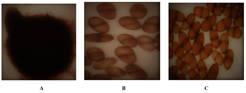
Figure 2.
Botryodiplodia theobromae conidium and conidiospores; A: Conidium, B: Scattered conidiospores, C: Clustered conidiospores.
Table 3.
Botryodiplodia theobromae conidiospore characteristics.
| Location | Isolates | Conidiospore characteristics |
|---|
Day of
production | Size in Diameter
(μm) | Arrangement | Colour | Septation |
|---|
| Length | Width |
|---|
| NIFOR Main Station | Bt.NGD
Bt.NOD
Bt.NYD
Bt.NRD | -
63
70
- | -
19.83
15.69
- | -
11.45
8.47
- | -
Scattered
Clustered
- | -
Light brown
Light brown
- | -
Septate/aseptate
Septate
- |
Coconut Garden,
Isihor | Bt.IGD
Bt.IOD
Bt.IYD
Bt.IRD | 70
70
63
- | 18.14
22.13
22.95
- | 10.89
12.33
12.95
- | Clustered
Scattered
Scattered
- | Light brown
Light brown
Light brown
- | Septate
Septate/aseptate
Septate/aseptate
- |
Molecular identification, bioinformatics and phylogenetic analysis of Botryodiplodia theobromae
The ITS1F/4R (universal primer) and Lt347-F/R (specific primer) successfully amplified the DNA of all eight isolates of B. theobromae in a PCR prior to diluting the DNA (Figure 4 and Figure 5). While Bt2aF/bR and EF1-728F/EF2-728R specific primers did not amplify the DNA of all eight isolates of B. theobromae in a PCR prior to diluting the DNA. The Bt2aF/bR primer amplified the DNA of isolates Bt.IGD, Bt.NGD, Bt.NYD, and Bt.NRD but was unable to amplify the DNA of other isolates in a PCR (Figure 6). While EF1-728F/EF2-728R amplified the DNA of isolates Bt. IGD, Bt.IYD, Bt. IRD, and Bt. NGD, it was unable to amplify the DNA of other isolates in a PCR (Figure 7). Furthermore, the sequence information of the amplified ITS1F/4R region (together with the Lt347-F/R primer) confirms the identity of each of the isolates as B. theobromae. The molecular analysis of the ITS1F/4R region also grouped the eight isolates of B. theobromae into five groups. Group 1 consists of three isolates: Bt.NYD, Bt.NGD, and Bt.IYD, with accession number MW774360.1; group 2 consists of two isolates: Bt.IGD and Bt.IRD, with accession number OM617921.1; group 3 to 5 consists of one isolate each (Bt.NOD, Bt.NRD, and Bt.IOD), with accession numbers MW282879.1, MZ502166.1, and MW478631.1. The identified isolates show 100% similarity with respect to each established accession of B. theobromae in the NCBI database or gene bank. Additionally, the percentage proportion of individual isolates with B. theobromae strains in the NCBI database shows that Group 1 isolates (representing 37.5% of the entire B. theobromae population) were more prevalent in both locations, followed by Group 2 isolates (25%), while Groups 3 to 5 represent 12.5%, respectively (Table 7). Isolates Bt.IOD has high level of genetic distance at site 2 (0.51896) and site 6 (0.61723) in the nucleotide sequence, isolates Bt. IYD has high level of genetic distance at site 6 (0.57171) in the nucleotide sequence, while isolate Bt.IRD also has high level of genetic distance at site 6 (0.56072) in the nucleotide sequence (Table 7). Other isolates (Bt.NOD, Bt.NRD, Bt.NYD, Bt.IGD, and Bt.NGD) have low genetic distance in the nucleotide sequence. The evolutionary history of each isolate was inferred using the ClustalW algorithm, as disclosed by Kumar et al. (2018). The optimal dendogram was constructed for each isolate as illustrated by Kumar et al. (2018), Saitou and Nei (1987), and Tamura et al. (2004) (Figure 8).
Table 4. Coconut fruit population and diseases’ incidence in both locations and growing seasons.
| Locations | Growing
seasons | No of coconut
fruits sampled | No of diseased
coconut fruits | PDO |
|---|
| | | | FR | PNF | |
|---|
| NIFOR Main Station | 2021 – 2022 | 1922 | 258 | 13.4 | 110 | 148 |
| 2022 – 2023 | 2536 | 419 | 195 | 224 | 16.5 |
|
Grand total
|
4458
|
677
|
305
|
372
|
15.2
|
| Coconut Garden, Isihor | 2021 – 2022 | 2238 | 266 | 128 | 138 | 11.9 |
| 2022 – 2023 | 2068 | 255 | 148 | 107 | 12.3 |
|
Grand total
|
4306
|
521
|
276
|
245
|
12.1
|
Table 5. Analysis of genomic DNA extracted from isolates of Botryodiplodia theobromae using nano drop spectrophotometer.
| SAMPLES | DNA CONC
(ng/µl) | A260 | A280 | A260/280 |
|---|
| Isolate 1 | 1273.3 | 26.446 | 14.395 | 1.837 |
| Isolate 2 | 1827.0 | 35.539 | 18.291 | 1.943 |
| Isolate 3 | 3068.0 | 61.360 | 31.041 | 1.980 |
| Isolate 4 | 1177.3 | 43.546 | 21.387 | 2.036 |
| Isolate 5 | 1032.5 | 40.651 | 21.985 | 1.849 |
| Isolate 6 | 1580.2 | 31.604 | 15.661 | 2.020 |
| Isolate 7 | 803.4 | 40.068 | 20.969 | 1.911 |
| Isolate 8 | 1533.7 | 30.675 | 15.285 | 2.007 |
Table 6. Grouping of eight Botryodiplodia theobromae isolates based on molecular identity.
| Groups | Isolates | Relationship to isolates in NCBI gene
bank | Accession
number | % proportion
of individual
strains |
|---|
| I | Bt.NYD
Bt.NGD
Bt.IYD | Botryodiplodia theobromae isolate BAL2 | MW774360 | 37.5 |
| II | Bt.IGD
Bt.IRD | Botryodiplodia theobromae isolate LSB-1 | OM617921 | 25 |
| III | Bt.NOD | Botryodiplodia theobromae isolate MRR-030 | MW282879 | 12.5 |
| IV | Bt.NRD | Botryodiplodia theobromae isolate MKMS 2.1.2 | MZ502166 | 12.5 |
| V | Bt.IOD | Botryodiplodia theobromae isolate C18 | MW478631 | 12.5 |
Table 7. Average genetic distance of eight Botryodiplodia theobromae from two locations.
| Isolates | Site 1 | Site 2 | Site 3 | Site 4 | Site 5 | Site 6 | Site 7 |
|---|
| Bt.NGD | 0.00000 | | | | | | |
| Bt.NOD | 0.00795 | | | | | | |
| Bt.NYD | 0.00000 | 0.00996 | | | | | |
| Bt.NRD | 0.00198 | 0.00998 | 0.00198 | | | | |
| Bt.IGD | 0.01392 | 0.01002 | 0.01793 | 0.00800 | | | |
| Bt.IOD | 0.57853 | 0.51896 | 0.57853 | 0.55689 | 0.61723 | | |
| Bt.IYD | 0.00198 | 0.01992 | 0.00198 | 0.00000 | 0.00798 | 0.57171 | |
| Bt.IRD | 0.01408 | 0.00203 | 0.01408 | 0.01411 | 0.02434 | 0.56072 | 0.02218 |
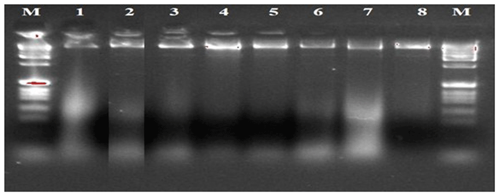
Figure 3. Gel image of genomic DNA of eight isolates.
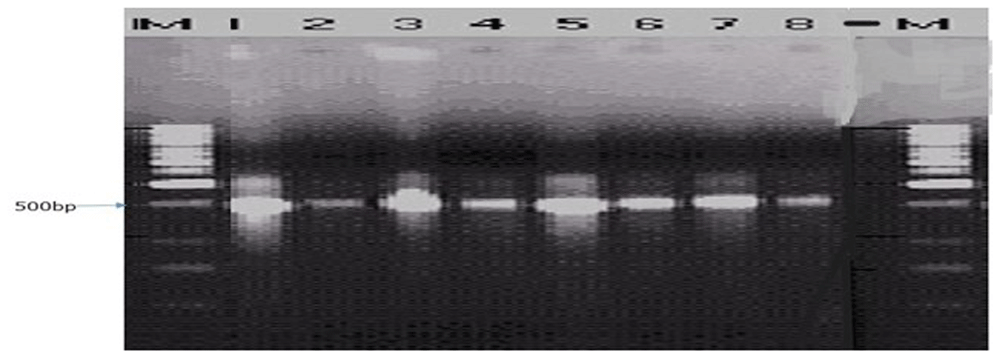
Figure 4. Picture of DNA amplicons of eight isolates using ITS1F/4R primers.
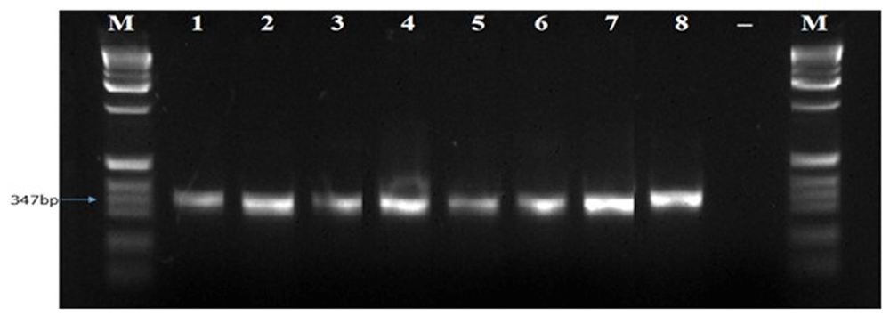
Figure 5. Picture of DNA amplicons of test isolates using Lt347-F/R primers.
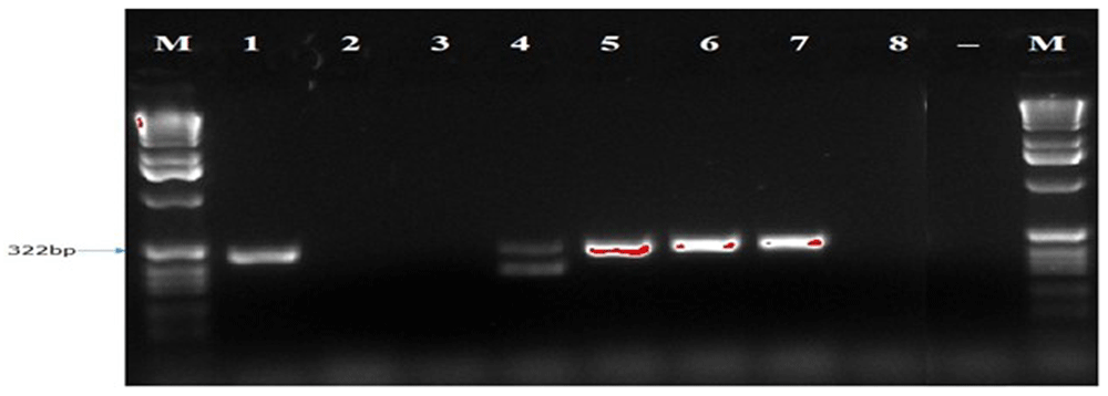
Figure 6. Picture of DNA amplicons of test isolates using Bt2a-F/bR primers.
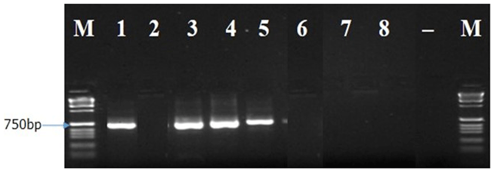
Figure 7. Image of DNA amplicons of test isolates using EF1-728F/EF2-728R primers.
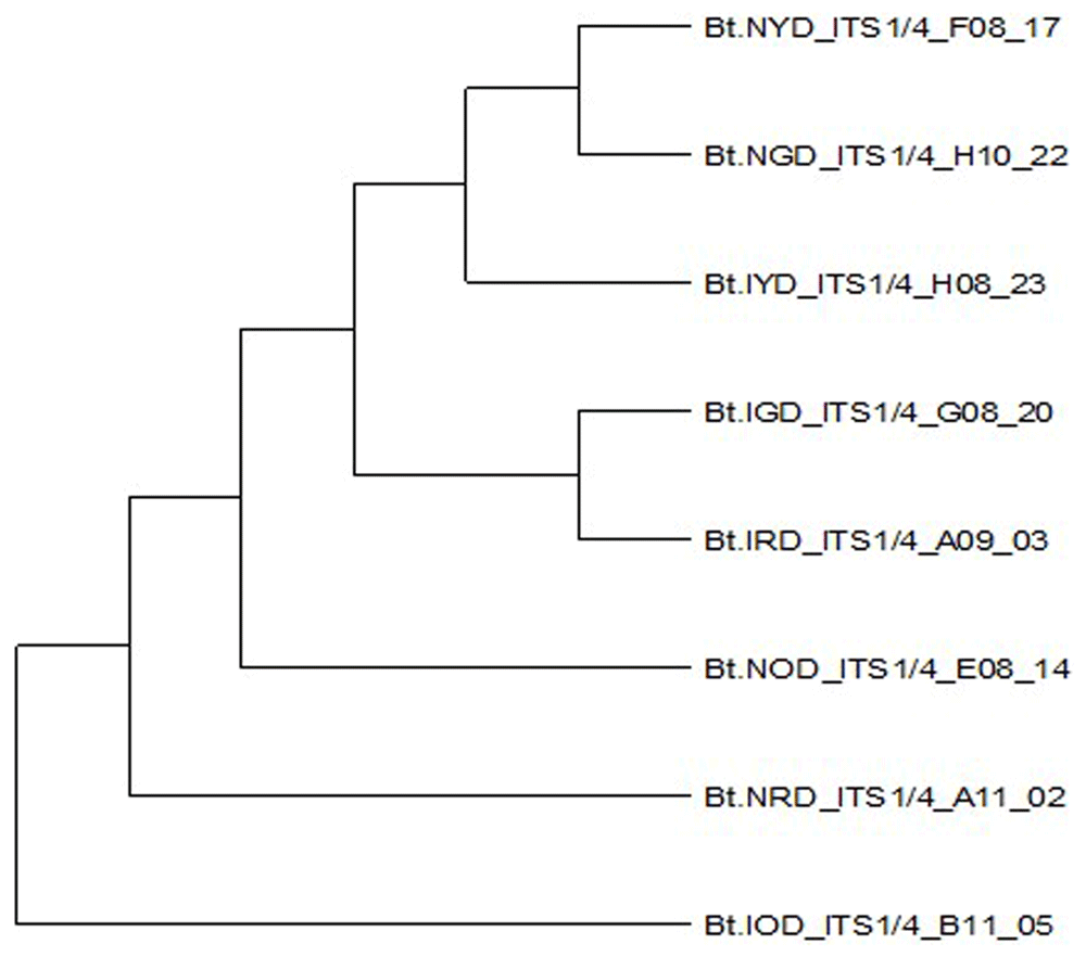
Figure 8. Dendrogram obtained from the combined data set of eight Botryodiplodia theobromae isolates.
Discussion
As previously mentioned, this study was conducted in two major coconut-growing locations in Edo State, Nigeria. The fungal pathogen was isolated from diseased coconut fruits showing signs of rot and fallen nuts (Phipps & Porter, 1998). The colony description of each fungal pathogen together with the microscopy observation were used for the presumptive identification of the eight isolates of B. theobromae (Ashokkumar et al., 2018). In addition, after 72 hours, the eight isolates of B. theobromae exhibit different colony pigments and textures according to the colonial morphological features that were determined in the study (Adeniyi et al., 2016). The colony pigments of B. theobromae isolates varied from white (Bt. NOD), to dull white changing to grey (Bt.NRD, Bt.NGD, Bt.NYD, and Bt.IYD), to greyish white (Bt.IOD, Bt.IRD, and Bt.IGD). The mycelia topography were either raised or flat, while the mycelia edge were either irregular or uniform. The colony pigments and textures grouped all eight isolates of B. theobromae into five categories (Table 2). Conidiospores arrangement were either clustered (Bt.NYD and Bt.IGD) or scattered (Bt.NOD, Bt.IOD and Bt.IYD). Conidiospores size in length ranges between 15.69 to 22.95 μm, while the width ranges between 8.47 to 12.95 μm. The conidiospores are light brown, initially hyaline, mostly sepatate, with few aseptate (Figure 2). Studies on the colonial morphological features align with the work of Adeniyi et al. (2016). The colony growth rate of the eight isolates was relatively rapid after 72 hours of the incubation process, suggesting that they had grown quickly (Table 1). After being incubated in PDA for 72 hours, all eight isolates covered the area of the Petri plate. It is postulated that B. theobromae fastidious nature makes them to colonize susceptible host tissues easily (Ashokkumar et al., 2018; Dheepa et al., 2018).
Table 2. Groupings of eight Botryodiplodia theobromae isolates based on colony texture and colour.
| Isolates | Colony texture and colour on PDA after 72 hours |
|---|
Bt.NGD
Bt.NYD
Bt.IYD | Large, flat, uniform, dull white changing to grey |
| Bt.NOD | Large, flat, uniform, white |
| Bt.NRD | Large, raised, irregular, greyish white |
| Bt.IOD | Large, flat, uniform, greyish white |
Bt.IGD
Bt.IRD | Large, raised, irregular, dull white changing to grey |
To confirm the identity of the eight B. theobromae isolates using molecular technique, one universal primer (ITS1F/4R) and three specific primers (Lt347-F/R, Bt2aF/bR, EF1-728F/EF2-728R) were employed to amplify the genes of B. theobromae isolates. The ITS1F/4R and Lt347-F/R primers successfully amplified the DNA of all eight B. theobromae isolates in a PCR. But the two other specific primers (Bt2aF/bR, EF1-728F/EF2-728R) were unable to amplify the DNA of all eight B. theobromae isolates in a PCR. Additionally, the amplified ITS1/4 region of the eight isolates was sequenced and blasted in the NCBI database, which shows 100% similarity with B. theobromae (accession numbers: MW774360.1, OM617921.1, MW282879.1, MZ502166.1, and MG778942.1) in the NCBI database. Also, the ability of the specific primer (Lt347-F/R) to amplify the DNA of B. theobromae confirms the identity of all eight isolates examined in the study. Hence, in this study, the eight fungal isolates responsible for the fruit rot and premature nut fall diseases of coconut were identified and confirmed as B. theobromae. The ITS1F/4R primer was successfully utilized to amplify the ITS1/4 gene and for the quick identification of various isolates of B. theobromae, which is in line with different investigations (Ashokkumar et al., 2018; Bharanideepan, 2013; Munirah et al., 2017). However, Xu et al. (2016) demonstrated that B. theobromae could also be identified using a species-specific (Lt347-F/R) primer. In addition, based on ITS1/4 sequence information, Bt.NGD, Bt.NYD, and Bt.IYD isolates are genetically similar, as all three isolates have similar ITS1/4 sequence information with B. theobromae isolate BAL2 with accession number MW774360.1. Two isolats (Bt.NGD, Bt.NYD) from the NIFOR location and one isolate (Bt.IYD) from Isihor were grouped into Group 1. This simply suggests these isolates do coexist and have a connection in both places. Because of this, they are more common in both places. While Bt.IGD and Bt.IRD isolates are genetically similar, the two isolates have similar ITS1/4 sequence information to B. theobromae isolate LSB-1 with accession number OM617921.1. Both isolates were in the same location (Isihor). This helps explain why they are so common in the Isihor area. Bt.NOD, Bt.NRD, and Bt.IOD isolates have no ITS1/4 similarity with each other. Also, they have no similarity (in terms of the ITS1/4 gene) to the other isolates mentioned previously. In addition, Bt.NOD isolate has similar ITS1/4 sequence information with B. theobromae isolate MRR-030 with accession number MW282879.1, Bt.NRD isolate has similar ITS1/4 sequence information with B. theobromae isolate MKMS 2.1.2 with accession number MZ502166.1, and Bt.IOD isolate has similar ITS1/4 sequence information with B. theobromae isolate C18 with accession number MG778942.1 (Table 7). The ITS1/4 sequence information and phylogenetic analysis grouped all eight isolates of B. theobromae into five categories (Table 7 and Figure 1). Coincidentally, the cultural and molecular techniques were able to group the eight isolates of B. theobromae into five groups. Group 1 consists of 3 isolates (Bt.NYD, Bt.NGD, and Bt.IYD with accession number MW774360.1); Group 2 consists of 2 isolates (Bt.IGD and Bt.IRD with accession number OM617921.1); and Groups 3 to 5 consist of 1 isolate each (Bt.NOD, Bt.NRD, and Bt.IOD with accession numbers MW282879.1, MZ502166.1, and MG778942.1, respectively). Group 1 isolates (37.5%) were more prevalent in both locations, followed by group 2 isolates (25%), while groups 3 to 5 represent 12.5%, respectively. The prevalence of Group 1 isolates in both locations indicates that these isolates are mostly responsible for the coconut fruit rot and premature nut fall diseases, followed by Group 2 isolates and Groups 3 – 5 isolates. Isolates Bt.IOD, Bt.IYD, and Bt.IRD have a high level of genetic distance at specific sites in their nucleotide sequence, while isolates Bt.NOD, Bt.NRD, Bt.NGD, Bt.IGD, and Bt.NYD have a low genetic distance in their nucleotide sequence. The substantial amount of inherited distance through genes is proportional to the extent of disparities in the nucleotide sequences in each of the three isolates, even when the substitution patterns of the nucleotide sequences are homogeneous among the lineages of each isolate.
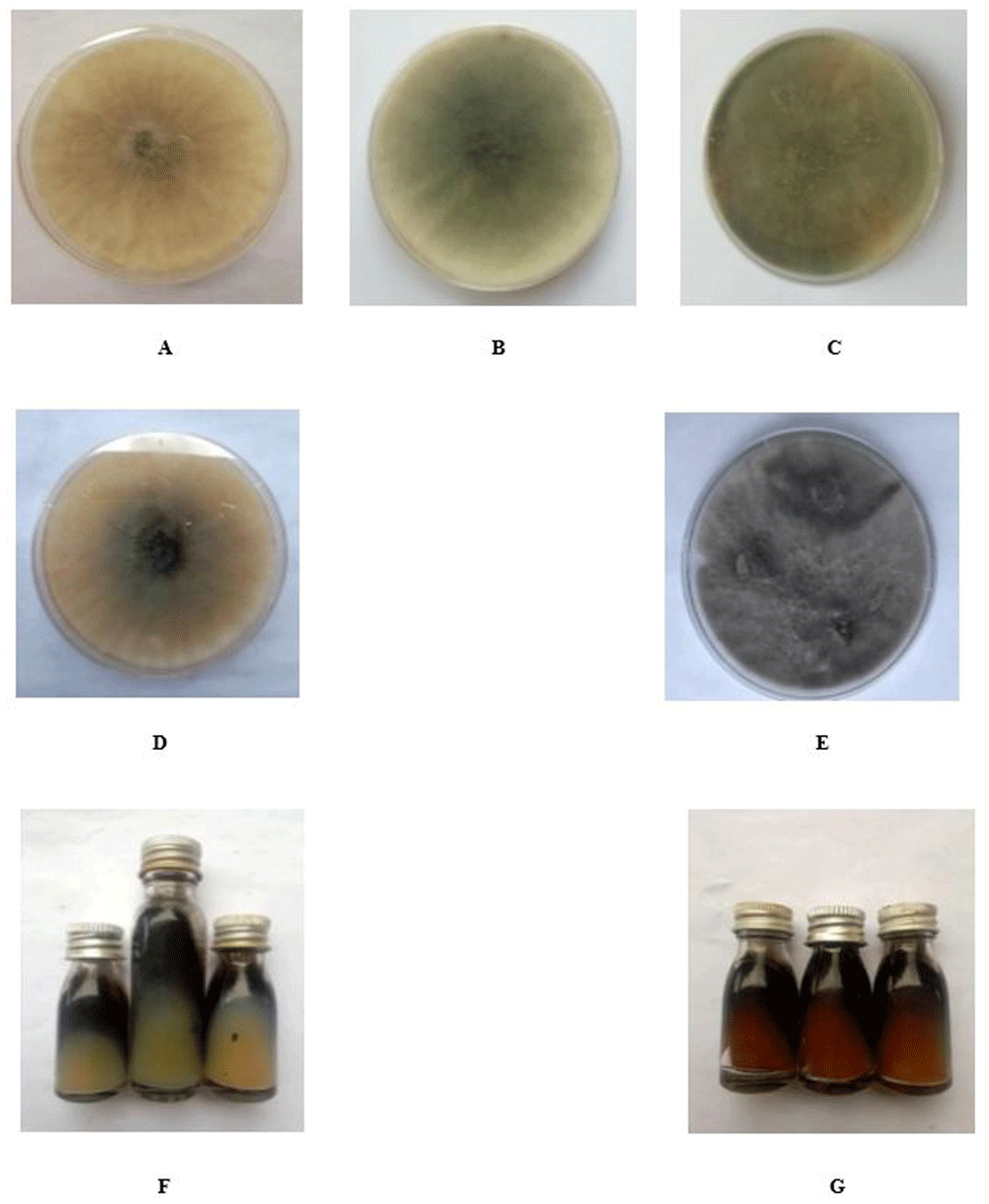
Figure 1.
A-G: Isolates of Botryodiplodia theobromae colony on potato dextrose agar in Petri dishes and slant.
Conclusion
Botryodiplodia theobromae causing the fruit rot and premature nut fall diseases of coconut was identified based on its cultural, morphological and molecular characteristics. Although B. theobromae isolates showed similarity and variation in their cultural, morphological and molecular characters. The colony colour, ITS1/4 gene sequence information and phylogenetic analysis grouped all eight isolates of B. theobromae into five categories, as these characters assisted in the identification and characterization of B. theobromae. The study has provided a guide for proper identification of B. theobromae required for the proper control of this pathogen in coconut producing areas.
Transparency
The authors state that the manuscript is honest, truthful, and transparent, that no key aspects of the investigation have been omitted, and that any differences from the study as planned have been clarified. This study followed all writing ethics.
Ethics and consent
Ethical approval and consent were not required.
Data availability
Underlying data
Mendeley: Cultural-morphology and molecular analysis of Botryodiplodia theobromae, a pathogen of coconut fruit. Doi: 10.17632/53ns2fcgps.1 (https://data.mendeley.com/datasets/53ns2fcgps/1) (Ekhorutomwen, 2025)
This project contains the following underlying data:
• Osayomore et al_Manuscript.docx
• Osayomore_Unedited_Data.docx
Data are available under the terms licensed under a Creative Commons Attribution 4.0 International licence.
Authors’ contributions
Osayomore Endurance Ekhorutomwen: Conceptualization, Methodology, Experimental Design, Writing of Manuscript. Oufensou S: Methodology. Nnamdi Ifechukwude Chidi: Conceptualization, Review and Editing of Manuscript. Migheli Quirico: Conceptualization, Review and Editing of Manuscript. Olalekan Hakeem Shittu: Conceptualization, Review and Editing of Manuscript.
Acknowledgements
I hereby confirm that I have received permission from those I acknowledged in this manuscript. Special thanks to the technical assistant received from Professor Virgilio Balmas, Dr. Giampiera Porqueddu and Ms. Stefania Oggiano at the Department of Agricultural Sciences and Desertification Research Centre (NRD), University of Sassari, Sassari, Italy. Also, special thanks to the growers of coconut plantations in NIFOR and Isihor Coconut Garden for their kind assistance during the field survey and sample collection.
Faculty Opinions recommendedReferences
- Adeniyi DO, Olufolaji DB, Joseph A:
Characteristic variations in Lasiodiplodia theobromae; pathogen of inflorescence dieback of cashew in growing ecologies of Nigeria.
Annu Res Rev Biol.
2016; 10(2): 1–6. Publisher Full Text
- Alves A, Crous PW, Correia A, et al.:
Morphological and molecular data reveal cryptic speciation in Lasiodiplodia theobromae.
Fungal Divers.
2008; 28: 1–13. Reference Source
- Ashokkumar P, Ushamalini C, Ramjegathesh R:
Variations in morphological and molecular characterization of Lasiodiplodia theobromae (Pat.) Griffon and Maubl associated with coconut leaf blight.
Madras Agric J.
2018; 105(1–3): 66–71. Publisher Full Text
- Asman A, Toru I, Ade R:
Effect of drought stress on dieback disease development under Lasiodiplodia theobromae infection in cocoa clone ‘MCC 02’.
Beverage Plant Res.
2024; 4(1): e034. Publisher Full Text
- Bharanideepan A:
Studies on die-back of acid lime (Lasiodiplodia theobromae) in acid lime. M. Sc. (Ag) Thesis. Tamil Nadu Agricultural University; Coimbatore. 2013; 45.
- Bourke RM, Harwood T:
Food and agriculture in Papua New Guinea. Australian National University, 2009. Publisher Full Text
- Cardoso JE, Wilkinson MJ:
Development and characterisation of microsatellite markers for the fungus Lasiodiplodia theobromae.
Summa Phytopathologica.
2008; 34(1): 55–57. Publisher Full Text
- Dheepa R, Goplakrishnan C, Kamalakannan A, et al.:
Coconut nut rot disease in India: prevalence, characterization of pathogen and standardization of inoculation techniques.
Int J Curr Microbiol Appl Sci.
2018; 7(2): 2046–2057. Publisher Full Text
- Disegha GC, Akani NP:
Bioinformatic method for fungi identification.
Journal of Biology and Genetic Research.
2019; 5(1): 1–14. Reference Source
- Ekhorutomwen OE, Chidi NI, Owoh EM, et al.:
Influence of varieties and growing seasons on the distribution of Botryodiplodia theobromae causing fruit rot disease of coconut.
International Journal of Sustainable Agricultural Research.
2024; 11(4): 179–189. Publisher Full Text
- Ekhorutomwen EO, Udoh ME, Esiegbuya DO:
A survey report on the incidence of fruit rot and premature nut fall diseases of coconut in Badagry, Lagos State, Nigeria. NIFOR Annual Report. 2019; 58: 157–158.
- Ekhorutomwen OE:
“Cultural-morphology and molecular analysis of Botryodiplodia theobromae, a pathogen of coconut fruit”. Mendeley Data, V1. 2025. Publisher Full Text
- El-Morsi MEA, Ibrahim IA:
Morphological, molecular and histopathological variation in different isolates of Botryodiplodia theobromae, the causative of leaf base rot disease of date palms in New Valley Governorate, Egypt.
Wudpecker Journal of Agricultural Research.
2012; 1(6): 215–222.
- Excoffier L, Laval G, Schneider S:
Arlequin (version 3.0): an integrated software package for population genetics data analysis.
Evol Bioinform Online.
2007; 1: 47–50. PubMed Abstract
| Free Full Text
- FAO: FAOStat, Rome. Food and Agriculture Organisation of the United Nations, 2014. Reference Source
- Gurr GM, Johnson AC, Ash GJ, et al.:
Coconut lethal yellowing diseases: a phytoplasma threat to palms of global economic and social significance.
Front Plant Sci.
2016; 7: 1521. PubMed Abstract
| Publisher Full Text
| Free Full Text
- Hernández-Ceja A, Loeza-Lara PD, Espinosa-García FJ, et al.:
In vitro antifungal activity of plant extracts on pathogenic fungi of blueberry (Vaccinium sp.).
Plants (Basel).
2021; 10(5): 852. PubMed Abstract
| Publisher Full Text
| Free Full Text
- Jacobs K, Bergdahl DR, Wingfield MJ, et al.:
Leptographium wingfieldii introduced into North America and found associated with exotic Tomicus piniperda and native bark beetles.
Mycol Res.
2004; 108(Pt 4): 411–418. PubMed Abstract
| Publisher Full Text
- Kang HW, Park DS, Go SJ, et al.:
Fingerprinting of diverse genomes using PCR with Universal Rice Primers generated from repetitive sequence of Korean weedy rice.
Mol Cells.
2002; 13(2): 281–287. PubMed Abstract
| Publisher Full Text
- Kumar S, Stecher G, Li M, et al.:
MEGA X: Molecular Evolutionary Genetics Analysis across computing platforms.
Mol Biol Evol.
2018; 35(6): 1547–1549. PubMed Abstract
| Publisher Full Text
| Free Full Text
- Machado AR, Pinho DB, Pereira OL:
Phylogeny, identification and pathogenicity of the Botryosphaeriaceae associated with collar and root rot of the biofuel plant Jatropha curcas in Brazil, with a description of new species of Lasiodiplodia.
Fungal Divers.
2014; 67: 231–247. Publisher Full Text
- Marques MW, Lima NB, Morais MA, et al.:
Species of Lasiodiplodia associated with mango in Brazil.
Fungal Divers.
2013; 61: 181–193. Publisher Full Text
- Munirah MS, Azmi AR, Yong SYC, et al.:
Characterization of Lasiodiplodia theobromae and Lasiodiplodia pseudotheobromae causing fruit rot on pre-harvest mango in Malaysia.
Plant Pathol Quar.
2017; 7(2): 202–213. Publisher Full Text
- Osagie IJ, Ojomo EE, Maduewesi AE:
Physiological studies on ten isolates of the oil palm pathogen, Fusarium oxysporum f. sp elaeidis.
Nigerian Journal of Agriculture, Food and Environment.
2013; 9(1): 33–39.
- Pandey KA, Sharma KH, Yadav S, et al.:
A new canker on Camellia sinensis (L.) Kuntze associated with Diaporthe longicolla and Lasiodiplodia theobromae in India.
Physiol Mol Plant Pathol.
2024; 134: 102–114. Publisher Full Text
- Phipps PM, Porter DM:
Collar rot of peanut caused by Lasiodiplodia theobromae.
Plant Dis.
1998; 82(11): 1205–1209. PubMed Abstract
| Publisher Full Text
- Rajapakse CNK, Wijesekara HTR, Norman AH:
A preliminary study on immature nut fall of coconut with reference to pest damage.
Cocos.
1995; 10: 53–54. Publisher Full Text
- Ramachandran K, Uyub AM, Latiffah Z:
Molecular characterization and pathogenicity of Erwinia spp. associated with pineapple [Ananas comosus (L.) Merr] and papaya (Carica papaya L.).
J Plant Prot Res.
2015; 55(4): 396–404. Reference Source
- Saitou N, Nei M:
The neighbor-joining method: a new method for reconstructing phylogenetic trees.
Mol Biol Evol.
1987; 4(4): 406–425. PubMed Abstract
| Publisher Full Text
- Santos PHD, Carvalho BM, Aguiar KP, et al.:
Phylogeography and population structure analysis reveals diversity by mutations in Lasiodiplodia theobromae with distinct sources of selection.
Genet Mol Res.
2017; 16(2): 1–14. PubMed Abstract
- Séré Y, Onasanya A, Afolabi A, et al.:
Genetic diversity of the blast fungus, Magnaporthe grisea (Hebert) Barr, in Burkina Faso.
Afr J Biotechnol.
2007; 6(22): 2568–2577. Publisher Full Text
- Shah MD, Verma KS, Singh K, et al.:
Morphological, pathological and molecular variability in Botryodiplodia theobromae (Botryosphaeriaceae) isolates associated with die-back and bark canker of pear trees in Punjab, India.
Genet Mol Res.
2010; 9(2): 1217–1228. PubMed Abstract
- Slippers B, Wingfield MJ:
Botryosphaeriaceae as endophytes and latent pathogens of woody plants: diversity, ecology and impact.
Fungal Biol Rev.
2007; 21(2–3): 90–106. Publisher Full Text
- Tamura K, Nei M, Kumar S:
Prospects for inferring very large phylogenies by using the neighbor-joining method.
Proc Natl Acad Sci U S A.
2004; 101(30): 11030–11035. PubMed Abstract
| Publisher Full Text
| Free Full Text
- Taylor J, Hyde KD:
Micro fungi of tropical and temperate palms.
Fungal Diversity Research Series.
2003; 12. : 1–459. Reference Source
- Wang TY, Wang L, Zhang JH, et al.:
A simplified universal genomic DNA extraction protocol suitable for PCR.
Genet Mol Res.
2011; 10(1): 519–525. PubMed Abstract
| Publisher Full Text
- White TJ, Bruns T, Lee SB, et al.:
Amplification and direct sequencing of fungal ribosomal RNA genes for phylogenetics. In: Innis MA, Gelfand DH, Sninsky JJ, White TJ (eds). PCR protocols: A guide to methods and applications. Academic press, San Diego. 1990. Reference Source
- Xia JQ, Correll JC, Lee FN, et al.:
Regional population diversity of Pyricularia grisea in Arkansas and the influence of host selection.
Plant Dis.
2000; 84(8): 877–884. PubMed Abstract
| Publisher Full Text
- Xie HH, Wei JG, Huang RS, et al.:
Genetic diversity analyses of Lasiodiplodia theobromae on Morus alba and Agave sisalana based on RAPD and ISSR molecular markers.
Mycology.
2016; 7(4): 155–164. PubMed Abstract
| Publisher Full Text
| Free Full Text
- Xu CN, Zhang HJ, Chi FM, et al.:
Species-specific PCR-based assays for identification and detection of Botryosphaeriaceae species causing stem blight on blueberry in China.
Journal of Integrative Agriculture.
2016; 15(3): 573–579. Publisher Full Text