Introduction
The family Zopheridae Solier, 1834, commonly referred to as ironclad beetles, is represented in the Canary Islands by thirty-one endemic species from the genus Tarphius (Emerson & Oromí, 2005). This makes Tarphius the seventh most species-rich arthropod genus within the archipelago, and a paradigm for diversification within forest habitats of the macaronesian region. Species within the family Zopheridae feed on rotting wood, or fungus associated with rotten wood. It seems likely that high species sympatry within Tenerife and other islands is facilitated by microhabitat and/or dietary partitioning, as appears to be the case between Tarphius canariensis and Tarphius simplex. On the island of Tenerife alone, the genus Tarphius is represented by 14 species that appear to have descended from a single common Tenerifean ancestor (Emerson & Oromí, 2005). The genus is considered an enigmatic radiation, as nearly all species occur almost exclusively within the geographically restricted cloud forest habitat of the islands, with extensive sympatry among species. Ecological data is limited for most species, but subtle ecological differentiation can be detected. Notably, the closely related sympatric sister species Tarphius canariensis and T. simplex differ in their vertical stratification, with T. canariensis being more commonly found in above-ground vegetation, while T. simplex is more soil associated.
Limited phylogenetic work to date based on mtDNA sequence data (Emerson et al., 2000; Emerson & Oromí, 2005) has revealed extensive mtDNA paraphyly coupled with limited mtDNA divergences among species representing extreme phenotypic variation within the genus. A recent analysis of the T. canariensis species complex within the island of Tenerife using genome-wide nuclear data has revealed hybridisation within the speciation history of the complex (Noguerales & Emerson, 2025), with evidence for genomic admixture between parental species as a potential driver of speciation.
The almost ubiquitous association between Canary Island Tarphius species and cloud forest habitat highlights strong niche constraints on speciation within the genus. It thus stands out as striking that a recent speciation event on the island of La Palma has involved a transition from lower elevation cloud forest habitat (600-1100 masl) to higher elevation alpine habitat (>2200 masl; Emerson et al., 2000). The Tarphius canariensis complex is distributed across three islands, and has its origins on the island of Tenerife (TF), from where the neighbouring islands of Gran Canaria (GC) and La Palma (LP) were colonised (Emerson & Oromí, 2005). There populations established into cloud forest habitat analogous to that of the founding population from Tenerife. The colonisation and establishment of T. canariensis on La Palma was then followed by the evolution of T. supranubius. Thus, divergence by founding events into similar habitat (cloud forest) among the three morphologically consistent island populations of T. canariensis is contrasted with peripatric or founder event divergence into a novel habitat (alpine), giving rise to a new ecologically and phenotypically distinct species. The T. canariensis complex thus provides an ideal opportunity to contrast (i) genomic change associated with a speciation event implicating divergent selection and adaptation (T. canariensis LP – T. supranubius LP), with (ii) genomic change where divergent selection is expected to play a much more limited role (T. canariensis TF – T. canariensis GC, and T. canariensis TF – T. canariensis LP).
Insular arthropod species radiations stand out when placed within the broader taxonomic context of insular diversification, because the majority of lineages within insular systems typically do not diversify, or do so to only a very limited extent (Patiño et al., 2017). Put another way, within a common environmental setting, some lineages have responded with extensive diversification, while others have not. Insular systems are thus fertile ground to explore intrinsic variables that may underpin why some lineages diversify, while others do not (Patiño et al., 2017). As such, the generation of a reference genome for T. canariensis will provide a valuable resource to advance our understanding of the genomic underpinnings of arthropod diversification.
The generation of this reference resource was coordinated by the European Reference Genome Atlas (ERGA) initiative’s Biodiversity Genomics Europe (BGE) project, supporting ERGA’s aims of promoting transnational cooperation to promote advances in the application of genomics technologies to protect and restore biodiversity (Mazzoni et al., 2023).
Materials & Methods
Sample and sampling information
Brent C Emerson and Víctor Noguerales sampled 10 specimens of Tarphius canariensis (Figure 1) from the Canary Islands, Tenerife, Spain on 20th February 2023. Specimens were identified by Víctor Noguerales based on morphology, and barcoding from one individual from the same location and sampling event. Sampling was performed under permission Sigma 133-23 AFF 17-23 issued by Servicio Técnico de Gestión Ambiental, Cabildo Insular de Tenerife. Sampling was performed at night via visual sampling and beating vegetation.
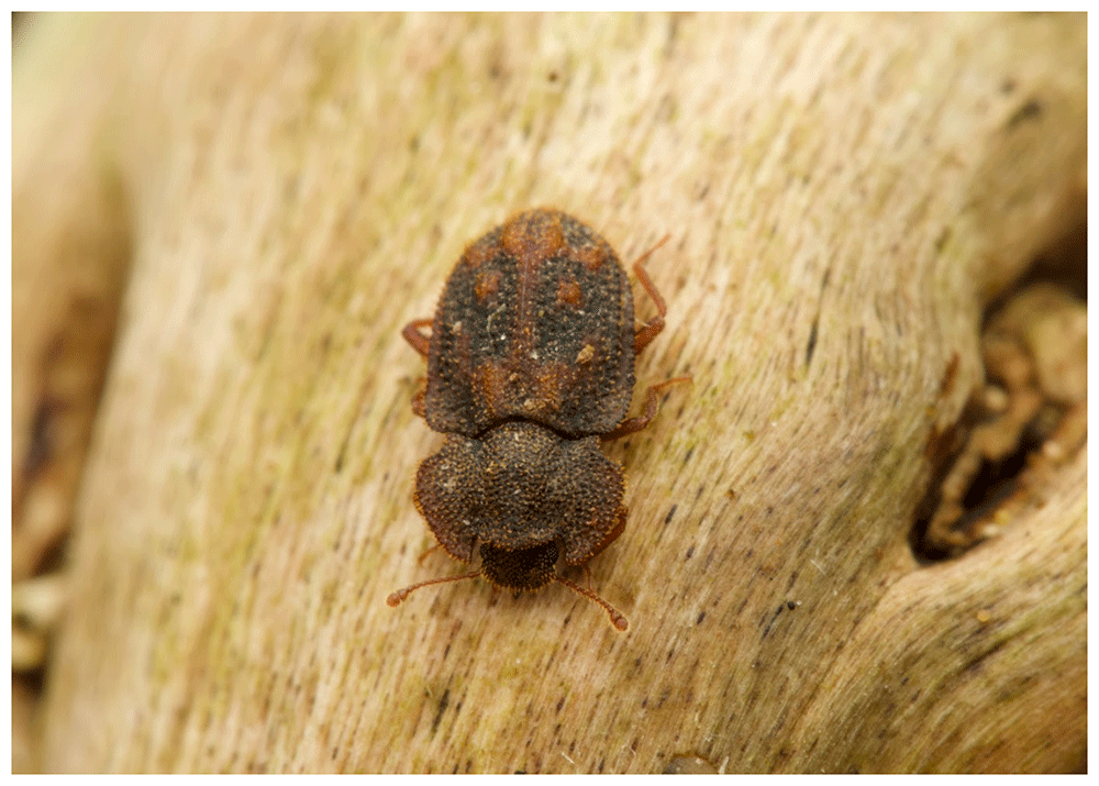
Figure 1. Image of a Tarphius canariensis specimen.
An example of a specimen of Tarphius canariensis. Photo credit: Mario Aponte.
Specimens were euthanized by placement on cold Petri dish and subsequently preserved in liquid nitrogen. Until DNA extraction, samples were preserved at -80°C.
Genetic information
The estimated genome size, based on ancestral taxa, is 450 Mb. This is a diploid genome with a haploid number of 10 chromosomes (2n=20) and unknown sex chromosomes. All information for this species was retrieved from Genomes on a Tree (Challis et al., 2023).
DNA/RNA processing
The workflow for high molecular weight (HMW) DNA extraction at the Wellcome Sanger Institute (WSI) Tree of Life Core Laboratory includes a sequence of procedures: sample preparation and homogenisation, DNA extraction, fragmentation and purification. Detailed protocols are available on protocols.io (Denton et al., 2023b). The icTarCana1 sample was prepared for DNA extraction by weighing and dissecting it on dry ice (Jay et al., 2023). Tissue from the whole organism was homogenised using a PowerMasher II tissue disruptor (Denton et al., 2023a). HMW DNA was extracted using version 3 of the Manual MagAttract protocol (Strickland et al., 2023). DNA was sheared into an average fragment size of 12–20 kb in a Megaruptor 3 system (Bates et al., 2023). Sheared DNA was purified by solid-phase reversible immobilisation, using AMPure PB beads to eliminate shorter fragments and concentrate the DNA (Oatley et al., 2023). The concentration of the sheared and purified DNA was assessed using a Nanodrop spectrophotometer and Qubit Fluorometer using the Qubit dsDNA High Sensitivity Assay kit. Fragment size distribution was evaluated by running the sample on the FemtoPulse system.
RNA was extracted from whole organism tissue of icTarCana3 in the Tree of Life Laboratory at the WSI using the RNA Extraction: Automated MagMax™ mirVana protocol (do Amaral et al., 2023). The RNA concentration was assessed using a Nanodrop spectrophotometer and a Qubit Fluorometer using the Qubit RNA Broad-Range Assay kit. Analysis of the integrity of the RNA was done using the Agilent RNA 6000 Pico Kit and Eukaryotic Total RNA assay.
Whole organism tissue from the icTarCana2 sample was processed at the WSI Scientific Operations core, using the Arima-HiC v2 kit. Tissue (stored at –80 °C) was fixed, and the DNA crosslinked using a TC buffer with 22% formaldehyde. After crosslinking, the tissue was homogenised using the Diagnocine Power Masher-II and BioMasher-II tubes and pestles. Following the kit manufacturer's instructions, crosslinked DNA was digested using a restriction enzyme master mix. The 5’-overhangs were then filled in and labelled with biotinylated nucleotides and proximally ligated. An overnight incubation was carried out for enzymes to digest remaining proteins and for crosslinks to reverse. A clean up was performed with SPRIselect beads prior to library preparation.
Library preparation and sequencing
Library preparation and sequencing were performed at the WSI Scientific Operations core.
At the minimum, samples were required to have an average fragment size exceeding 8 kb and a total mass over 400 ng to proceed to the low input SMRTbell Prep Kit 3.0 protocol (Pacific Biosciences, California, USA), depending on genome size and sequencing depth required. Libraries were prepared using the SMRTbell Prep Kit 3.0 (Pacific Biosciences, California, USA) as per the manufacturer's instructions. The kit includes the reagents required for end repair/A-tailing, adapter ligation, post-ligation SMRTbell bead cleanup, and nuclease treatment. Following the manufacturer’s instructions, size selection and clean up was carried out using diluted AMPure PB beads (Pacific Biosciences, California, USA). DNA concentration was quantified using the Qubit Fluorometer v4.0 (Thermo Fisher Scientific) with Qubit 1X dsDNA HS assay kit and the final library fragment size analysis was carried out using the Agilent Femto Pulse Automated Pulsed Field CE Instrument (Agilent Technologies) and gDNA 55kb BAC analysis kit. Prepared libraries were normalised to 2nM and 15μL used for making complexes. For libraries below 2nM all 10uL was used for making complexes. Primers were annealed and polymerases were hybridised to create circularised complexes according to manufacturer’s instructions. The complexes were purified with the 1.2X clean up with SMRTbell beads. The purified complexes were then diluted to the Revio loading concentration, in the range of 200–300 pM, and spiked with a Revio sequencing internal control.
Samples were sequenced using the Revio system on Revio 25M SMRT cells (Pacific Biosciences, California, USA). The SMRT link software, a PacBio web-based end-to-end workflow manager, was used to set-up and monitor the run, as well as perform primary and secondary analysis of the data upon completion.
For Hi-C library preparation, DNA was fragmented to a size of 400 to 600 bp using a Covaris E220 sonicator. The DNA was then enriched, barcoded, and amplified using the NEBNext Ultra II DNA Library Prep Kit following manufacturers’ instructions. The Hi-C sequencing was performed using paired-end sequencing with a read length of 150 bp on an Illumina NovaSeq X instrument.
Poly(A) RNA-Seq libraries were constructed using the NEB Ultra II RNA Library Prep kit, following the manufacturer’s instructions. RNA sequencing was performed on the Illumina NovaSeq X instrument.
Genome assembly methods
The HiFi reads were first assembled using Hifiasm v0.19.8-r603 (Cheng et al., 2021) with the --primary option. The Hi-C reads were mapped to the primary contigs using bwa-mem2 (Vasimuddin et al., 2019). The contigs were further scaffolded using the provided Hi-C data (Rao et al., 2014) in YaHS v1.2a.2 (Zhou et al., 2023) using the --break option for handling potential misassemblies. The scaffolded assemblies were evaluated using Gfastats (Formenti et al., 2022), BUSCO (Manni et al., 2021) and MERQURY.FK (Rhie et al., 2020).
The mitochondrial genome was assembled using MitoHiFi v3 (Uliano-Silva et al., 2023), which runs MitoFinder (Allio et al., 2020) and uses these annotations to select the final mitochondrial contig and to ensure the general quality of the sequence.
The assembly was decontaminated using the Assembly Screen for Cobionts and Contaminants (ASCC) pipeline. Flat files and maps used in curation were generated in TreeVal (Pointon et al., 2023). Manual curation was primarily conducted using PretextView (Harry, 2022), with additional insights provided by JBrowse2 (Diesh et al., 2023) and HiGlass (Kerpedjiev et al., 2018). Scaffolds were visually inspected and corrected as described by Howe et al. (2021). Any identified contamination, missed joins, and mis-joins were corrected, and duplicate sequences were tagged and removed. The curation process is documented at https://gitlab.com/wtsi-grit/rapid-curation.
Summary analysis of the released assembly was performed using the ERGA-BGE Genome Report ASM Galaxy workflow (https://workflowhub.eu/workflows/1104?version=2).
Genome annotation methods
A gene set was generated using the Ensembl Gene Annotation system (Aken et al., 2016), primarily by aligning publicly available short-read RNA-seq data from BioSample: SAMEA113595922 to the genome. Gaps in the annotation were filled via protein-to-genome alignments of a select set of clade-specific proteins from UniProt (The UniProt Consortium, 2019), which had experimental evidence at the protein or transcript level. At each locus, data were aggregated and consolidated, prioritising models derived from RNA-seq data, resulting in a final set of gene models and associated non-redundant transcript sets. To distinguish true isoforms from fragments, the likelihood of each open reading frame (ORF) was evaluated against known metazoan proteins. Low-quality transcript models, such as those showing evidence of fragmented ORFs, were removed. In cases where RNA-seq data were fragmented or absent, homology data were prioritised, favouring longer transcripts with strong intron support from short-read data. The resulting gene models were classified into two categories: protein-coding, and long non-coding. Models that did not overlap protein-coding genes, and were constructed from transcriptomic data were considered potential lncRNAs. Potential lncRNAs were further filtered to remove single-exon loci due to their unreliability. Putative miRNAs were predicted by performing a BLAST search of miRBase (Kozomara et al., 2019) against the genome, followed by RNAfold analysis (Gruber et al., 2008). Other small non-coding loci were identified by scanning the genome with Rfam (Kalvari et al., 2018) and passing the results through Infernal (Nawrocki & Eddy, 2013). Summary analysis of the released annotation was carried out using the ERGA-BGE Genome Report ANNOT Galaxy workflow (https://doi.org/10.48546/workflowhub.workflow.1096.1).
Results
Genome assembly
The genome assembly has a total length of 310,741,076 bp in 13 scaffolds including the mitogenome (Figure 2 & Figure 3), with a GC content of 34.73%. The assembly has a contig N50 of 14,492,433 bp and L50 of 8 and a scaffold N50 of 31,130,618 bp and L50 of 5. 99.97% of the assembled sequences are localised to the 10 pseudochromosomal sequences. The assembly has a total of 37 gaps, totaling 7.4 kb in cumulative size. The single-copy gene content analysis using the Endopterygota_odb10 database with BUSCO (Manni et al., 2021) resulted in 99.6% completeness (98.9% single and 0.7% duplicated). 57.01% of read k-mers were present in the assembly and the assembly has an average base accuracy Quality Value (QV) of 67.5 as calculated by Merqury (Rhie et al., 2020).
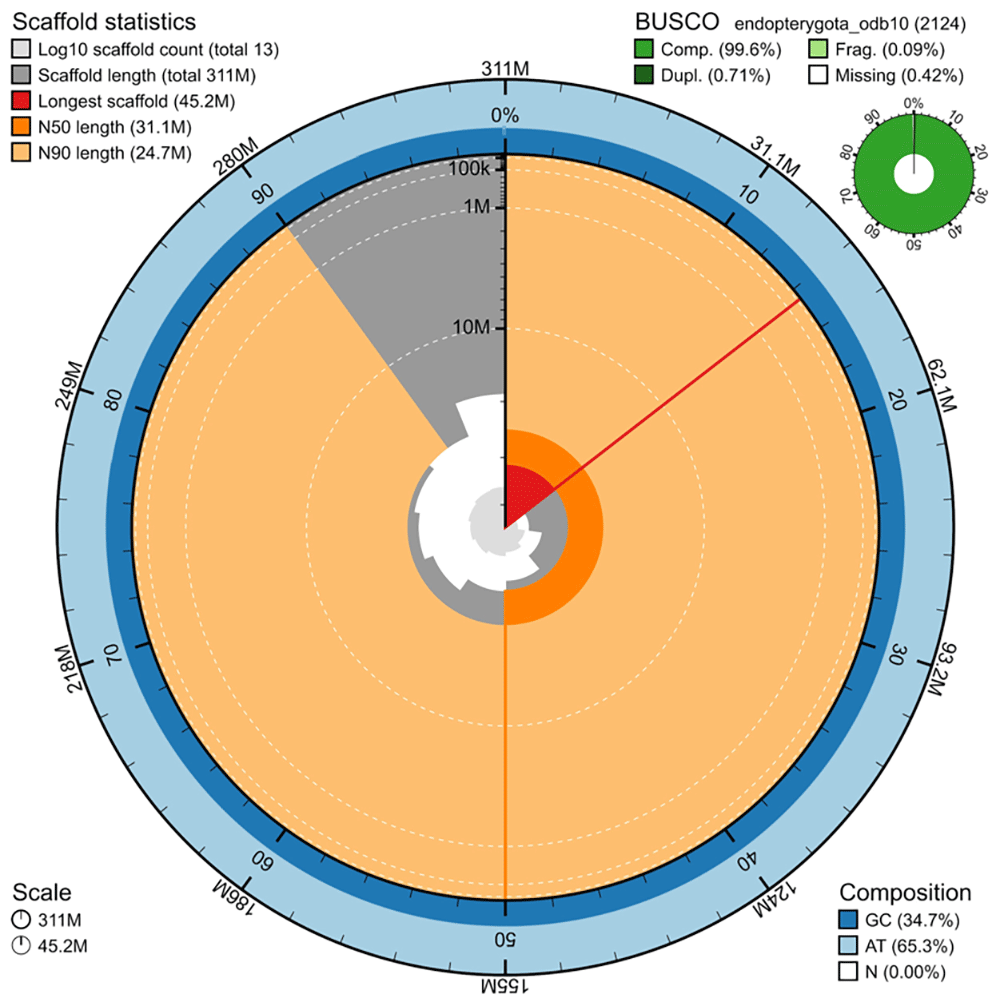
Figure 2. Snail plot summary of assembly statistics.
The main plot is divided into 1,000 size-ordered bins around the circumference, with each bin representing 0.1% of the 310,741,076 bp assembly including the mitochondrial genome. The distribution of sequence lengths is shown in dark grey, with the plot radius scaled to the longest sequence present in the assembly (45,241,133 bp, shown in red). Orange and pale-orange arcs show the scaffold N50 and N90 sequence lengths (31,130,618 and 24,717,953 bp), respectively. The pale grey spiral shows the cumulative sequence count on a log-scale, with white scale lines showing successive orders of magnitude. The blue and pale-blue area around the outside of the plot shows the distribution of GC, AT, and N percentages in the same bins as the inner plot. A summary of complete, fragmented, duplicated, and missing BUSCO genes found in the assembled genome from the endopterygota database (odb10) is shown in the top right.
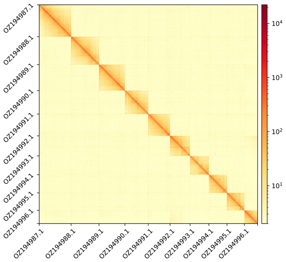
Figure 3. Hi-C contact map showing spatial interactions between regions of the genome.
The diagonal corresponds to intra-chromosomal contacts, depicting chromosome boundaries. The frequency of contacts is shown on a logarithmic heatmap scale. Hi-C matrix bins were merged into a 100 kb bin size for plotting.
Genome annotation
The genome annotation consists of 14,119 protein-coding genes with an associated 21,504 transcripts, in addition to 2,278 non-coding genes (Table 1). Using the longest isoform per transcript, the single-copy gene content analysis using the Endopterygota_odb10 database with BUSCO resulted in 96.7% completeness. Using the OMAmer Endopterygota database for OMArk (Nevers et al., 2025) resulted in 93.95% completeness and 79.84% consistency (Table 2).
Table 1. Statistics from assembled gene models.
| No.
genes | No.
transcripts | Mean gene
length (bp) | No. single-
exon genes | Mean exons
per transcript |
|---|
| mRNA | 14,119 | 21,504 | 10,688 | 380 | 5.7 |
| snoRNA | 20 | 20 | 120 | 20 | 1 |
| lncRNA | 1,950 | 2,123 | 6,255 | 3 | 2.2 |
| snRNA | 31 | 31 | 142 | 31 | 1 |
| rRNA | 39 | 39 | 1,333 | 39 | 1 |
| scRNA | 1 | 1 | 130 | 1 | 1 |
| tRNA | 235 | 235 | 74 | 235 | 1 |
Table 2. Annotation completeness and consistency scores calculated by BUSCO run in protein mode (endopterygota_odb10) and OMArk (Endopterygota).
| Complete | Singular | Duplicated | Fragmented | Missing |
|---|
| BUSCO | 2,054 (96.7%) | 2,036 (95.9%) | 18 (0.8%) | 8 (0.4%) | 62 (2.9%) |
| OMArk | 4,475 (93.95%) | 4,258 (89.40%) | 217 (4.56%) | - | 288 (6.05%) |
| Consistent | Inconsistent | Contaminants | Unknown |
|---|
| OMArk | 11,272 (79.84%) | 484 (3.43%) | 0 (0.00%) | 2,363 (16.74%) |
Data availability
Tarphius canariensis and the related genomic study were assigned to Tree of Life ID (ToLID) icTarCana1 and all sample, sequence, and assembly information are available in INSDC databases under the umbrella BioProject PRJEB76560. The sample information is available at the following BioSample accessions: SAMEA113595920, SAMEA113595921, SAMEA113595922, and SAMEA113595923. The genome assembly is accessible from INSDC databases under accession number GCA_964277315.1 and the annotated genome is available through the Ensembl ERGA-BGE project page (https://projects.ensembl.org/erga-bge/) and and Ensembl website (https://beta.ensembl.org). Sequencing data produced as part of this project are available at the following accessions: ERX13553711, ERX13020688, ERX12671954, and ERX12671953. Documentation related to the genome assembly and curation can be found in the ERGA Assembly Report (EAR) document available at https://github.com/ERGA-consortium/EARs/tree/main/Assembly_Reports/Tarphius_canariensis/icTarCana1. Further details and data about the project are hosted on the ERGA portal at https://portal.erga-biodiversity.eu/data_portal/12480
Author contributions
BCE and VN collected the species, VN identified the species, BCE and VN sampled and preserved biological material and provided metadata, RM, AB, JPG and RF provided sampling and metadata support and management, MC and AR carried out barcoding and deposited specimens for vouchering biobanking, the WSI ToL Samples and Laboratory teams and WSI Scientific Operations teams extracted DNA, prepared libraries, and performed sequencing, the WSI ToL Informatics teams performed genome assembly and curation, LH performed genome annotation under the supervision of FM. TB generated the analysis and report. All authors contributed to the writing, review, and editing of this genome note and read and approved the final version.
Acknowledgements
We would like to acknowledge the assembly reviewer, Jean-Marc Aury, from Genoscope.
Faculty Opinions recommendedReferences
- Aken BL, Ayling S, Barrell D, et al.:
The ensembl gene annotation system.
Database (Oxford).
2016; 2016: baw093. PubMed Abstract
| Publisher Full Text
| Free Full Text
- Allio R, Schomaker-Bastos A, Romiguier J, et al.:
MitoFinder: efficient automated large-scale extraction of mitogenomic data in target enrichment phylogenomics.
Mol Ecol Resour.
2020; 20(4): 892–905. PubMed Abstract
| Publisher Full Text
| Free Full Text
- Bates A, Clayton-Lucey I, Howard C:
Sanger Tree of Life HMW DNA fragmentation: diagenode Megaruptor®3 for LI PacBio.
protocols.io.
2023. Publisher Full Text
- Challis R, Kumar S, Sotero-Caio C:
Genomes on a Tree (GoaT): a versatile, scalable search engine for genomic and sequencing project metadata across the eukaryotic Tree of Life [version 1; peer review: 2 approved].
Wellcome Open Res.
2023; 8: 24. PubMed Abstract
| Publisher Full Text
| Free Full Text
- Cheng H, Concepcion GT, Feng X, et al.:
Haplotype-resolved de novo assembly using phased assembly graphs with hifiasm.
Nat Methods.
2021; 18(2): 170–175. PubMed Abstract
| Publisher Full Text
| Free Full Text
- Denton A, Oatley G, Cornwell C, et al.:
Sanger Tree of Life sample homogenisation: PowerMash.
protocols.io.
2023a. Publisher Full Text
- Denton A, Yatsenko H, Jay J, et al.:
Sanger Tree of Life wet laboratory protocol collection V.1.
protocols.io.
2023b. Publisher Full Text
- Diesh C, Stevens GJ, Xie P, et al.:
JBrowse 2: a modular genome browser with views of synteny and structural variation.
Genome Biol.
2023; 24(1): 74. PubMed Abstract
| Publisher Full Text
| Free Full Text
- do Amaral RJV, Bates A, Denton A, et al.:
Sanger Tree of Life RNA extraction: automated MagMaxTM mirVana.
protocols.io.
2023. Publisher Full Text
- Emerson BC, Oromí P, Hewitt GM:
Tracking colonization and diversification of insect lineages on islands: mitochondrial DNA phylogeography of Tarphius canariensis (Coleoptera: Colydiidae) on the Canary Islands.
Proc Biol Sci.
2000; 267(1458): 2199–2205. PubMed Abstract
| Publisher Full Text
| Free Full Text
- Emerson BC, Oromí P:
Diversification of the forest beetle genus Tarphius on the Canary Islands, and the evolutionary origins of Island endemics.
Evolution.
2005; 59(3): 586–598. PubMed Abstract
| Publisher Full Text
- Formenti G, Abueg L, Brajuka A, et al.:
Gfastats: conversion, evaluation and manipulation of genome sequences using assembly graphs.
Bioinformatics.
2022; 38(17): 4214–4216. PubMed Abstract
| Publisher Full Text
| Free Full Text
- Gruber AR, Lorenz R, Bernhart SH, et al.:
The Vienna RNA Websuite.
Nucleic Acids Res.
2008; 36(suppl_ 2): W70–W74. PubMed Abstract
| Publisher Full Text
| Free Full Text
- Harry E:
PretextView (Paired REad TEXTure Viewer): a desktop application for viewing pretext contact maps. 2022. Reference Source
- Howe K, Chow W, Collins J, et al.:
Significantly improving the quality of genome assemblies through curation.
GigaScience.
2021; 10(1): giaa153. PubMed Abstract
| Publisher Full Text
| Free Full Text
- Jay J, Yatsenko H, Narváez-Gómez JP, et al.:
Sanger Tree of Life sample preparation: triage and dissection.
protocols.io.
2023. Publisher Full Text
- Kalvari I, Nawrocki EP, Argasinska J, et al.:
Non-coding RNA analysis using the Rfam database.
Curr Protoc Bioinformatics.
2018; 62(1): e51. PubMed Abstract
| Publisher Full Text
| Free Full Text
- Kerpedjiev P, Abdennur N, Lekschas F, et al.:
HiGlass: web-based visual exploration and analysis of genome interaction maps.
Genome Biol.
2018; 19(1): 125. PubMed Abstract
| Publisher Full Text
| Free Full Text
- Kozomara A, Birgaoanu M, Griffiths-Jones S:
miRBase: from microRNA sequences to function.
Nucleic Acids Res.
2019; 47(D1): D155–D162. PubMed Abstract
| Publisher Full Text
| Free Full Text
- Manni M, Berkeley MR, Seppey M, et al.:
BUSCO update: novel and streamlined workflows along with broader and deeper phylogenetic coverage for scoring of eukaryotic, prokaryotic, and viral genomes.
Mol Biol Evol.
2021; 38(10): 4647–4654. PubMed Abstract
| Publisher Full Text
| Free Full Text
- Mazzoni CJ, Ciofi C, Waterhouse RM:
Biodiversity: an atlas of European reference genomes.
Nature.
2023; 619(7969): 252. PubMed Abstract
| Publisher Full Text
- Nawrocki EP, Eddy SR:
Infernal 1.1: 100-fold faster RNA homology searches.
Bioinformatics.
2013; 29(22): 2935–5. PubMed Abstract
| Publisher Full Text
| Free Full Text
- Nevers Y, Warwick Vesztrocy A, Rossier V, et al.:
Quality assessment of gene repertoire annotations with OMArk.
Nat Biotechnol.
2025; 43(1): 124–133. PubMed Abstract
| Publisher Full Text
| Free Full Text
- Noguerales V, Emerson BC:
Arthropod mtDNA paraphyly: a case study of introgressive origin.
J Evol Biol.
2025; 38(2): 272–283. PubMed Abstract
| Publisher Full Text
- Oatley G, Sampaio F, Howard C:
Sanger Tree of Life fragmented DNA clean up: automated SPRI.
protocols.io.
2023. Publisher Full Text
- Patiño J, Whittaker RJ, Borges PAV, et al.:
A roadmap for Island biology: 50 fundamental questions after 50 years of the theory of Island biogeography.
J Biogeogr.
2017; 44(5): 963–983.
Publisher Full Text
- Pointon DL, Eagles W, Sims Y, et al.:
sanger-tol/treeval v1.0.0 – Ancient Atlantis. 2023. Publisher Full Text
- Rao SSP, Huntley MH, Durand NC, et al.:
A 3D map of the human genome at kilobase resolution reveals principles of chromatin looping.
Cell.
2014; 159(7): 1665–1680. PubMed Abstract
| Publisher Full Text
| Free Full Text
- Rhie A, Walenz BP, Koren S, et al.:
Merqury: reference-free quality, completeness, and phasing assessment for genome assemblies.
Genome Biol.
2020; 21(1): 245. PubMed Abstract
| Publisher Full Text
| Free Full Text
- Strickland M, Moll R, Cornwell C, et al.:
Sanger Tree of Life HMW DNA extraction: manual MagAttract.
protocols.io.
2023. Publisher Full Text
- The UniProt Consortium: UniProt: a worldwide hub of protein knowledge.
Nucleic Acids Res.
2019; 47(1): D506–D515. PubMed Abstract
| Publisher Full Text
| Free Full Text
- Uliano-Silva M, Ferreira JGRN, Krasheninnikova K, et al.:
MitoHiFi: a python pipeline for mitochondrial genome assembly from PacBio high fidelity reads.
BMC Bioinformatics.
2023; 24(1): 288. PubMed Abstract
| Publisher Full Text
| Free Full Text
- Vasimuddin M, Misra S, Li H, et al.:
Efficient architecture-aware acceleration of BWA-MEM for multicore systems. In: 2019 IEEE International Parallel and Distributed Processing Symposium (IPDPS). IEEE, 2019; 314–324. Publisher Full Text
- Zhou C, McCarthy SA, Durbin R:
YaHS: Yet another Hi-C Scaffolding tool.
Bioinformatics.
2023; 39(1): btac808. PubMed Abstract
| Publisher Full Text
| Free Full Text