Introduction
Over the past two decades, functional magnetic resonance imaging (fMRI) in rhesus macaques has become a key tool in understanding the neural mechanisms underlying auditory perception, from basic — tonotopy (Joly et al., 2014; Tanji et al., 2010), auditory cortical fields mapping (Petkov et al., 2006; Schönwiesner et al., 2015), temporal and spectral modulations (Erb et al., 2019) — to higher-level processes — complex sounds (Kikuchi et al., 2014), sound location (Ortiz-Rios et al., 2017), multimodal integration (Froesel et al., 2022; Kayser et al., 2005).
Furthermore, fMRI with macaques has contributed to our understanding of the origins of speech by revealing cortical regions that are sensitive to voices and vocalizations in our close relatives (Bodin et al., 2021; Joly et al., 2012; Ortiz-Rios et al., 2015; Petkov et al., 2008), as well as to guide electrophysiological recordings to those regions (Esposito et al., 2025; Giamundo et al., 2024; Perrodin et al., 2010; Perrodin et al., 2011). Such studies have shown that conspecific vocalizations selectively activate higher-level areas of the auditory cortex, particularly in an anterior portion of the temporal gyrus, analogous to the anterior temporal voice area (aTVA) observed in humans (Aglieri et al., 2018; Pernet et al., 2015). Furthermore, vocalization-sensitive regions have been reported in areas beyond the temporal gyrus, specifically in the ventral premotor and prefrontal cortices (Bodin et al., 2021; Ortiz-Rios et al., 2015).
Auditory fMRI studies in macaques have essentially been performed in awake, behaving animals. Awake fMRI in macaques is particularly valuable because it captures neural responses under naturalistic conditions, in fully engaged, behaviorally active subjects. In addition to the findings related to voice selectivity, awake fMRI has also demonstrated its efficacy in other modalities, particularly in detecting face-selective areas (Ku et al., 2011; Pinsk et al., 2009; Tsao et al., 2003). However, awake monkey fMRI poses substantial challenges. The animal requires extensive behavioural training to acclimate to the scanner environment, to remain motionless during long sessions of image acquisition, and to perform an auditory task (Goense et al., 2010; Vanduffel & Farivar, 2014). The acquisition of awake fMRI data from as few as two naïve monkeys can easily take over a year. Consequently, the number of animals used in awake fMRI studies is limited, with only two to three animals per study due to feasibility issues. In addition, and despite the behavioral training, inevitable head and body movements occur during acquisition, even under head restraint. This results in large residual motion artifacts that degrade data quality and increase the amount of data that needs to be collected (Goense et al., 2010). As a point of comparison, it has been determined that up to sixty times the amount of data acquired in humans is required to match response reliability in macaque fMRI (Norman-Haignere et al., 2019).
In contrast to awake imaging, anesthetized fMRI protocols in rhesus macaques significantly simplify data acquisition. Anesthesia nearly eradicates head motion, thereby enabling the collection of higher-quality data. Most importantly, anesthesia eliminates the need for long-term behavioral training to ensure that the animal sits still for extended periods in the scanner, a hostile environment for the animal (Goense et al., 2010), and the surgical procedures concomitant with head fixation. These advantages offer the potential for acquiring data on cohort sizes that are nearly equivalent to those of human fMRI studies, and at a significantly reduced time frame when compared to the whole process employed in studies involving only two awake monkeys.
However, anesthetized fMRI comes with its own set of drawbacks. Anesthetic agents used to induce sedation can markedly alter neural activity and neurovascular coupling (Slupe & Kirsch, 2018; Zhang, 2022), thereby potentially attenuating or even distorting the fMRI BOLD signal that reflects the underlying neuronal response to auditory stimuli (Guldenmund et al., 2017; Uhrig et al., 2016). To maximize the outcome of anesthetized fMRI, the anesthesia protocol must be optimized to induce a state that provides immobilization while minimally interfering with the underlying neural processes that support auditory perception. Various combinations of anesthetic agents have been used to achieve this goal. These “near-awake” protocols include: remifentanyl, an opioid analgesic, associated with mivacurium, a neuromuscular-blocking drug (Ku et al., 2011; Logothetis et al., 1999; Petkov et al., 2006); moderate propofol sedation, with (Barttfeld et al., 2015) or without (Uhrig et al., 2016) muscle blocking agent (cisatracrium); sufentanil or propofol, supplemented with low-dose isoflurane (Shi et al., 2021); the combination of low-dose halogenated ether (isoflurane or sevoflurane) alongside either nitrous oxide (Obliger-Debouche et al., 2025; Sadagopan et al., 2015) or dexmedetomidine (Autio et al., 2020), a sedative that has been shown to relatively preserve functional connectivity in humans (Guldenmund et al., 2017).
The objective of the present study was to map the cerebral (subcortical and cortical) response to complex natural sounds, including vocalizations, in a large group of anesthetized macaques. Eleven rhesus macaques were exposed to macaque vocalizations and non-vocal complex sounds during fMRI acquisitions, while under a near-awake anesthesia protocol involving low-dose sevoflurane combined with dexmedetomidine. Our results show robust activation of core and belt auditory cortices, suggesting that much of the fundamental neural circuitry for auditory perception remains active under this anesthesia protocol. However, in contrast with awake fMRI studies, vocalization selectivity was not observed in higher-order, anterior auditory cortices, but was found in the ventral premotor and prefrontal cortex as well as in the posterior middle temporal gyrus. This suggests different involvement of the ventral and dorsal auditory pathways during anesthesia. We conclude that near-awake anesthesia fMRI is appropriate for studying basic auditory functions in a large cohort of subjects, enabling efficient and reproducible mapping of core auditory areas without the extensive training required for awake imaging, while it may obscure the neural mechanisms underlying complex auditory processing, such as the selective response to conspecific vocalizations.
Methods
Animals
Eleven rhesus monkeys (Macaca mulatta), 7 females and 4 males, aged between 1 and 17 years (median = 6) and weighing between 1 and 8 kg (median = 5), were scanned. The animals were sourced from the same colony and were selected based on their small head size, which was required to be compatible with the MRI head coil. All experimental procedures were in compliance with the National Institutes of Health’s Guide for the Care and Use of Laboratory Animals and approved by the Ethical board of Institut de Neurosciences de la Timone (ref 2016060618508941).
Animal preparation and anesthesia
All scans were performed under anesthesia with a protocol inspired by a previous study (Autio et al., 2025). Anesthesia was induced with an intramuscular injection of glycopyrrolate (10 µg/kg), dexmedetomidine (4.5 μg/kg) and ketamine (6 mg/kg), followed by 2% sevoflurane gas administration through tracheal intubation. Two intravenous catheters were implanted into both saphenous veins to provide fluid support for dexmedetomidine and contrast agent delivery separately. Each animal was then secured in the supine position in an MRI testing chair (Rogue Research, Canada) slided feet-first in the MRI scanner. Prior to MRI acquisitions, respiratory control with mechanical ventilation was initiated, as well as continuous delivery of dexmedetomidine (2–4.5 μg/kg/hr). During anatomical scanning, the concentration of sevoflurane gas was lowered to 1–1.5% (depending on the animal), and during functional scanning, it was reduced to 0.5–1.2%. Ferrous oxide contrast agent (monocrystalline iron oxide nanoparticle, MION, 11 mg/kg) was administered at the end of anatomical scanning, some minutes before functional scanning. The animal's physiological characteristics, which includes heart and respiratory rate, capnometry, and oximetry, were monitored during scanning to ensure its health and well-being, as well as to adjust anesthesia to levels close to awakening. A warming blanket was employed to prevent hypothermia.
Stimuli & audio apparatus
Sound stimuli were a subset of the open access PrimaVoice audio stimulus set (Bodin et al., 2025) and comprised two main categories: macaque vocalizations and non-vocal sounds. Each category contained 24 stimuli, for a total of 48 sound stimuli. Each main category was divided into 4 subcategories of 6 stimuli, forming 8 subcategories in total (see Figure 1). Macaque vocalizations were provided by Marc Hauser (Hauser, 1991) and included both positive (coos, n = 6, grunts, n = 6) and negative (barks, n = 6, screams, n = 6) calls. Non-vocal sounds included both natural (living, n = 6, non-living, n = 6) and artificial sounds (human actions, n = 6, or not, n = 6) from previous studies from our group (Belin et al., 2000; Capilla et al., 2013) or kindly provided by Christopher Petkov (Petkov et al., 2008) and Elia Formisano (Moerel et al., 2012). Stimuli were adjusted in duration, resampled at 48828 Hz and normalized by root mean square amplitude. Finally, a 10-ms cosine ramp was applied to the onset and offset of all stimuli. During experiments, stimuli were digitally processed by a TDT System 3 RM1 (Tucker Davis Technologies, Alachua, FL, USA) and delivered via three MRI-compatible piezoelectric speakers (Sonitron, Sint-Niklaas, Belgium). The sound was essentially delivered by bone conduction. Two smaller speakers (frequency response ranging from 1 kHz to 20 kHz) were placed on or near the animal's ears inside the antenna and gently pressed against its skull. A third, larger speaker (frequency response ranging from 300 Hz to 20 kHz), was placed against the dorsal part of the animal’s head through the antenna’s hole designed to house a head post.
MRI data acquisition
Images were acquired at the MRI Center of the Institut de Neurosciences de la Timone (Marseille, France) using a 3-Tesla PRISMA scanner (Siemens, Erlangen, Germany) with a 24-channels macaque head coil (Rogue Research, Canada) (Autio et al., 2025). High-resolution T1 & T2-weighted anatomical volumes were first acquired for coregistration and normalization purposes (T1w: MPRAGE sequence, TR = 2.2 s, TE = 2.23 ms, TI = 900 ms flip angle: 8°, matrix size = 224 × 256 × 256, resolution 0.5 × 0.5 x 0.5 mm3, bandwidth 270 Hz/Pixel, GRAPPA = 2, sagittal orientation; T2w: SPACE sequence, TR = 3.2 s, TE = 562 ms, matrix size = 224 × 256 × 256, resolution 0.5 × 0.5 x 0.5 mm3, bandwidth 723 Hz/Pixel, turbo factor 314, echo train length 1234 ms, GRAPPA = 2, sagittal orientation).
Fieldmaps B0 field-maps were estimated using a pair of spin-echo EPI images with one opposite phase encoding directions (Andersson et al., 2003) (LR and RL directions, 1.5 mm isotropic, TE 46.2 ms, TR 7.06 s). The spin-echo EPI scans were matched with geometry and distortion properties of fMRI acquisitions (FOV 96 × 96 mm, matrix 64 × 64, number of slices 39, phase partial Fourier 65%, bandwidth 1240 Hz/pixel, echo spacing = 0.93 ms, GRAPPA = 2, fat suppression, flip-angle = 90°, averages 3).
Functional volumes were acquired with the same coil using multiband EPI sequences obtained from the Center for Magnetic Resonance Research (CMRR, University of Minnesota), version R016a (multiband acceleration factor = 3, TA = 646 ms, TE = 16.6 ms, matrix size = 64 × 64 × 39, resolution 1.5 x 1.5 x 1.5 mm3, flip angle = 50°, multiband LeakBlock kernel, GRAPPA = 2 with EPI calibration scan with 18 reference lines, bandwidth = 1240 Hz/Pixel, echo spacing = 0.93 ms, phase partial Fourier = 75%, fat suppression).
Functional imaging procedure
To avoid interference from the scanner noise during stimulus presentation, clustered sparse sampling of functional acquisitions was employed (Schmidt et al., 2008; Trapeau et al., 2024; Zaehle et al., 2007). Clusters of seven fMRI volumes (4.6 s) were acquired after a 3.5 s acquisition-free period, which allowed stimuli to be presented when the scanner was silent. During each 7-minute functional run, 32 blocks of stimuli were presented (16 blocks of each of the two main categories), interleaved with 16 silent blocks. Each block contained six stimuli (ISI = 50 ms) belonging to one of the eight subcategories. The time interval between the end of an acquisition cluster and the start of a block of stimuli was randomly jittered from 100 ms to 250 ms. A total of 188 runs were performed among the 11 subjects (one session per subject except for M1 who underwent six sessions; median of 12 runs per subject).
fMRI data preprocessing
Brain extraction and tissue segmentation was performed on the structural scans using a custom-made pipeline which included: bias field correction using the product of the T1w and T2w images (Glasser et al., 2013); denoising (spatially adaptive nonlocal means, SPM); first brain extraction (FSL); registration of the NIMH Macaque Template v1.2 (Seidlitz et al., 2018) to the anatomical scan, in order to provide priors to SPM’s old_segment algorithm; second brain extraction from the segmented tissues. Transformation matrices between anatomical and functional data were computed using non-linear registration (SyN, ANTS). Additionally, white matter and cerebrospinal fluid masks were eroded using a 3 mm cubic kernel in order to provide a mask that contains no region of interest to a principal component analysis of the functional data (see fMRI data analysis). Transformation matrices between functional, anatomical and template volumes were computed using non-linear registration (SyN, ANTS). These transformation matrices were used to register the segmented tissues to the functional data as well as to register the functional results to individual high-resolution anatomical scans and to the template.
Preprocessing of the functional data included motion correction, spatial distortion reduction using field maps, inter-runs registration and spatial smoothing. For each subject, every functional volume was realigned and unwarped (FSL) to a reference volume that was spatially the closest to the average of all runs. Spatial smoothing (SPM) was done with a full-width half-maximum 3-dimensional Gaussian kernel that was twice the size of the functional voxels (i.e., 3 mm).
fMRI data analysis
For each subject, general linear model (GLM) estimates of responses to all sounds versus silence (“Sound vs. Silence” contrast) and to macaque vocalizations against non-vocal sounds (“Macaque vs. Non-Vocal” contrast) were computed with fMRISTAT (Worsley et al., 2002) using MION-based response function, manually designed to have a reversed sign, a long tail and no undershoot, similar to previous descriptions (Leite et al., 2002). The GLM included covariates of no interest: the first principal components of two principal component analyses performed on eroded masks of the functional voxels identified as containing white matter and cerebrospinal fluid. The number principal components included as covariates of no interest was chosen to optimize the “Sound vs. Silence” contrast in each subject (6 for white matter, 4 for cerebrospinal fluid, on average).
Following this first level-analysis, fixed and random effects group maps were computed for both contrasts, using the template registered data from all subjects and all runs. Additionally, the group map for the “Macaque vs. Non-Vocal” contrast was computed on a selection of runs that exhibited a positive whole-brain influence on this contrast. The contribution of each run to the contrast was assessed on a per-subject basis using a jackknife procedure. This procedure systematically excluded one run from the dataset of each subject and returned the maximum t-value elicited by the remaining runs. Then, the resulting t-values of a subject were compared with the maximum t-value elicited by all runs of this subject. A lower maximum t-value after run removal meant that the run had a positive contribution to the contrast and vice versa. Note that no topographical constraints were imposed on where these maximum t-values should be, as they were computed using the whole-brain t-maps. Only the runs that showed a positive contribution to the “Macaque vs. Non-Vocal” contrast of each subject were kept, leaving 72 runs in total (median of 6 runs per subject).
T-values of each subcategory were extracted for each subject in several bilateral regions of interest (ROI): the primary auditory cortex (A1), which was defined, for each subject, as the intersection of the A1 auditory field identified by (Petkov et al., 2006) and the “Sound vs. Silence” contrast of the subject; the inferior colliculus (IC), based on peak activations in this anatomically defined region from the random effects group map of the “Sound vs. Silence” contrast; the posterior middle temporal gyrus (pMTG) and the rostral portion of the ventral premotor cortex (F5), both based on peak activations in this region from the fixed effects group map of the “Macaque vs. Non-Vocal” contrast; and the anterior temporal voice area (aTVA) that was previously observed in three macaques during an awake fMRI study by our group (Bodin et al., 2021). Each ROI size was 19 voxels in the functional space and a diameter of 4.5 mm.
Results
Auditory activations
Second-level random effects group analysis revealed strong activations in the primary auditory cortex (A1) and nuclei of the ascending auditory pathway (inferior colliculus, IC; medial geniculate body, MGB) in response to all sounds (“Sound vs. Silence” contrast, Figure 1). Cortical activation extended to the rostral field of the core auditory cortex (R), but also to belt areas (CM, CL, ML, AL, RTL, MM, RM, RTM, Figure 1A). The parabelt and higher auditory fields were marginally activated, except for the right rostral parabelt (RPB) and the temporalis superior 2 (Ts2), which showed significant activation in some voxels.
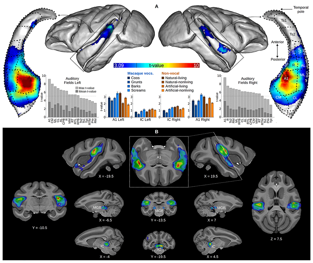
Figure 1. Group-averaged auditory activations.
Surface projection and volume map of the “Sound vs. Silence” contrast in a second-level random effects group analysis. For both surface and volume, the contrast map was thresholded at p < 0.05 and corrected for multiple comparisons by cluster size (p < 0.05). The dotted line on the color bar indicates peak threshold corrected for multiple comparisons (t = 4.93) using Gaussian random field theory (Worsley et al., 1996) at p < 0.05. (A) The contrast map is projected onto the surface of the NIMH macaque template (NMT) as well as onto isolated left and right superior temporal gyri viewed from above. Isolated superior temporal gyri include projections of the macaque auditory fields, which were provided by Christopher Petkov’s group (Petkov et al., 2006; Petkov et al., 2008). Maximum and mean voxel t-values of each auditory field from the random effects group map are displayed next to the temporal gyri, the horizontal dotted line indicates peak threshold. The panel also shows t-values averaged across individuals (error bars indicate the standard error of the mean) of each subcategory of sound versus silence, extracted from the left and right A1 and IC ROIs (see Methods). (B) The contrast map is overlaid on axial, longitudinal and sagittal slices of the NMT. Top central image is an oblique slice along the superior temporal gyrus. A1: primary auditory cortex; IC: inferior colliculus; MGB: medial geniculate body.
At the individual level, all subjects had significant auditory responsive clusters in IC and in the auditory cortex (Table 1). ROI t-value extraction showed that all subcategories of sounds elicited positive auditory activations in both IC and A1. (Figure 1A).
Table 1. Individual activations in IC and AC.
Peak t-value and number of significant voxels at cluster threshold (t = 3.09) in the inferior colliculi (IC) and auditory cortices (AC) of each subject.
| Subject | IC peak t-value | IC extent (voxels) | AC peak t-value | AC extent (voxels) |
|---|
| M1 | 12.87 | 105 | 8.04 | 271 |
| M2 | 5.22 | 27 | 4.2 | 23 |
| M3 | 5.14 | 20 | 6.78 | 110 |
| M4 | 4.07 | 15 | 6.06 | 257 |
| M5 | 4.43 | 13 | 8.45 | 344 |
| M6 | 7.61 | 40 | 11.26 | 367 |
| M7 | 4.69 | 13 | 9.91 | 361 |
| M8 | 6.79 | 45 | 3.6 | 11 |
| M9 | 5.69 | 19 | 8.39 | 278 |
| M10 | 6.92 | 32 | 8.34 | 235 |
| M11 | 6.74 | 33 | 8.2 | 198 |
Vocalization-specific activations
We could not reproduce our previous findings of voice-selective areas in the anterior temporal lobe (aTVA) of awake macaques (Bodin et al., 2021) in a second-level, random effects group analysis computed on all runs. However, a fixed effects analysis revealed activations in the rostral ventral premotor cortex (F5) and area 8a of the right hemisphere (white outline in Figure 2).
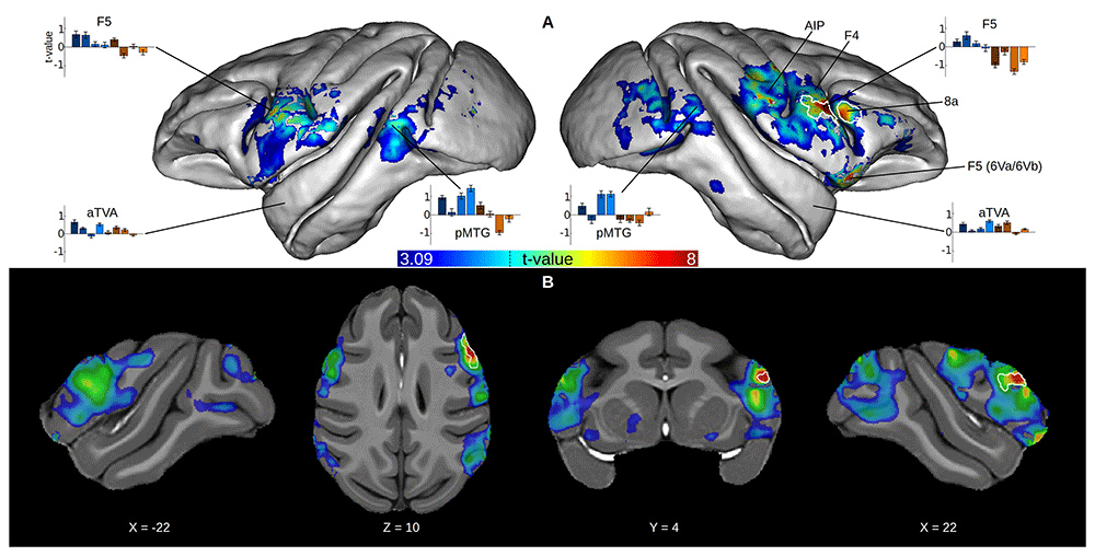
Figure 2. Group-averaged activations to macaque vocalizations.
Surface projection and volume map of the “Macaque vs. Non Vocal” contrast in a second-level fixed effects group analysis, computed on a selection of runs that had a positive whole-brain influence on this contrast (see Methods). The white outlines indicate significant areas (p < 0.05, cluster size corrected) of the same analysis computed on all runs. For both surface and volume, the contrast map was thresholded at p < 0.05 and corrected for multiple comparisons by cluster size (p < 0.05). The dotted line on the color bar indicates peak threshold corrected for multiple comparisons using Gaussian random field theory (Worsley et al., 1996) at p < 0.05. (A) The contrast map is projected on the surface of the NMT. The panel also shows t-values averaged across individuals (error bars indicate the standard error of the mean) of each subcategory of sound versus silence, extracted from the several ROIs (see Methods). (B) The contrast map is overlaid on axial, longitudinal and sagittal slices of the NMT. F5: rostral ventral premotor cortex; F4: caudal ventral premotor cortex; AIP: anterior intraparietal area; pMTG: posterior middle temporal gyrus; aTVA: anterior temporal voice area.
Next, we conducted the same analyses on a selection of runs that had a positive whole-brain contribution to the contrast (see fMRI data analysis). The random effects group map computed from this selection of 72 runs was very similar to the fixed effects group map computed from all 188 runs, showing activations in areas F5 and 8a in the right hemisphere. The fixed effects analysis on the selection of runs revealed much broader activations: F5 bilaterally, right anterior intraparietal area (AIP) and caudal ventral premotor cortex (F4), right 6Va/6Vb, posterior part of the middle temporal gyrus (pMTG) bilaterally.
ROI t-value extraction showed showed that auditory activations for each subcategory of sounds in F5 and pMTG were mainly positive for macaque vocalizations and negative for non-vocal sounds (Figure 2A). This difference between vocalizations and non-vocal sounds was not observed in the awake macaques aTVA, where auditory activations were mainly positive for all subcategories.
Discussion
This study aimed to map the auditory response to complex natural sounds in a large group of rhesus macaques under a near-awake anesthesia protocol involving low-dose sevoflurane and dexmedetomidine. Using a sizable sample of eleven macaques, which approaches the median of twelve subjects employed in human fMRI studies (Szucs & Ioannidis, 2020), we have obtained the first random-effects group map of auditory activation in this species. The results revealed robust responses in the core and belt areas of the auditory cortex, as well as in subcortical structures, including the midbrain (inferior colliculi) and thalamus (medial geniculate body). This suggests that essential neural circuitry supporting auditory perception remains functional under this anesthesia regimen. These findings demonstrate the efficacy of anesthetized macaque fMRI in facilitating efficient and high-quality mapping of subcortical and core auditory regions across larger cohorts, thereby circumventing the logistical challenges associated with awake imaging.
Vocalization-specific activations revealed a different picture. To our disappointment, our results revealed a conspicuous absence of vocalization selectivity in higher-order, anterior auditory cortices. This result diverges from previous findings of our group, which highlighted the crucial involvement of the aTVA in processing the same set of stimuli (Bodin et al., 2021), as well as from earlier awake macaque studies (Ortiz-Rios et al., 2015; Petkov et al., 2008). This outcome was somewhat unexpected, given that aTVA activation had been previously reported even in anesthetized macaques (Petkov et al., 2008). Nevertheless, a critical distinction exists between this study and ours with respect to the pharmaceutical agents utilized for immobilizing the animals during experimentation. In their study, Petkov and colleagues used remifentanyl and mivacurium, which is the combination of an analgesic with a neuromuscular-blocking drug. However, the absence of hypnotic agents, even at sedation levels, in this cocktail suggests that the animals remained conscious during the experiment. Conversely, we opted for a general anesthesia protocol, involving a sedative agent combined with a hypnotic agent at low dose, with meticulous physiologic monitoring and adjustment of drug levels throughout the experiment to bring the animal close to awakening. Our protocol, involving sevoflurane and dexmedetomidine, prioritized animal well-being and minimized the risk of a non pleasant mnesic experience, though possibly at the cost of reduced activation in anterior auditory regions. The inclusion of each animal in a singular session, in which some completed less than ten trials, might have also been suboptimal for detecting vocalization sensibility in higher-order auditory cortices.
Despite the absence of observed vocalization selectivity in the anterior temporal gyrus, our findings revealed the presence of voice-selective regions in the ventral premotor (F5) and prefrontal cortex (8a). The involvement of these regions in vocalization processing has been previously observed in some awake macaque fMRI studies (Bodin et al., 2021; Ortiz-Rios et al., 2015) and they have been identified as part of the dorsal auditory pathway (Arnott & Alain, 2011; Petrides & Pandya, 2009), which connects the caudal parabelt auditory regions to the ventral inferior parietal cortex (Lewis & Van Essen, 2000), and up to the frontal eye fields (Hackett et al., 1999; Romanski et al., 1999). These findings suggest that under our anesthesia regimen, the dorsal auditory stream may be preferentially engaged, speculatively as a resilient backbone for action planning, over the ventral pathway, which is more dedicated to the identification of auditory objects (Kuśmierek et al., 2012). More surprisingly, we found additional bilateral vocalization sensitivity in the posterior part of the middle temporal gyrus, a region previously identified as a site of audio-visual integration in humans (Beauchamp et al., 2004; Callan et al., 2004; Xu et al., 2009), macaques (Froesel et al., 2022) and marmosets (Dureux et al., 2024), but, to our knowledge, not reported in auditory only experiments.
In conclusion, while awake fMRI remains indispensable for probing the full complexity of the cortical processing of natural sounds such as conspecific vocalizations, our findings demonstrate that anesthetized fMRI is a powerful and efficient alternative for large-scale mapping of basic auditory function in non-human primates. Our results confirm that primary auditory pathways remain responsive under near-awake anesthesia. However, they also reveal that anesthesia, even at low levels, induces a dampening of higher-order, anterior auditory cortices, where voice-selective responses were previously identified with awake fMRI studies. Nevertheless, vocalization selectivity was observed in the ventral premotor and prefrontal cortex as well as in the posterior middle temporal gyrus, which suggests a different involvement of the ventral and dorsal auditory pathways during anesthesia. These regions have received relatively little attention in the context of vocalization processing, whether in fMRI or electrophysiological studies, where the focus has traditionally been on anterior temporal areas. In light of their pronounced involvement during anesthesia, future research endeavors should consider conducting a more thorough investigation of their specific role in the auditory processing of vocal signals.
Ethics and consent
All experimental procedures were in compliance with the National Institutes of Health’s Guide for the Care and Use of Laboratory Animals and approved by the Ethical board of Institut de Neurosciences de la Timone (ref 2016060618508941). Efforts were made to ameliorate the suffering of animals using endpoints, environmental enrichment, and careful monitoring of health and well-being.
Data availability statement
Faculty Opinions recommendedReferences
- Aglieri V, Chaminade T, Takerkart S, et al.:
Functional connectivity within the voice perception network and its behavioural relevance.
NeuroImage.
2018; 183: 356–365. PubMed Abstract
| Publisher Full Text
| Free Full Text
- Andersson JLR, Skare S, Ashburner J:
How to correct susceptibility distortions in spin-echo echo-planar images: application to diffusion tensor imaging.
NeuroImage.
2003; 20(2): 870–888. PubMed Abstract
| Publisher Full Text
- Arnott SR, Alain C:
The auditory dorsal pathway: orienting vision.
Neurosci Biobehav Rev.
2011; 35(10): 2162–2173. PubMed Abstract
| Publisher Full Text
- Autio JA, Glasser MF, Ose T, et al.:
Towards HCP-Style macaque connectomes: 24-channel 3T multi-array coil, MRI sequences and preprocessing.
NeuroImage.
2020; 215: 116800. PubMed Abstract
| Publisher Full Text
| Free Full Text
- Autio JA, Yoshida A, Kawabata Y, et al.:
Development of a 24-channel 3T phased-array coil for fMRI in awake monkeys - mitigating spatiotemporal artifacts in ferumoxytol-weighted functional connectivity estimation.
bioRxiv.
2025–04. Publisher Full Text
- Barttfeld P, Uhrig L, Sitt JD, et al.:
Signature of consciousness in the dynamics of resting-state brain activity.
Proc Natl Acad Sci U S A.
2015; 112(3): 887–892. PubMed Abstract
| Publisher Full Text
| Free Full Text
- Beauchamp MS, Lee KE, Argall BD, et al.:
Integration of auditory and visual information about objects in superior temporal sulcus.
Neuron.
2004; 41(5): 809–823. PubMed Abstract
| Publisher Full Text
- Belin P, Zatorre RJ, Lafaille P, et al.:
Voice-selective areas in human auditory cortex.
Nature.
2000; 403(6767): 309–312. PubMed Abstract
| Publisher Full Text
- Bodin C, Trapeau R, Belin P:
PrimaVoice: a cross-species stimulus set to study voice processing in primates [Audio recording].
Zenodo.
April 28, 2025. http://www.doi.org/10.5281/zenodo.15297317
- Bodin C, Trapeau R, Nazarian B, et al.:
Functionally homologous representation of vocalizations in the auditory cortex of humans and macaques.
Curr Biol.
2021; 31(21): 4839–4844.e4. PubMed Abstract
| Publisher Full Text
| Free Full Text
- Callan DE, Jones JA, Munhall K, et al.:
Multisensory integration sites identified by perception of spatial wavelet filtered visual speech gesture information.
J Cogn Neurosci.
2004; 16(5): 805–816. PubMed Abstract
| Publisher Full Text
- Capilla A, Belin P, Gross J:
The early spatio-temporal correlates and task independence of cerebral voice processing studied with MEG.
Cereb Cortex.
2013; 23(6): 1388–1395. PubMed Abstract
| Publisher Full Text
- Dureux A, Zanini A, Everling S:
Mapping of facial and vocal processing in common marmosets with ultra-high field fMRI.
Commun Biol.
2024; 7(1): 317. PubMed Abstract
| Publisher Full Text
| Free Full Text
- Erb J, Armendariz M, De Martino F, et al.:
Homology and specificity of natural sound-encoding in human and monkey auditory cortex.
Cereb Cortex.
2019; 29(9): 3636–3650. PubMed Abstract
| Publisher Full Text
- Esposito Y, Boë LJ, Trapeau R, et al.:
Dynamic norm-based coding of vocal identity in the primate auditory cortex.
bioRxiv.
2025–01. Publisher Full Text
- Froesel M, Gacoin M, Clavagnier S, et al.:
Socially meaningful visual context either enhances or inhibits vocalisation processing in the macaque brain.
Nat Commun.
2022; 13(1): 4886. PubMed Abstract
| Publisher Full Text
| Free Full Text
- Giamundo M, Trapeau R, Thoret E, et al.:
A population of neurons selective for human voice in the monkey brain.
Proc Natl Acad Sci U S A.
2024; 121(25): e2405588121. PubMed Abstract
| Publisher Full Text
| Free Full Text
- Glasser MF, Sotiropoulos SN, Wilson JA, et al.:
The minimal preprocessing pipelines for the human connectome project.
NeuroImage.
2013; 80: 105–124. PubMed Abstract
| Publisher Full Text
| Free Full Text
- Goense JBM, Whittingstall K, Logothetis NK:
Functional magnetic resonance imaging of awake behaving macaques.
Methods.
2010; 50(3): 178–188. PubMed Abstract
| Publisher Full Text
- Guldenmund P, Vanhaudenhuyse A, Sanders RD, et al.:
Brain functional connectivity differentiates dexmedetomidine from propofol and natural sleep.
Br J Anaesth.
2017; 119(4): 674–684. PubMed Abstract
| Publisher Full Text
- Hackett TA, Stepniewska I, Kaas JH:
Prefrontal connections of the parabelt auditory cortex in macaque monkeys.
Brain Res.
1999; 817(1–2): 45–58. PubMed Abstract
| Publisher Full Text
- Hauser MD:
Sources of acoustic variation in Rhesus Macaque (Macaca mulatta) vocalizations.
Ethology.
1991; 89(1): 29–46. Publisher Full Text
- Joly O, Baumann S, Balezeau F, et al.:
Merging functional and structural properties of the monkey auditory cortex.
Front Neurosci.
2014; 8: 198. PubMed Abstract
| Publisher Full Text
| Free Full Text
- Joly O, Pallier C, Ramus F, et al.:
Processing of vocalizations in humans and monkeys: a comparative fMRI study.
NeuroImage.
2012; 62(3): 1376–1389. PubMed Abstract
| Publisher Full Text
- Kayser C, Petkov CI, Augath M, et al.:
Integration of touch and sound in auditory cortex.
Neuron.
2005; 48(2): 373–384. PubMed Abstract
| Publisher Full Text
- Kikuchi Y, Horwitz B, Mishkin M, et al.:
Processing of harmonics in the Lateral Belt of macaque auditory cortex.
Front Neurosci.
2014; 8: 204. PubMed Abstract
| Publisher Full Text
| Free Full Text
- Ku SP, Tolias AS, Logothetis NK, et al.:
fMRI of the face-processing network in the ventral temporal lobe of awake and anesthetized macaques.
Neuron.
2011; 70(2): 352–362. PubMed Abstract
| Publisher Full Text
- Kuśmierek P, Ortiz M, Rauschecker JP:
Sound-identity processing in early areas of the auditory ventral stream in the macaque.
J Neurophysiol.
2012; 107(4): 1123–1141. PubMed Abstract
| Publisher Full Text
| Free Full Text
- Leite FP, Tsao D, Vanduffel W, et al.:
Repeated fMRI using iron oxide contrast agent in awake, behaving macaques at 3 Tesla.
NeuroImage.
2002; 16(2): 283–294. PubMed Abstract
| Publisher Full Text
- Lewis JW, Van Essen DC:
Corticocortical connections of visual, sensorimotor, and multimodal processing areas in the parietal lobe of the macaque monkey.
J Comp Neurol.
2000; 428(1): 112–137. PubMed Abstract
| Publisher Full Text
- Logothetis NK, Guggenberger H, Peled S, et al.:
Functional imaging of the monkey brain.
Nat Neurosci.
1999; 2(6): 555–562. PubMed Abstract
| Publisher Full Text
- Moerel M, Martino FD, Formisano E:
Processing of natural sounds in human auditory cortex: tonotopy, spectral tuning, and relation to voice sensitivity.
J Neurosci.
2012; 32(41): 14205–14216. PubMed Abstract
| Publisher Full Text
| Free Full Text
- Norman-Haignere SV, Kanwisher N, McDermott JH, et al.:
Divergence in the functional organization of human and macaque auditory cortex revealed by fMRI responses to harmonic tones.
Nat Neurosci.
2019; 22(7): 1057–1060. PubMed Abstract
| Publisher Full Text
| Free Full Text
- Obliger-Debouche M, Trapeau R, Robinet L, et al.:
Investigating the cortical mechanisms of conspecific voice perception in anesthetized marmosets using 3T fMRI [version 1; peer review: 2 approved with reservations].
Open Res Eur.
2025; 5: 127. Publisher Full Text
- Ortiz-Rios M, Azevedo FAC, Kuśmierek P, et al.:
Widespread and opponent fMRI signals represent sound location in macaque auditory cortex.
Neuron.
2017; 93(4): 971–983.e4. PubMed Abstract
| Publisher Full Text
| Free Full Text
- Ortiz-Rios M, Kuśmierek P, DeWitt I, et al.:
Functional MRI of the vocalization-processing network in the macaque brain.
Front Neurosci.
2015; 9: 113. PubMed Abstract
| Publisher Full Text
| Free Full Text
- Percie Du Sert N, Hurst V, Ahluwalia A, et al.:
The ARRIVE guidelines 2.0: updated guidelines for reporting animal research.
J Cereb Blood Flow Metab.
2020; 40(9): 1769–1777. PubMed Abstract
| Publisher Full Text
| Free Full Text
- Pernet CR, McAleer P, Latinus M, et al.:
The human voice areas: spatial organization and inter-individual variability in temporal and extra-temporal cortices.
NeuroImage.
2015; 119: 164–174. PubMed Abstract
| Publisher Full Text
| Free Full Text
- Perrodin C, Kayser C, Logothetis NK, et al.:
A brain region consisting of neurons with moderate sensitivity for voices. 2010. Reference Source
- Perrodin C, Kayser C, Logothetis NK, et al.:
Voice cells in the primate temporal lobe.
Curr Biol.
2011; 21(16): 1408–1415. PubMed Abstract
| Publisher Full Text
| Free Full Text
- Petkov CI, Kayser C, Augath M, et al.:
Functional imaging reveals numerous fields in the monkey auditory cortex.
PLoS Biol.
2006; 4(7): e215. PubMed Abstract
| Publisher Full Text
| Free Full Text
- Petkov CI, Kayser C, Steudel T, et al.:
A voice region in the monkey brain.
Nat Neurosci.
2008; 11(3): 367–374. PubMed Abstract
| Publisher Full Text
- Petrides M, Pandya DN:
Distinct parietal and temporal pathways to the homologues of Broca’s area in the monkey.
PLoS Biol.
2009; 7(8): e1000170. PubMed Abstract
| Publisher Full Text
| Free Full Text
- Pinsk MA, Arcaro M, Weiner KS, et al.:
Neural representations of faces and body parts in macaque and human cortex: a comparative fMRI study.
J Neurophysiol.
2009; 101(5): 2581–2600. PubMed Abstract
| Publisher Full Text
| Free Full Text
- Romanski LM, Bates JF, Goldman-Rakic PS:
Auditory belt and parabelt projections to the prefrontal cortex in the Rhesus monkey.
J Comp Neurol.
1999; 403(2): 141–157. PubMed Abstract
| Publisher Full Text
- Sadagopan S, Temiz-Karayol NZ, Voss HU:
High-field functional magnetic resonance imaging of vocalization processing in marmosets.
Sci Rep.
2015; 5(1): 10950. PubMed Abstract
| Publisher Full Text
| Free Full Text
- Schmidt CF, Zaehle T, Meyer M, et al.:
Silent and continuous fMRI scanning differentially modulate activation in an auditory language comprehension task.
Hum Brain Mapp.
2008; 29(1): 46–56. PubMed Abstract
| Publisher Full Text
| Free Full Text
- Schönwiesner M, Dechent P, Voit D, et al.:
Parcellation of human and monkey core auditory cortex with fMRI pattern classification and objective detection of tonotopic gradient reversals.
Cereb Cortex.
2015; 25(10): 3278–3289. PubMed Abstract
| Publisher Full Text
| Free Full Text
- Seidlitz J, Sponheim C, Glen D, et al.:
A population MRI brain template and analysis tools for the macaque.
NeuroImage.
2018; 170: 121–131. PubMed Abstract
| Publisher Full Text
| Free Full Text
- Shi S, Xu AG, Rui YY, et al.:
Infrared neural stimulation with 7T fMRI: a rapid in vivo method for mapping cortical connections of primate amygdala.
NeuroImage.
2021; 231: 117818. PubMed Abstract
| Publisher Full Text
| Free Full Text
- Slupe AM, Kirsch JR:
Effects of anesthesia on cerebral blood flow, metabolism, and neuroprotection.
J Cereb Blood Flow Metab.
2018; 38(12): 2192–2208. PubMed Abstract
| Publisher Full Text
| Free Full Text
- Szucs D, Ioannidis JP:
Sample size evolution in neuroimaging research: an evaluation of highly-cited studies (1990–2012) and of latest practices (2017–2018) in high-impact journals.
NeuroImage.
2020; 221: 117164. PubMed Abstract
| Publisher Full Text
- Tanji K, Leopold DA, Ye FQ, et al.:
Effect of sound intensity on tonotopic fMRI maps in the unanesthetized monkey.
NeuroImage.
2010; 49(1): 150–157. PubMed Abstract
| Publisher Full Text
| Free Full Text
- Trapeau R, Belin P:
Original code—functionally homologous representation of vocalizations in the auditory cortex of humans and macaques. [Computer software]. Zenodo. 2021.
http://www.doi.org/10.5281/zenodo.5075675
- Trapeau R, Belin P:
Anesthetized macaque fMRI data—vocalizations and non-vocal sounds. [Dataset]. Zenodo. 2025. http://www.doi.org/10.5281/zenodo.16941259
- Trapeau R, Sein J, Obliger-Debouche M, et al.:
Comparison of auditory fMRI protocols for a voice localizer [version 1; peer review: 1 approved, 1 approved with reservations].
Open Res Eur.
2024; 4: 261. Publisher Full Text
- Tsao DY, Freiwald WA, Knutsen TA, et al.:
Faces and objects in macaque cerebral cortex.
Nat Neurosci.
2003; 6(9): 989–995. PubMed Abstract
| Publisher Full Text
| Free Full Text
- Uhrig L, Janssen D, Dehaene S, et al.:
Cerebral responses to local and global auditory novelty under general anesthesia.
NeuroImage.
2016; 141: 326–340. PubMed Abstract
| Publisher Full Text
| Free Full Text
- Vanduffel W, Farivar R:
Functional MRI of awake behaving macaques using standard equipment. In: Advanced Brain Neuroimaging Topics in Health and Disease-methods and Applications. IntechOpen, 2014. Publisher Full Text
- Worsley KJ, Liao CH, Aston J, et al.:
A general statistical analysis for fMRI data.
NeuroImage.
2002; 15(1): 1–15. PubMed Abstract
| Publisher Full Text
- Worsley KJ, Marrett S, Neelin P, et al.:
A unified statistical approach for determining significant signals in images of cerebral activation.
Hum Brain Mapp.
1996; 4(1): 58–73. PubMed Abstract
| Publisher Full Text
- Xu J, Gannon PJ, Emmorey K, et al.:
Symbolic gestures and spoken language are processed by a common neural system.
Proc Natl Acad Sci U S A.
2009; 106(49): 20664–20669. PubMed Abstract
| Publisher Full Text
| Free Full Text
- Zaehle T, Schmidt CF, Meyer M, et al.:
Comparison of “silent” clustered and sparse temporal fMRI acquisitions in tonal and speech perception tasks.
NeuroImage.
2007; 37(4): 1195–1204. PubMed Abstract
| Publisher Full Text
- Zhang X:
Effects of anesthesia on cerebral blood flow and functional connectivity of Nonhuman Primates.
Vet Sci.
2022; 9(10): 516. PubMed Abstract
| Publisher Full Text
| Free Full Text









