Introduction
Brain lesions are one of the major causes of death and adult physical disability in the world. Focusing specifically on stroke, there are around 5.5 million deaths a year, and it is responsible for approximately 100 million disability-adjusted life years annually (Johnson et al., 2019). These figures indicate the importance of research on brain lesions, their consequential impact, and the brain’s subsequent response. Brain lesions are defined as regions of tissue damaged due to disease or injury in the brain (Whishaw & Sholomiski, 2020). Common examples include stroke, traumatic brain injury, tumours, and even diseases like Alzheimer’s and Parkinson’s disease can be characterised as brain lesions (de la Monte et al., 1989). Starting in the nineteenth century, studies on brain lesions have revealed complex cognitive functions, such as language, and how these functions depend on interactions among distinct brain areas (Broca, 1861; Wernicke, 1874). Subsequently, research suggested that damages caused by, for example, Traumatic Brain Injury (TBI) or stroke, often result in structural and functional reorganisation (Feydy et al., 2002; Saur et al., 2006). However, given the brain’s dense and highly interconnected network, these disruptions and subsequent adaptations likely extend well beyond the immediate vicinity of the lesion (Carrera & Tononi, 2014). It is therefore key to gaining a better understanding of how reorganisation works, identifying its mechanisms, and determining its impact on brain dynamics.
WBMs offer a powerful computational framework for simulating large-scale brain dynamics and have been successfully applied in diverse neurological conditions, including Alzheimer’s disease, disorders of consciousness, stroke, and seizure propagation (Pathak et al., 2022). Simulating brain dynamics with high fidelity is challenging due to the number of neurons and the complex structural connectivity, which is largely unknown. However, significant progress has been achieved through the development of WBMs, which capture critical aspects of the complex behaviour and key functional features of the brain. These WBMs aim to find a balance between the complexity of our brain and computational feasibility by utilising fundamental principles from statistical physics, which indicate that large-scale systems can exhibit consistent laws independently of many mesoscopic details (Haken, 1975). Beyond their demonstrated role in basic neuroscience, WBMs hold great potential for clinical applications, particularly in predicting recovery trajectories and identifying markers of functional reorganisation after stroke. By integrating patient-specific structural connectivity data, these models allow researchers and clinicians to simulate post-stroke brain dynamics, offering insights into how lesions disrupt functional networks over time. WBMs can also help predict recovery trajectories, evaluate the effectiveness of therapeutic interventions, and identify biomarkers for prognosis (Yang et al., 2025). Given the heterogeneous nature of stroke and its impact on brain networks, these models provide a powerful framework for personalising rehabilitation strategies and advancing our understanding of post-stroke neuroplasticity.
This review provides a comprehensive introduction to WBMs, critically examines four widely studied computational frameworks—the Kuramoto (Kuramoto, 1975), Stuart-Landau (Andronov et al., 1966), Dynamic Mean Field (Deco et al., 2014), and Wilson-Cowan models (Wilson & Cowan, 1972), and assesses their utility within stroke research contexts. Additionally, the review highlights promising pathways toward clinical translation, including patient-specific modeling approaches and emerging concepts such as digital twins. By discussing recent findings, existing challenges, and potential solutions, this review aims to bridge computational neuroscience towards clinical practice, demonstrating how Whole Brain Modeling can substantially enhance stroke prognosis, inform clinical decisions, and advance personalised rehabilitation strategies.
General concepts
WBMs are defined as coupled stochastic dynamical systems in which brain regions, as nodes, communicate through connections, as edges, which represent the structural connectivity of the brain (Ponce-Alvarez & Deco, 2024). Each node is governed by a set of differential equations that interact through a connectivity matrix, which represents the edges and thus determines how nodes influence one another. The general differential equation of WBM appears in the following form:
Here, xi(t) represents the activity of the brain region, in terms of the overall spike rate of multiple subpopulations, but is sometimes reduced to the phase of the dominant oscillatory component. The first component of the equation reflects the influence of the node on its future self, where f(xi(t)) is the function that depends on the model that is used. The second component depicts the interaction with the other nodes, where c is the global coupling strength, wij is the connectivity matrix, and τij is the time delay between the nodes i and j. The final term η adds noise to the model to model the observed trial-to-trial variability. Figure 1, adapted from Cabral et al. (2022), illustrates how global coupling strength and conduction delays shape whole-brain dynamics, influencing the emergence of metastable oscillatory modes (MOMs) (described in more detail later). Specifically, the figure characterises MOMs in terms of their duration (consecutive periods where neural oscillatory power remains above threshold), size (number of simultaneously active brain regions), and occupancy (overall proportion of simulation time exhibiting significant oscillatory activity). The visualisation demonstrates that identical structural connectivity can produce markedly different spatiotemporal patterns of brain activity depending on global system parameters (Cabral et al., 2022). Within optimal parameter ranges, alpha-band oscillations are dominant and spatially extensive. Conversely, increased global coupling strength promotes widespread synchronised delta oscillations, analogous to neural dynamics observed in reduced states of consciousness. Such simulations provide critical insights into how parameter variations affect global brain dynamics, further informing the use of WBMs for studying brain states and lesion-induced alterations.
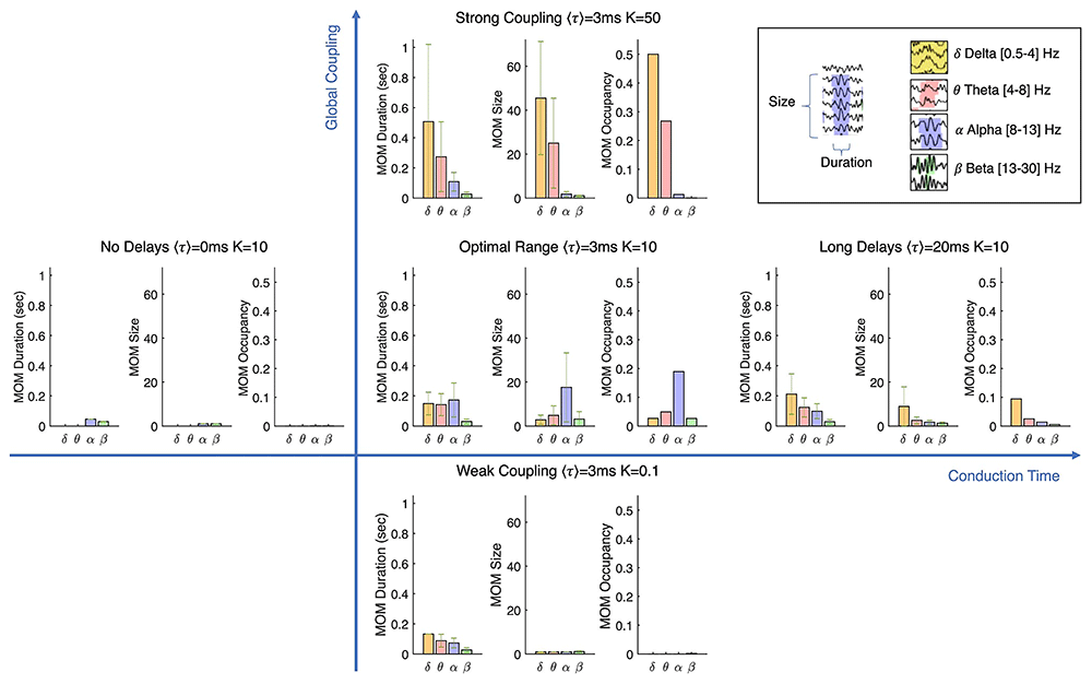
Figure 1. Evolution of global brain dynamics across varying global coupling strength (K) and mean conduction delays (〈τ〉).
Depending on these parameters, the same structural connectome gives rise to distinct spatiotemporal oscillatory patterns across frequency bands. Within a critical range (Optimal Range), alpha-band activity becomes more prominent and spatially distributed, while stronger coupling promotes globally synchronised delta oscillations, resembling states of reduced consciousness. Dynamics are quantified by their temporal persistence, spatial extent, and frequency of occurrence. Adapted from Cabral et al. (2022) (Cabral et al., 2022).
The Kuramoto and Stuart-Landau models, categorised as a coupled oscillator framework, provide a relatively simple yet effective representation of brain dynamics. The Kuramoto model captures phase synchronisation across interacting nodes, whereas the Stuart-Landau model extends this framework by including both amplitude and phase dynamics. These models allow interactions between oscillators governed by the underlying structural network (Csaba & Porod, 2020). In contrast, more biologically grounded models—such as the Wilson-Cowan and Dynamic Mean Field (DMF) models—explicitly simulate the interplay between excitatory and inhibitory neural populations, offering greater physiological realism (Breakspear, 2017; Pathak et al., 2022). Such models are particularly valuable for studying how focal lesions disrupt excitation-inhibition balance across networks, thereby influencing global brain dynamics. Importantly, these whole-brain models, especially coupled oscillator frameworks like the Stuart-Landau model, can give rise to emergent phenomena such as metastable oscillatory modes (MOMs) (Cabral et al., 2022). These MOMs are transient episodes of spatially distributed synchronisation, often within specific frequency bands such as alpha. They manifest as brief bursts of band-limited activity across multiple brain areas, resembling patterns observed in resting-state electrophysiological recordings, and provide a mechanistic link between structural connectivity and large-scale neural dynamics. An example of alpha-band MOMs emerging in a Stuart-Landau simulation is illustrated in Figure 2.
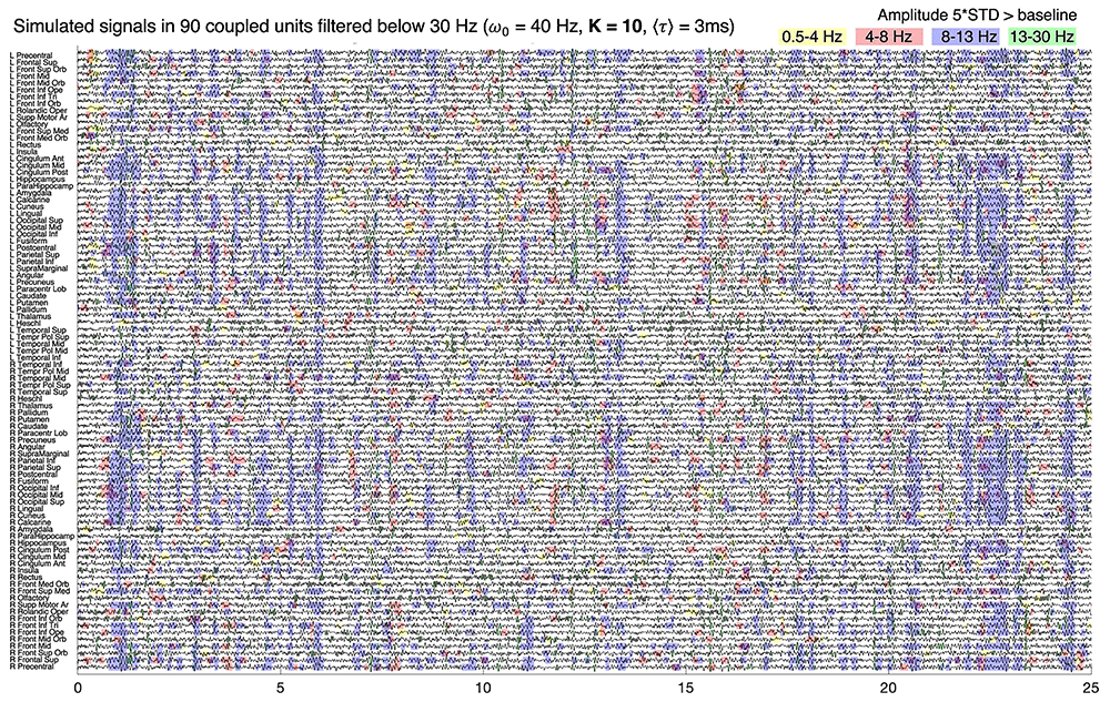
Figure 2. Simulated whole-brain activity illustrating metastable oscillatory modes (MOMs).
Signals from 90 brain areas (parcellated units) are shown over a 25-second segment, filtered below 30 Hz to highlight sub-gamma activity detectable with MEG. Coloured shades mark transient episodes of increased power in delta (yellow), theta (red), alpha (blue), and beta (green) bands, reflecting the emergence of frequency-specific MOMs across space and time. Adapted from Cabral et al. (2022) (Cabral et al., 2022).
Structural connectivity
The selection of an appropriate parcellation scheme is a fundamental consideration in Whole Brain Modeling (WBM), as it defines the nodes (brain regions) and edges (connectivity patterns) within the model. Parcellation schemes can be classified into structural and functional approaches. Structural parcellations are based on anatomical properties, such as gross morphology, gene expression profiles, or chemoarchitecture (Nowinski, 2021). In contrast, functional parcellations are derived from resting-state or task-related functional connectivity, grouping regions based on correlated neural activity patterns (James et al., 2016).
The choice of parcellation has significant implications for both computational efficiency and interpretability. Higher spatial resolution, achieved by increasing the number of nodes, results in a more detailed representation of brain dynamics but also substantially raises computational demands due to the increased number of differential equations that must be solved. Additionally, high-resolution parcellations can introduce ambiguity in source reconstruction, complicating the accurate mapping of neural activity to anatomical structures (Tait et al., 2021).
Therefore, the selection of a parcellation scheme requires a trade-off between spatial granularity and computational feasibility. Several commonly used parcellation schemes in WBMs include the Desikan-Killiany atlas (Desikan et al., 2006), a widely used low-resolution anatomical atlas; the Schaefer parcellation (Schaefer et al., 2018), which combines local functional connectivity gradients with global similarity clustering to define functionally homogeneous regions; and the Glasser atlas (Glasser et al., 2016), a high-resolution functional parcellation designed to capture fine-scale cortical organisation. Figure 3 illustrates the differences between the Desikan-Killiany and Glasser parcellations, highlighting their varying levels of spatial granularity and application in whole-brain modeling.
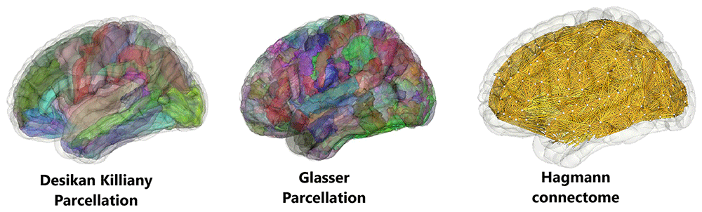
Figure 3. Illustration of different brain parcellation schemes used in Whole Brain Modeling.
The left panel depicts the Desikan-Killiany parcellation (Desikan et al., 2006), a functional atlas comprising 68 cortical regions. The middle panel shows the Glasser parcellation (Glasser et al., 2016), which provides a higher-resolution functional atlas with 360 cortical areas. The right panel represents the Hagmann connectome (Hagmann et al., 2008), a structural connectivity atlas based on white matter tractography. The figures were generated using BrainX3 (Yang et al., 2025).
Theoretical frameworks for whole brain dynamics
This section outlines the mathematical foundations of different Whole Brain Models (WBMs). In Figure 4, an overview is displayed that categorises various models according to their specific dynamics. While each model follows a general framework, variations in equations may arise depending on the specific research application. Key variables and parameters are introduced here to facilitate a deeper understanding of each model’s dynamics.
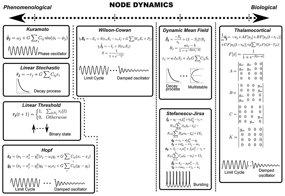
Figure 4. Various whole-brain models are categorised according to their dynamical characteristics - such as oscillatory behaviour, multistability, or bursting.
Adapted from Pathak et al. (2022). For models beyond the scope of this review, readers are referred to Pathak et al. (2022) for further detail (Pathak et al., 2022).
Kuramoto model
The Kuramoto model is a widely used phase oscillator model that represents neural dynamics in terms of phase evolution. It captures the transition between subcritical (asynchronous) and supercritical (synchronous) states, and it can exhibit critical-like dynamics such as avalanches (Villegas et al., 2014). The original formulation (Kuramoto, 1975) assumes all-to-all coupling, where every oscillator is connected to all others. The model is defined by the following equation:
Here θi represents the phase of the ith oscillator (with N oscillator in total), λ is the coupling strength and ωi is the natural frequency.
The adjacency matrix Cij can be incorporated into the coupling term to represent the connectivity between nodes, where each element is 1 if there is a connection from node j to node i, and 0 otherwise. This matrix can also be weighted to reflect the strength of structural coupling between nodes. Lastly, τij represents the delay that occurs between node i and j, typically derived from the tract length and conduction velocity.
Stuart-Landau model
The model is based on the Stuart-Landau equation (eq [Equation 2]) (Cabral et al., 2022), which describes the behaviour of a nonlinear oscillating system near an Andronov-Hopf bifurcation - a critical point where a system transitions from a stable fixed point to a limit cycle, giving rise to sustained oscillations (Andronov et al., 1966). Each node in the model follows the Stuart-Landau equation, incorporating structural connectivity through coupling strength, Cnp, and conduction delays, τnp, between each pair of nodes n and p.
Here Zn(t) is a complex variable that describes the state of nth oscillator at time t. The first term of the equation describes the internal dynamics of the oscillator, where ω = 2πff is the angular frequency, with ff as the fundamental frequency. The parameter a determines the oscillator’s distance from the limit cycle, influencing its stability.
The second term of the equation represents the total input received from other oscillators, Zp. This input is scaled by parameter K, which sets the strength of all network interactions concerning the intrinsic node dynamics. The last term of the equation introduces uncorrelated white noise, adding stochasticity to the model and increasing biological plausibility. For more information on the model, refer to Cabral et al. (2022) (Cabral et al., 2022).
Wilson-Cowan model
The Wilson-Cowan model (Wilson & Cowan, 1972) is a biophysically inspired framework that describes the firing rate dynamics of two recurrently connected neuronal populations: excitatory (rE) and inhibitory (rI) neurons. This model enables the investigation of hypotheses related to neural population interactions, such as disruptions in the excitatory-inhibitory balance, which have been implicated in neurological and psychiatric conditions, including schizophrenia (Beneyto & Lewis, 2011; Glantz & Lewis, 2000). The model is governed by the following equations:
Equations [Equation 3]–[Equation 4] represent the reduced form of the Wilson–Cowan model commonly adopted in whole-brain simulations, where long-range inhibitory-to-inhibitory coupling and self-inhibition (cII, Cnp in the inhibitory population) are omitted for stability and simplicity (Castaldo et al., 2023).
Here, τE and τI denote the characteristic time constants of the excitatory and inhibitory populations, respectively. The parameter cxy defines the coupling strength between populations x and y. Similar to the Stuart-Landau model, K and Cnp represent the global coupling strength and the connectivity between nodes, respectively. The function F follows a sigmoidal form, governing the nonlinear transformation of neural activity. External inputs are given by P(t) for the excitatory population and Q(t) for the inhibitory one; in line with previous work, Q(t) is often set to zero so that only excitatory populations receive constant drive (Castaldo et al., 2023). Finally, ξ(t) denotes additive Gaussian noise, which is implemented as independent and uncorrelated for each node and population to capture stochastic fluctuations.
Dynamic Mean Field model
Like the Wilson-Cowan model, the Dynamic Mean-Field model is a biophysical framework that assumes each node consists of two interacting neural populations: excitatory and inhibitory. However, a distinguishing feature of the Dynamic Mean-Field model is that these populations are interconnected through NMDA and AMPA (excitatory) as well as GABA (inhibitory) synapses. This synaptic specificity allows the model to be used for investigating not only macroscopic brain dynamics but also synaptic-level processes, i.e. the effect of neuromodulation and genetic variants affecting synchrony & coupling.
The model employs a mean-field approximation, representing neural populations through a set of coupled stochastic differential equations. The fundamental principle of the mean-field theory is to simplify a high-dimensional system of randomly interacting elements, such as neurons, by approximating their collective behaviour as independent entities (Idesis et al., 2024b). These entities are then treated as nodes within the model, enabling large-scale simulations of brain activity. The following functions describe the simulated neural dynamics.
Here, represents the firing rate of the excitatory or inhibitory population in node i, while denotes the average excitatory or inhibitory synaptic gating variable within the local area i. The input currents are given by , with Iexternal representing external stimulation used to simulate task-evoked activity. The parameter Cij defines the connectivity strength between nodes i and j, and G denotes the global coupling strength.
In equation [Equation 6], λ takes a value of either 1 or 0, indicating whether long-range feed-forward inhibition is considered. In equations [Equation 7] and [Equation 8], H represents the input-output function, which transforms incoming signals into firing rates. Equations [Equation 9]–[Equation 10] describe the evolution of the synaptic gating variables , with the factor γ in equation [Equation 9] representing the kinetic rate constant of NMDA receptors. This parameter modulates the opening rate of excitatory synapses in response to presynaptic activity, ensuring biologically realistic NMDA time courses (Deco et al., 2014). Stochastic variability is introduced in the model through the noise term σ. For a detailed description of the parameters and constants in these equations, refer to Deco et al. (2014) (Deco et al., 2014).
Modeling stroke
WBMs have been widely employed to simulate stroke and its effects on brain dynamics. For instance, they have been used to demonstrate that lesions can induce widespread network-level alterations in neural activity (Alstott et al., 2009; Honey & Sporns, 2008). The underlying principle is straightforward: a network resembling the brain’s structural connectivity is constructed, often based on empirical data. Simulating a stroke involves the removal or perturbation of one or more nodes, leading to a significant disruption in the system’s dynamical equilibrium. Subsequently, activity-dependent plasticity processes can be modeled to examine recovery trajectories and guide therapeutic interventions. A graphical overview adapted from Váša et al. (2015) of simulating a lesion is shown in Figure 5.
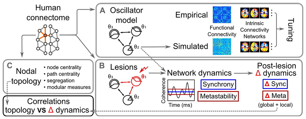
Figure 5. Overview of the modelling framework used to study lesion effects in whole-brain dynamics, adapted from Váša et al. (2015).
(A) The Kuramoto model with time delay is fit to empirical Functional MRI (fMRI) data. (B) The lesion is modeled by removing individual nodes and their connections, allowing the assessment of global measures such as synchrony and metastability. (C) Outlines the graph-theoretical properties used to characterise network nodes, which were tested for their predictive value in explaining changes in network dynamics (Váša et al. 2015).
This section examines recent studies that have utilised stroke simulations to investigate the impact of lesions on brain function and network reorganisation.
Coupled oscillators
Numerous studies have employed WBMs based on coupled oscillators to simulate stroke and investigate its effects on brain dynamics (Chen & Yang, 2025; Gollo et al., 2017; Sangdani & Tavakolpour-Saleh, 2025; Sanz Perl et al., 2021). Coupled oscillator models are particularly well-suited for examining network-level phenomena such as synchrony and metastability, which are critical for understanding functional brain dynamics (Cabral et al., 2017; Deco et al., 2017). As a result, these models have been widely employed to explore changes in functional connectivity following stroke.
Among coupled oscillator models, the Stuart-Landau model has seen increasing popularity in recent years for simulating whole-brain dynamics, particularly due to its ability to capture both amplitude and phase properties of neural oscillations. In contrast, fewer studies have applied the Kuramoto model in stroke modeling, despite recent extensions that enhance its capabilities—such as incorporating noise, delays, or coupling heterogeneity—to better reflect brain dynamics (Clusella et al., 2022). Given this disparity in usage and clinical application, we restricted our review of Stuart-Landau model studies to the past five years to focus on the most recent and relevant developments. In contrast, a broader timeframe was considered for the Kuramoto model to capture foundational and earlier influential studies, given its longer history of use in whole-brain modeling and its foundational role in the field. Relevant literature was identified through systematic searches in Google Scholar, using the keywords “Whole Brain Model,” followed by the specific model name (“Stuart-Landau model” or “Kuramoto model”), and “stroke.” Among the candidates identified, the studies were selected based on their scientific relevance and effective use of the model.
Kuramoto model in stroke research
Váša et al. (2015) applied the Kuramoto model with time delays, calibrated to empirical Functional MRI (fMRI) data, to explore how the structural connectome shapes two key metrics of brain dynamics: synchrony (global coherence) and metastability (dynamic flexibility). They showed that removing nodes with high eigenvector centrality or those acting as inter-modular hubs, regions that link otherwise segregated network modules, significantly decreased global synchrony and increased global metastability. At the regional level, five nodal graph measures predicted how local lesioning impacted synchrony and metastability, underscoring the importance of local network topology (Váša et al., 2015). Alderson et al. (2015) also offered a computational perspective using a connectome-based model of traumatic brain injury to reveal that reductions in metastability (observed empirically via resting-state fMRI) correlated with decreased cognitive flexibility and slower information processing, mediated by structural damage to frontal networks (Alderson et al., 2018). These studies collectively indicate that focal lesions that affect the central nodes not only alter large-scale neural coherence but also impair the brain’s ability to transition between functional states, an effect exhibited behaviourally as reduced cognitive flexibility.
Building on these insights, Alderson et al. (2018) (Alderson et al., 2018) used the Kuramoto model to investigate how cognitive processes emerge from large-scale neural dynamics and how these processes are disrupted by neurological impairments. The study focused on patients with mild cognitive impairment (MCI)—individuals who exhibit cognitive deficits but do not yet meet the diagnostic criteria for dementia. By comparing simulated brain activity with empirical data from MCI and Alzheimer’s disease (AD) patients, the study demonstrated a strong association between metastability, cognitive performance, and structural integrity of the connectome. Notably, introducing macroscopic structural damage into the model produced reductions in metastability comparable to those observed in empirical AD data, further reinforcing the link between structural disconnection and functional impairment. Although the clinical condition studied was MCI/AD, the findings provide a mechanistic framework that directly translates to stroke: in both cases, focal structural damage alters global brain dynamics by reducing metastability, which is associated with cognitive dysfunction. This supports the view that stroke-related lesions impair not only local function but also disrupt global network dynamics. Models like the Kuramoto framework thus help link structural damage to behavioural outcomes across brain disorders, including stroke.
Stuart-Landau model in stroke research
A more recent study by Idesis et al. (2022) (Idesis et al., 2022) utilised the Stuart-Landau model to develop a generative, subject-specific framework for exploring post-stroke brain dynamics. The study emphasised the critical role of white matter disconnection, the interruption of structural pathways due to stroke-induced lesions, which disrupts interregional communication and significantly alters functional reorganisation. By incorporating individual-specific structural disconnection masks derived from lesion maps, the authors were able to simulate patient-specific changes in resting-state dynamics without introducing additional parameters. This approach successfully reproduced empirical alterations in functional connectivity and significantly improved the model’s ability to predict stroke severity, highlighting the value of structural connectivity data in computational stroke modeling. Complementing these modeling insights, Tuladhar et al. (2013) (Tuladhar et al., 2013) reported reduced functional connectivity within the default mode network (DMN) in stroke patients, particularly in the posterior cingulate cortex, medial prefrontal cortex, and medial temporal lobe. These reductions were associated with memory impairments, supporting the idea that focal lesions can induce widespread disruptions in large-scale brain networks. Figure 6 illustrates the DMN, a set of regions typically active during rest.
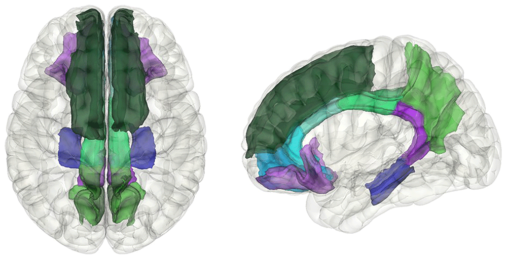
Figure 6. Visualisation of the default mode network (DMN) using the Desikan-Killiany parcellation.
The DMN is a network of interconnected brain regions that are particularly active during resting states. It includes the isthmus cingulate, medial orbitofrontal, posterior cingulate, precuneus, rostral anterior cingulate, lateral orbitofrontal, and parahippocampal regions. Post-stroke alterations in the functional connectivity of the DMN have been associated with cognitive impairments, highlighting its significance in post-stroke recovery and rehabilitation.
A study by Martínez-Molina et al. (2024) also applied the Stuart-Landau model to simulate post–traumatic brain injury (TBI) dynamics and explore their potential relevance to stroke (Martínez-Molina et al., 2024). They introduced the concept of turbulent brain dynamics, where the healthy brain operates near a critical state representing a delicate balance between stability and flexibility, characterised by amplitude turbulence (fluctuations in local synchrony), information cascade flow (the hierarchical transfer of information across scales), and information transfer (the exchange of information between regions) (Deco & Kringelbach, 2020). Their framework showed that after structural disconnection (simulating lesions via disconnection masks), the brain shifts away from this healthy turbulent regime, with reductions in global dynamics and susceptibility to external perturbations.
They suggested that turbulence-based biomarkers (e.g., metrics that capture global and regional disruptions in turbulent activity) can aid in assessing both diffuse and focal damage in TBI—and, by extension, could be applied to stroke, where similar principles of structural disconnection and altered dynamical states apply (Martínez-Molina et al., 2024).
To illustrate structural disconnection effects, Martínez-Molina et al., used in-silico perturbations to identify regions whose connectivity loss most strongly disrupted turbulent dynamics, potentially indicating optimal stimulation targets (for techniques such as deep brain stimulation (DBS) or transcranial Temporal Interference Stimulation (tTIS)), localised based on where perturbations had the greatest dynamical impact.
Further advancing the clinical applications of WBMs, Idesis et al. (2024a) (Idesis et al., 2024a) developed a data-driven generative model trained on healthy subjects, capable of predicting post-stroke functional connectivity (FC) using patient-specific structural disconnection connectivity (SDC) data. The SDC matrix summarised the percentage of spared streamlines between each pair of regions after lesioning, serving as a proxy for connection strength. Notably, even with fixed model parameters, simulated dynamics reproduced key FC alterations observed in stroke patients: reduced intra-hemispheric homotopic (connections between mirror cortical regions in both hemispheres) FC, disrupted inter-hemispheric connectivity, and altered network modularity. These results indicate that lesion-informed structural disconnection is sufficient to predict major functional reorganisation patterns, reinforcing their potential for clinical translation.
However, while these whole-brain models offer a powerful framework for linking structural disconnection to large-scale functional outcomes, they rely on coarse-grained representations of brain structure and dynamics. They do not account for the underlying microcircuit-level neurobiology, such as neural excitability, synaptic interactions, or neurotransmitter dynamics. These limitations may constrain their ability to capture the full complexity of post-stroke reorganisation. To address this gap, biophysically informed models, such as Wilson-Cowan and dynamic mean-field approaches, provide a complementary avenue. By explicitly modeling excitation–inhibition balance and local circuit dynamics, these models allow for a more mechanistic understanding of how stroke disrupts brain function and recovery.
Biophysical modeling
Biophysical models are frequently employed to investigate the effects of diseases and pharmacological interventions on brain function (Herzog et al., 2023; Jobst et al., 2017; Patow et al., 2023). However, they are also increasingly used for predictive modeling, offering insight into how brain dynamics may evolve over time in relation to disease progression or treatment response (Good et al., 2022; Jung et al., 2023). To assess the role of biophysical WBMs in stroke research, a systematic literature search was conducted using Google Scholar with the keywords “Whole Brain Model,” followed by “Wilson-Cowan model” or “Dynamic Mean Field Model,” and “stroke.” The retrieved studies were subsequently evaluated based on their methodological rigour and relevance, with a focus on those used in stroke research.
A recent study by Schmitt et al. (2024) (Schmitt, 2024) implemented the Wilson-Cowan WBM in rat stroke models to examine stimulus-response alterations in a structurally perturbed connectome. Simulations showed that subcortical lesions, despite being physically smaller, caused more severe functional disruptions than equivalent cortical lesions. This was evidenced by increased co-activation between lesioned regions and motor/spatial learning networks and marked hippocampal hypoactivation, which correlated with behavioural deficits in maze-based spatial memory tasks. Functional impairment was quantified using co-activation strength, signal propagation changes, and network graph metrics, linking structural damage to dynamic functional consequences. Moreover, the study identified key hub regions whose integrity was critical for recovery, suggesting targeted network-based interventions could support post-stroke rehabilitation outcomes.
Another study investigated the role of excitatory-inhibitory homeostasis in the recovery of functional connectivity (FC) after stroke, using the Wilson-Cowan model (Páscoa dos Santos et al., 2023). Homeostasis is a fundamental mechanism that stabilises neural activity and connectivity patterns, for instance by convergence to a specific set point for the firing rate (Turrigiano, 2011). They found that E–I homeostasis drives partial restoration of static functional connectivity (FC) properties, including network modularity and small-worldness, once plasticity has stabilised (“after converged plasticity”). However, dynamic FC patterns, such as temporal fluctuations in synchrony, did not fully return to pre-lesion levels, indicating that the plasticity of inhibition alone is insufficient to fully recover all functional dynamics. Moreover, the model revealed that E–I homeostasis led to widespread increases in excitability, with region-specific patterns that varied by lesion location. Some excitability changes aligned with known biomarkers of post-stroke side-effects—such as heightened excitability in hippocampal regions linked to increased seizure risk—while other regions showed either no change or reduced excitability (Páscoa dos Santos et al., 2023).
Supporting this notion, an earlier study by Vattikonda et al. (2016) (Vattikonda et al., 2016) used the DMF model to examine global functional recovery following stroke. The findings indicated that local homeostatic balance contributes to functional restoration by re-establishing excitation-inhibition balance in affected regions. While this mechanism supports the stability and complexity of resting-state dynamics, Páscoa dos Santos et al. (2023) (Páscoa dos Santos et al., 2023) extended this work by analysing changes in excitability across the brain, uncovering more detailed region-specific increases, particularly in hippocampal areas, linked to symptom emergence such as seizures and depression (Fu et al., 2021; Towfighi et al., 2017).
The Dynamic Mean Field model has also been utilised to study the interplay between local structural damage and global functional alterations following stroke (Idesis et al., 2024b). This study revealed the emergence of sleep-like slow waves in the awake brain, particularly in regions adjacent to the lesion. These waves propagated across the brain via long-range structural connections, affecting distant areas. Using (f)MRI-derived FC and structural disconnection connectivity (SDC) masks, the WBM successfully replicated stroke-induced FC patterns. Crucially, a comparison between models with and without sleep-like slow waves demonstrated that incorporating these waves significantly improved model performance, quantified using Generative Effective Connectivity, a model-based measure that evaluates how well the simulated causal interactions reproduce empirical functional connectivity (Kringelbach et al., 2023), providing a causal explanation for the role of sleep-like slow waves in shaping post-stroke brain dynamics.
A related study by Adhikari et al. (2015) (Adhikari et al., 2015) applied the DMF model to examine the impact of early-life stroke on cognitive function. The study sought to explain why certain brain regions maintain near-normal cognitive function despite prenatal or perinatal lesions. Their findings indicated that early brain injury does not have a sustained impact on the functional connectivity of affected areas. Instead, resting-state FC in stroke-affected children closely matched those of typically developing children. This finding suggests that developmental neuroplasticity enables the brain to reorganise its functional architecture, allowing alternative pathways or network configurations to emerge during critical periods of development. As a result, the brain may converge on a functionally viable state, even when structural integrity is compromised early in life.
Another study used the DMF model to explore how stroke-induced lesions affect the temporal balance between integration and segregation, that is, the brain’s dynamic ability to alternate between globally connected and locally modular states during rest (Wei et al., 2022). This was measured using the Integrated State Occurrence Rate, capturing how frequently each state occurs over time. Virtual lesions in nodes across the visual, somatomotor, limbic, and default mode networks significantly disrupted this balance, making the brain network less flexible. Importantly, the effect of these disruptions could be predicted from anatomical heterogeneity, meaning that regions with higher graph-theoretical measures, such as degree, centrality and cortical hierarchy derived from T1w/T2w maps, had a disproportionately larger impact on global dynamics.
Discussion
The studies reviewed here demonstrate how WBMs are applied practically and highlight their potential to advance our understanding of brain dynamics in stroke patients. Each model reviewed offers significant strengths alongside notable limitations. Given the inherent complexity of WBMs, several challenges remain. This section highlights key pitfalls and discusses future directions, especially focusing on how patient-specific modeling, digital twins, and integration into clinical infrastructures may overcome current limitations.
Pitfalls and future directions
WBMs rely heavily on connectomes to define node interactions. However, existing imaging techniques, such as diffusion-weighted imaging, still face limitations, particularly in accurately tracking interhemispheric white-matter tracts within complex anatomical regions like the centrum semiovale (Hagmann et al., 2008). This limitation often leads to an underrepresentation of homotopic connectivity, resulting in less accurate models. Importantly, homotopic connectivity is a critical biomarker of stroke recovery (Corbetta et al., 2018), making its accurate determination crucial. Advances in white-matter tractography, combined with patient-specific imaging, could significantly enhance the precision and clinical relevance of WBMs.
Another challenge involves adequately incorporating subcortical areas, which frequently suffer strokes and profoundly affect cortical dynamics (Corbetta et al., 2015). Recent approaches embedding subcortical networks into cortical mean-field models offer promising solutions (Meier et al., 2022). These frameworks enable simulations based on empirical patient data, potentially supporting personalised predictions about the impact of lesions and aiding in the creation of digital twin models of the brain. However, methods such as those used by Idesis et al. (2022) (Idesis et al., 2022), which select specific structural lesions as drivers of empirical dynamics, must be carefully evaluated, as they may overlook alternative explanations, such as patient-specific variability in neural excitability, compensatory reorganisation, or differences in model parameters, all of which could equally account for the empirical data.
Selecting accurate and physiologically meaningful model parameters remains a central challenge, particularly when lesions alter the brain’s structural connectivity. As reported by Idesis et al. (2024b), stroke-induced slow-wave propagation in neighbouring regions may shift critical model parameters such as bifurcation thresholds (Idesis et al., 2024b), potentially undermining the robustness of whole-brain models (WBMs). Parameters must therefore retain clear physiological interpretations, particularly when structural changes (e.g., lesions) constrain conduction paths. For instance, conduction delays, computed from tract lengths and conduction velocities, have been empirically linked to dominant oscillatory frequencies, such as the alpha band. Recent lifespan EEG modeling shows that estimated global conduction delays are negatively correlated with peak alpha frequencies (i.e., longer delays correspond to lower dominant alpha frequency) (Reyes et al., 2025). Aligning your model’s conduction delays with such measurable neural properties not only grounds the simulation in anatomical realism but also enhances interpretability and clinical relevance. If model parameters lack such physiological anchoring, the translational and patient-specific value of WBMs becomes questionable. Addressing this issue through biologically informed parameterisation will strengthen the predictive power of WBMs and clinical utility.
Furthermore, it is essential to recognise the heterogeneity of stroke types in clinical studies. Ischaemic strokes, resulting from arterial blockage, typically cause focal disruptions in connectivity, leading to localised alterations in node activity and subsequent compensatory reorganisation within nearby brain networks. Conversely, haemorrhagic strokes, caused by bleeding within the brain tissue, often produce more diffuse disruptions due to increased intracranial pressure, inflammation, and damage to adjacent neural structures, resulting in widespread changes in network dynamics and connectivity patterns. Given that ischaemic and haemorrhagic strokes differ in their network-level impacts, comprehensive datasets capturing these distinct pathological processes are thus crucial, especially when developing predictive WBMs intended for clinical prognosis. Similarly, age-associated changes in structural connectivity, including cortical thinning and white matter degradation, are linked to changes in functional connectivity and declines in executive function (Raz et al., 2005; Schulz et al., 2022). These changes can influence stroke outcomes, underscoring the importance of incorporating age-related brain changes, such as in inter-regional coordination and network communication latency, into stroke modeling to enhance predictive accuracy and clinical relevance (Chakraborty et al., 2025). Given that stroke incidence rises markedly with age, overlooking these changes could lead to biased or inaccurate clinical predictions and misinterpretations of neurophysiological data in older populations.
Finally, the choice of empirical data used for model validation—fMRI versus MEG/EEG—can significantly influence model performance, especially in clinically predictive scenarios. Functional MRI (fMRI) has limited temporal resolution and captures neural activity indirectly through the nonlinear BOLD response (Robinson et al., 2006), whereas MEG/EEG provide superior temporal resolution at the cost of lower spatial accuracy. Importantly, model outputs must be appropriately transformed to match the resolution and dynamics of the empirical data, for instance, by applying a low-pass filter or a haemodynamic response model (Buxton et al., 1998) to compare simulated signals with fMRI data. Selecting appropriate modalities or multimodal integration strategies will be critical for enhancing WBMs’ predictive power and clinical relevance.
Whole brain modeling in clinical practice
The WBMs discussed in this review have already demonstrated substantial utility in clinical neuroscience, particularly for simulating brain dynamics, predicting recovery trajectories, and identifying biomarkers. Their practical utility can be assessed through their suitability for patient-specific modeling.
The Kuramoto model, a simple coupled oscillator approach, demands minimal computational resources while providing theoretical insights into large-scale synchronisation phenomena and predicting global measures of brain dynamics, such as metastability and coherence (Bhowmik & Shanahan, 2012). However, its reliance on sinusoidal coupling and omission of anatomical factors such as tract length (Cabral et al., 2014) may restrict detailed patient-specific simulations. Nevertheless, it remains valuable for proof-of-concept studies and early-stage predictive clinical modeling and also allows for analytical approaches and theoretical insights.
The Stuart-Landau model, operating in the complex domain, provides enhanced flexibility by incorporating axonal delays and variable oscillatory damping, facilitating more anatomically accurate, patient-specific simulations. Although slightly more computationally demanding, it remains straightforward and efficient for clinical exploration. Its primary drawback is the limited biological plausibility, suggesting that its findings should be validated with more biologically grounded models for robust clinical application.
The Wilson-Cowan model, as a representative biophysical model, explicitly includes excitatory and inhibitory interactions, thus more accurately mimicking physiological brain dynamics. Despite greater complexity, it remains computationally manageable and can generate clinically relevant insights into the excitatory-inhibitory balance crucial for understanding stroke recovery dynamics. Recent advances have introduced systematic strategies for deriving homeostatic parameter settings, including synaptic gains, inhibitory strength, and neuronal excitability, that ensure the model operates within desired dynamical regimes (e.g., near a Hopf bifurcation) while maintaining stable firing rates across input conditions (Páscoa dos Santos & Verschure, 2025). By incorporating multiple modes of excitation–inhibition homeostasis, such approaches enhance the model’s capacity for reliable personalisation, reinforcing its utility in clinical prediction and intervention (Froudist-Walsh et al., 2021).
Finally, the Dynamic Mean Field model, the most complex model reviewed here, offers unparalleled detail by capturing multiple neuronal variables and cell-type-specific interactions. Its complexity and specificity make it ideally suited for clinical applications that require precise, personalised simulation, such as pharmacological intervention modeling (Burt et al., 2021) or detailed rehabilitation planning following stroke. Despite its computational demands, its potential for patient-specific clinical prediction through digital twin technology is substantial.
The integration of WBMs into digital neuroscience platforms such as The Virtual Brain Cloud and eBRAINS Health (Schirner et al., 2022) presents a transformative opportunity for clinical translation. These infrastructures enable the construction of personalised “digital twins” by combining patient-specific neuroimaging with mechanistic whole-brain simulations. Clinicians can use these models to simulate recovery trajectories, evaluate intervention strategies, and predict functional outcomes—all based on the individual’s structural and functional brain architecture. In silico simulations offer a virtual testbed to assess the effects of neuromodulation (e.g., transcranial magnetic stimulation [TMS] or transcranial alternating current stimulation [tACS]), pharmacological therapies, or lesion impact on network-level dynamics before applying interventions in vivo. Moreover, model-derived biomarkers—such as metastability, synchrony, or excitation–inhibition balance—can aid in stratifying patients, monitoring recovery, and identifying targets for personalised rehabilitation. By linking computational models to clinical workflows through user-friendly interfaces and secure data exchange pipelines, these platforms have the potential to transform WBMs from theoretical tools into practical clinical decision-support systems. Ultimately, WBMs offer a unique bridge between systems neuroscience and precision medicine, empowering clinicians to understand and treat the complex dynamics of stroke with unprecedented mechanistic and patient-specific insight.
Conclusion
This review explored several types of WBMs—Kuramoto, Stuart-Landau, Wilson-Cowan, and Dynamic Mean Field—and highlighted their utility, challenges, and future potential in stroke research. By assessing the strengths and limitations of each model, the review has demonstrated WBMs’ considerable promise for bridging theoretical neuroscience and clinical application.
Simple models like the Kuramoto and Stuart-Landau remain valuable for preliminary clinical predictions with low-cost simulations. Though the challenge remains of the effective parameterisation, including selecting physiologically meaningful parameters that accurately reflect stroke-induced changes in brain dynamics. Meanwhile, biophysical models such as Wilson-Cowan and Dynamic Mean Field, although more complex, offer increased biological realism, essential for accurate patient-specific modeling. Integrating these models with patient-derived data enables personalised simulation frameworks, aligning with the concept of digital twins. These digital twins could dramatically advance personalised stroke rehabilitation, improve prognosis accuracy, and offer deeper insights into post-stroke neuroplasticity.
Recent advances in artificial intelligence (AI) are likely to substantially influence future developments in WBMs. AI-driven algorithms could assist in more accurate parameter fitting, improved structural connectivity reconstruction, and the identification of predictive biomarkers from large-scale empirical datasets.
By addressing current limitations—such as structural connectivity accuracy, parameter determination, stroke heterogeneity, and empirical data selection—WBMs can more effectively inform clinical decision-making and treatment planning. Ultimately, Whole Brain Modeling represents a critical tool for transforming stroke research and clinical practice, moving closer towards individualised medicine and improved patient outcomes globally.
Ethics and consent
Ethical approval and consent were not required.







