Introduction
Animal forests as a microbial habitat
In November 2024, several leading microbiologists called for urgent and decisive emergency actions to safeguard the planet for future generations as the climate crisis intensified (Peixoto et al., 2024). This call to action centred on the power microbes possess as biogeochemical engines, playing a key role in regulating biogeochemical cycles, as well as serving as a source of genetic information with potential effects on the fitness of their hosts, ranging from humans to corals. Indeed, microbes play a key role in both detrital (production based on recycled organic matter) and microbial food webs (Shakya & Allgeier, 2023). Through the remineralization and production of organic matter, they feed higher trophic levels, and the diversity of their communities drives their key metabolic pathways and these processes. The symbiotic host-microbiome relationship has been proven to be essential for the health of animal forests, contributing to their persistence in the face of climate changes, which threatens Marine Animal Forests (MAFs) with extinction at a rate that could be witnessed in a lifetime (Knowlton, 2021; Lee et al., 2022; Peixoto & Voolstra, 2023).
In 1994, the molecular biologist Richard Jefferson introduced the term hologenome at Cold Spring Harbor Laboratory, proposing that the material once dismissed as “contamination” in DNA samples was actually an integral component of an organism’s genome. Building on this insight, Eugene Rosenberg and Ilana Zilber Rosenberg formalised the Hologenome Theory of Evolution in 2008, after observing Vibrio shiloi-mediated bleaching of the coral Oculina patagonica, framing the host and its entire microbiota as the holobiont, a single evolutionary unit. This paradigm change results from the partnership between the host and its microbiota, which has brought organisms’ symbiosis with microorganisms into the spotlight, highlighting the roles microbes play in host evolution and considering them (host + microbiota) as evolutionary units (Bordenstein & Theis, 2015; Zilber-Rosenberg & Rosenberg, 2008). This concept is essential for multiple disciplines, including developmental and evolutionary genetics, as well as ecology, and is of utmost interest in the face of global environmental changes.
MAFs are inhabited by different types of aquatic holobionts, whose symbiotic microorganisms play a pivotal role in their ecology as they face diverse biotic and abiotic stressors. In this chapter, these symbiotic microorganisms will be referred to as microbiomes of marine animal forests. Herein, the term microbiome refers to the characteristic microbial community occupying a well-defined habitat, with distinct physico-chemical properties, encompassing the microorganisms and their “theatre of activity”, which results in the formation of specific ecological niches (definition originally proposed by Whipps et al., 1988, and re-visited by Berg et al., 2020). These micro-ecosystems are dynamic, interactive, and prone to change in time and space, being integrated into macro-ecosystems, including eukaryotic hosts, and are crucial for host functioning and health.
MAFs’ holobionts engage in a multitude of symbioses, the relationship between zooxanthellate corals and Symbiodiniaceae being the most notable. However, there are many more microorganisms associated with forest-forming marine animals, such as Bacteria, Archaea, Fungi, viruses, protists, and other microeukaryotes that together form their microbiomes. The resulting set of relationships is quite complex, as some symbioses recapitulate the phylogeny of their host with evidence of phylosymbiosis. Others are subjected to change according to environmental status, age, season, or stress. Moreover, these relationships are very versatile among the different types of holobionts, and not all holobionts rely on their microbiome members equally, nor do the microbiome members contribute equally to their host (Voolstra & Ziegler, 2020).
Using omics methods for studying MAF microbiomes
To date, most studies on MAF’s microbiomes have employed metabarcoding techniques to focus on the bacterial assemblage structure and composition. Assemblages have been proven to be diverse and flexible, closely related to host health and disease status (Zaneveld et al., 2017). Recent efforts have also focused on increasing the number of microbial isolates for mechanistic and manipulation experiments, as well as biotechnological approaches, and on metagenomics to elucidate their metabolic potential (Botté et al., 2019; Voolstra et al., 2024). The study of these elements has been proven essential for a better understanding of ongoing and future adaptation, conservation, and restoration of MAFs.
Many tools have been used to study these microbiomes, expanding the knowledge of the taxonomy of microbial assemblages, their environmental interactions, plasticity, and physiology, as well as how the host acquires them and how they respond to environmental impacts. Most of this knowledge is derived from studies using molecular tools, as the number of unculturable microbes is orders of magnitude higher than that of culturable microbes. Nevertheless, despite the great plate count anomaly (Staley & Konopka, 1985) highlighted the shortcomings of cultivation-based assessments (culturomics) of microbial communities, exploring the taxonomy and functionality of cultural microbiomes is essential to fully characterize and elucidate the roles these microorganisms play in shaping host health. Such characterization can provide valuable baseline information for biotechnological and experimental applications, such as the use of beneficial microorganisms as probiotics (Ushijima et al., 2023; Zhang et al., 2021). Nevertheless, the cultivation of marine microbes remains a challenge, as these microbes have specific conditions and nutritional requirements that are difficult to reproduce, including members of not only Bacteria but also Archaea, Fungi, protists, unicellular phytoplankton, and viruses (Stulberg et al., 2016). Thus, molecular methods have become a crucial tool to dive into the structure and function of the microbiomes within MAFs, also providing insights into how to improve cultivation (Schultz et al., 2022).
In the present chapter, we concentrate on methodologies for characterizing MAFs’ microbiomes, because emerging evidence links microbial composition and activity to animal health and stress resilience. Such knowledge is strategic in a rapidly changing biosphere, underpinning nature-based solutions and improving our capacity to predict ecosystem responses from local to global scales.
Herein, we will focus on those DNA-based molecular tools and how they are helpful to explore taxonomic, phylogenetic, and functional characteristics of MAFs' microbiomes and provide suggestions to enhance comparisons between different environmental matrices and animal hosts across time, space, and environmental conditions. These tools are designed to improve the study of some emblematic holobionts, such as corals and sponges. (Figure 1)
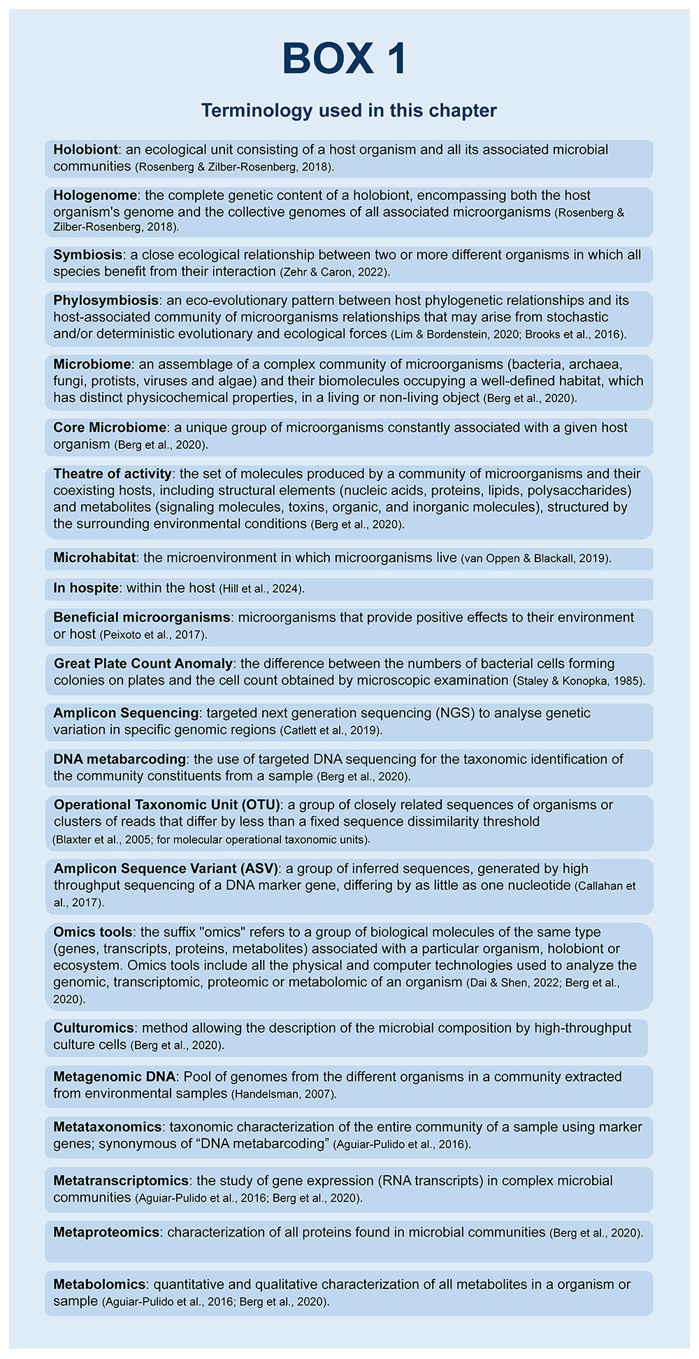
Figure 1. Terminology used in the chapter.
Sampling, preservation, and processing
The microbial community associated with a marine animal host is best conceived as a context-dependent consortium composed of resident (core) and transient (episodic) taxa that fluctuate across space, time, and host life stages. Thus, the host-associated microbiome can be a small subset of the microbial species of the surrounding environment or can be transmitted vertically from parent to offspring. Additionally, in this association, a complex set of potential interactions occurs between the host and each microbe (directly and indirectly), as well as amongst members of the microbial community (Carrier & Reitzel, 2017). The resulting multipartite network is therefore intrinsically complex and requires an explicit, carefully designed methodological framework to disentangle causes from consequences.
For the success of any ecological study, an appropriate sampling design is crucial, considering scientific questions, the type and number of samples (e.g., soft vs hard tissues, number of biological replicates), logistics, upstream analysis, and available budget. Furthermore, adequate documentation of critical aspects of specimen collection, preservation, processing, and storage has the potential to foster collaboration and standardization (Figure 2).
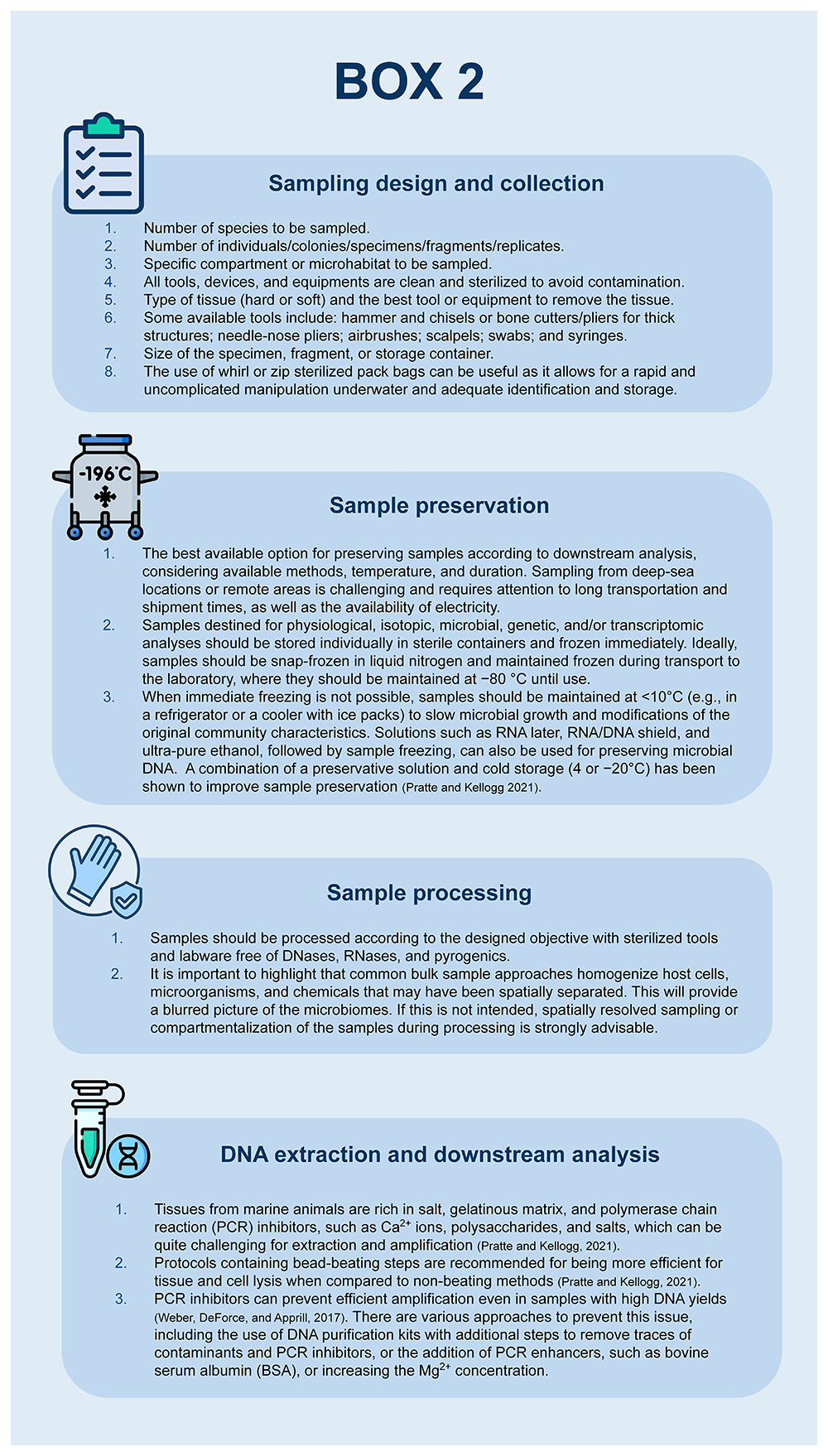
Figure 2. Main methodological aspects to consider when studying the microbiome associated with marine animal forests using molecular techniques.
Scientific questions and objectives
Recent technological advances have enabled an explosion of microbial ecology research, increasing our ability to study these systems and better understand how microorganisms interact with one another, other organisms, and their environment. However, all these research venues need to be based on research questions that guide the methodological delineation designed to answer them. Microbiomes associated with marine forest-forming animals are complex and dynamic communities that fluctuate in response to diverse factors, including host genotype, developmental stage, diet, and physiological and health changes, among others (Antwis et al., 2017). To understand such interactions and their consequences, a proper study design will evaluate the available techniques and select the most appropriate one to be employed, depending on the study's scientific questions and objectives.
For example, suppose the research question is “What factors influence the coral microbiome and lead to changes in coral health?”, the study design should compare healthy and unhealthy corals, taking into consideration the influences of host genetics, age, and size, as well as the influence of environmental conditions, such as water temperature and nutrients availability. Based on this, the sampling will be centred on the coral tissues alongside measurements of environmental parameters and metadata collection. In the case that the study objective is to characterize the colonization process of a host by symbiotic microbes, a proper approach is to collect samples of both host tissues and environmental matrices during a significative time (see, for instance, Tignat-Perrier et al., 2025). Sponge sampling, for example, should consider both the outer layers and the mesohyl, and by comparing these compartments, one can shed light on the sponge-microbiome selection process (Selvin et al., 2010). These general delineations can involve multiple analyses to gain a deeper understanding of the molecular mechanisms underlying them.
Logistics and budget
Logistics and financial issues imposed several challenges on the study of microbiomes associated with marine animal forests. For sampling, researchers must first consider the available technical and monetary resources to reach the planned sampling sites. Oceanic sites are only accessible by marine vessels or aircraft, often requiring diving or the use of remotely operated devices to collect samples. The deeper the location, the more difficult it is to reach; thus, more financial resources and technical expertise are involved. Sample preservation adds a second layer of complexity, especially at remote or deep-sea locations, which involves freezing, the use of sample preservatives, or even in situ extraction and analysis (e.g., Oxford Nanopore MinION in situ sequencing). The time between sampling and processing or long-term storage has a profound effect on the research outcomes regarding the associated microbiomes. Moreover, the capacity to maintain aseptic conditions in the field to avoid sampling contamination is a key issue to be taken into consideration as well (Vandeputte et al., 2017).
Sampling delineation
The outcomes of a research endeavour strongly depend on the sampling procedures, which are linked to the study's questions and objectives. Several approaches can be followed, which is critical for the research outcomes. The sampling techniques and approaches will depend on the site location and depth, as well as the target species, tissue characteristics, and the availability of equipment, with varying degrees of invasiveness (i.e., the potential harm to the animal). Animals that constitute marine forests, such as corals (soft or hard), sponges, and zoantharians, among others, exhibit a range of body sizes and tissues of varying hardness and composition, often covered by mucus in different amounts. Regarding the sample’s size, i.e., tissue aliquots or the whole animal, the sampling strategy must consider whether the investigation is tissue or compartment-specific and the potential adverse effects on the target holobiont (i.e., the destructive potential). Thus, before sampling, one should consider whether removing large animal portions is necessary. If such an approach alters host physiology and causes extensive damage that may lead to mortality, it should be avoided. Sampling methods that minimize negative consequences while maintaining accuracy should be prioritized (Silva et al., 2023). Sampling strategies should also consider the number of samples, replicates, and the spatial or temporal distribution of the samples, with attention to the distribution of the target species, which can be distributed heterogeneously. Replicates are crucial to balance issues related to undersampling, i.e., samples that represent only a small portion of the ecosystem (Zinger et al., 2012).
Another key aspect regards the holobiont anatomy, as different microbial symbionts might inhabit different tissues or compartments of an animal host. This is important to consider, depending on the research question and hypothesis, as inadequate or bulk samples (i.e., homogenized samples composed of host cells, microorganisms, and chemicals that may have been spatially separated, blurring the whole picture) could lead to error in microbiome characterization. This tissue compartmentalization creates distinct physical-chemical microhabitats or compartments that can differ in pH, light, dissolved oxygen, and chemical composition even in “structurally simple” reef-building corals. For instance, some corals displayed the surface mucus layer, ectoderm, mesoglea, endoderm, and gastrovascular cavity as distinct microhabitats, which harbour different microbes. This type of spatially resolved sampling revealed that the notorious Symbiodiniaceae inhabit the endoderm layer (Hughes et al., 2022). This also allowed the discovery of in hospite intracellular association of bacteria (e.g., Hyphomicrobium, Methylobacterium, and Sphingomonas) with Symbiodiniaceae, as well as compacted tissue-aggregates. Thus, spatially resolved sampling can be helpful for the accurate characterization of host microbiomes, providing an image free from discrepancies and variabilities that can come from using bulk samples, and a better understanding of the roles played by those animal microbiomes.
Besides tissue samples, water and sediment samples may also be collected if the investigation aims to study the regional pool of microbial species from which local host-associated communities can be assembled. It is important to consider the spatial distribution and persistence time of microbes and their genetic material in each environmental sample type. In the water column, for example, vertical stratification due to thermoclines and haloclines affects the mixing of microbes and DNA; therefore, it is essential to collect water samples exactly from the target depth, within and/or near a MAF (Jeunen et al., 2020). On the other hand, sediments exhibit less temporal variation, with genetic material persisting for prolonged periods, allowing for the recovery of even ancient sedimentary environmental DNA (sedaDNA) (Corinaldesi et al., 2018).
For the sampling strategy and methodological aspects to consider when studying the microbiome of MAFs using molecular techniques, four main aspects should be considered. They are summarized in Figure 2.
Sampling
The methods and equipment to be used in the field will depend directly on the type of sample to be collected. Remotely Operated underwater Vehicles (ROVs) and/or scuba diving can both be used for underwater sampling, depending on the location, sampling depth, and available resources. For water or sediment samples, several tools can be used, from Van Veen dredges and Niskin bottles to shovels and syringes (De Brauwer et al., 2023). Additionally, semi-autonomous submersible filtering devices can be used. For instance, one of these tools has been developed by the CNRS and the SA MAREPOLIS (Figure 3), and recently tested and applied for MAFs water sampling below 20 m depth and up to 100 m deep, along with the setting up of a combined protocol allowing to study both microbial and eukaryotic diversity (teleost fish and elasmobranch in the context of the related BNP Paribas Foundation funded research project DEEPLIFE, https://underthepole.org). For each sampling replicate, 20 L of seawater is filtered using a device equipped with a 300 µm strainer (to eliminate larger particles and prevent clogging) and two hydrophilic polycarbonate membranes to collect eDNA and microbial cells (142mm diameter, pore sizes: 1.2 µm and 0.2 µm, Merck Millipore). The membranes are installed in a two-layer cartridge and are supported by a Teflon support and drainage grid (Merck Millipore). Once the 20L water filtration is complete, the device automatically turns off and can be returned to the surface. The filters are then removed, placed in individual 5 mL tubes filled with DNA/RNA preservative, and frozen at -20 °C or -80 °C until further analysis. To prevent contamination between samples or replicates, the device and cartridge undergo a series of alternating washes with bleach, sterile water, and 70% ethanol. Sampling blanks are made with pre-filtered 0.22 μm water before each sampling activity and used as control samples.
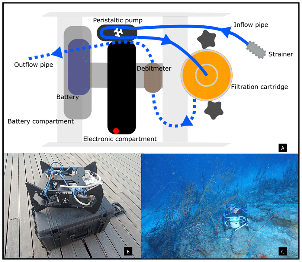
Figure 3. Semi-autonomous device for water sampling in Marine Animal Forests (MAFs).
A) Schematic view. The system enables the simultaneous collection of eukaryotic environmental DNA and microbial cells, thanks to the presence of two collection membranes (1.2 µm and 0.22 µm) in the filtration cartridge. B–C) Photos of the device, also in action within a MAF.
It is crucial to guarantee sterile conditions when working with microbes to avoid contamination with microbes other than those associated with the sample of interest. Sterilization of sampling tools can generally be performed traditionally by steam sterilization in an autoclave, although it is not very efficient for degrading nucleic acids. Specific protocols can be implemented for the decontamination of DNA on surfaces, materials, and equipment in both the laboratory and the field. A low-cost, complete, and efficient decontamination can be performed with 1%–5% (v/v) sodium hypochlorite, which is highly effective in degrading DNA (Prince & Andrus, 1992). This can be achieved using a 1:8 diluted commercial bleach solution (6%–8% sodium hypochlorite) for ~15 minutes, being careful not to let it sit for an extended period, as this can be challenging to remove. After this, the sodium hypochlorite solution must be completely removed to prevent residual contamination and sample degradation. Complete removal can be achieved by rinsing all equipment with sterile distilled or sample water (Patin & Goodwin, 2023). This protocol can also be used to avoid sample cross-contamination during sampling or laboratory procedures.
Most studies focus on holobiont tissues (fragments ranging from 1 to 8 cm²) or whole specimens. An advantage of using omics tools is the possibility of sampling only very small fragments, such as those with diameters of 2 mm, which can be increased in size depending on the other associated analyses to be conducted. The collection tools might vary depending on the hardness and thickness of the material, ranging from the collector’s hand, snips, and cutters for softer and delicate materials (e.g., sponges and plating corals), to hammer, chisel, and needle-nose pliers (e.g., scleractinian corals and thick branched corals) (Apprill, 2017; Silva et al., 2023).
When sampling and manipulating samples, it is essential to minimize contamination by using gloves and changing tools to prevent cross-contamination. Different control samples can be included during sampling and analysis procedures. A good practice involves filtering sterile seawater to assess contamination throughout the sampling or processing process. Additionally, contamination controls can be included at different steps, such as negative controls and positive controls of known microbial composition (mock communities) for nucleic acid extraction and PCR. It is worth highlighting the value of recording secondary environmental data in MAF studies, such as sampling coordinates, temperature, pressure, pH, oxygen levels, depth, luminosity, and chlorophyll concentration, among others (Taberlet et al., 2018).
Sample preservation
After the sample collection, to maintain the integrity of the investigated microbiome, the material should be, as quick as possible, stored in sterile and adequately identified containers, such as tubes or sealed bags, and be snap-frozen with liquid nitrogen, at -20 °C or maintained at <10°C when freezing is not available. For water samples, it is recommended to filter them within a maximum of six hours, preferably without freezing. However, if this is not possible, it is possible to add a DNA preservative to the sample, such as a mixture of 3M sodium acetate and 95% ethanol. The DESS buffer (DMSO, EDTA, and saturated NaCl) has also been used to preserve samples obtained in remote locations without freezing. DESS is commonly used as a storage buffer for coral samples; however, several alternatives are available, including DNA/RNA Shield, RNA Later, Longmire buffer, and other solutions with varying use and effectiveness among different species (Bruce et al., 2021; Patin & Goodwin, 2023). Tissue samples can also be stored in ethanol. In the case of using 96–100% ethanol, the sample volume should not exceed 20% of the alcohol volume, while when using 70% ethanol, the sample volume should not exceed 30% of that of the alcohol (Taberlet et al., 2018).
There are many protocols and reagents available for preserving DNA samples, as minimization of degradation is a crucial issue when dealing with this type of sample, which can be challenging to obtain, involving many times traveling by boat to remote locations, and usually presents a low concentration of the microbial symbionts when compared to that of the host.
There is no universal approach for the collection and processing methods designed to study marine animal-associated microbiomes, as microbial communities are highly complex and variable. However, as challenging as it may be, standardizing methodological approaches would enhance cooperation, comparability, and facilitate meta-analysis, such as comparisons across environments, species, and conditions (Samuel et al., 2021; Voolstra et al., 2021).
Sample processing and DNA extraction
In the dry laboratory, before starting sample processing, frozen samples must be thawed with caution or processed frozen, as thawing and freezing cycles can contribute to the degradation of the available DNA, which may have a low concentration, depending on the target and considering the low microbe-to-host DNA concentration ratio. All equipment should be sterilized and free of DNases, RNases, and pyrogens, and all surfaces should have been previously disinfected with ethanol 70%. If available, a separate space should be used for DNA extraction to avoid the co-extraction of contaminants. This room should be separate from the rooms designated for DNA or RNA manipulation, such as for PCR and library preparation.
Regarding water samples, small volumes can be extracted directly without filtration, while bigger volumes can be filtered before the DNA extraction process. There are many filtering methods, like open and enclosed filters. The choice of the best method must align with the project's necessities, budget, and availability. Also, various filters are available on the market, like cellulose nitrate (CN), polyethersulfone (PES), polyvinylidene difluoride (PVDF), glass fiber (GF), and polycarbonate track-etched (PCTE). Especially for microbiome studies, the most indicated are CN and GF ones, with porosity equal to or less than 0.22 μm (Bruce et al., 2021).
The DNA contained in the holobiont tissue, water, filters, or sediment samples, among other types, can be extracted using appropriate commercial kits or in-house protocols. The efficiency of these methods can vary, yet steps involving bead-beating and removal of contaminants and PCR inhibitors are highly recommended (Pratte & Kellogg, 2021). Numerous commercial kits are available on the market, providing a suitable balance between DNA yield quantity and quality. The use of kits is preferred to ensure efficient sample extraction and streamline the workflow. However, if it is not possible, established in-house DNA extraction protocols, such as those using phenol-chloroform-isoamyl alcohol (Mahon & Jerde, 2016), can be employed to match the available budget and may be suitable for more complex samples, such as those rich in mucus and pigments. Much of the DNA extracted from a tissue sample will originate from the host. Thus, it is necessary to perform an enrichment of the microbial symbionts' DNA.
Molecular techniques
Many molecular techniques have been employed to characterize the microbiomes associated with MAFs, including DNA fingerprinting (e.g., Denaturing Gradient Gel Electrophoresis (DGGE) and Terminal Restriction Fragment Length Polymorphism (T-RFLP)), which is now rarely used, quantitative PCR (qPCR), whole genome shotgun sequencing (metagenomics), and marker gene amplicon sequencing, complemented recently by metatranscriptomics, metaproteomics, and metabolomics.
DNA Fingerprinting (e.g, DGGE and T-RFLP) and marker gene sequencing (firstly using Sanger sequencing) were the first techniques used to approach the uncultivated diversity of marine prokaryotes (DeLong, 1992; Olsen et al., 1986) and eukaryotes (Díez et al., 2001; Moon-Van Der Staay et al., 2001; Rappé et al., 1997). The first results revealed the taxonomic diversity of the studied zooxanthellae (Symbiodiniaceae family), opening the eyes of the scientific community not only on the two-partite relationship between corals and microbes but also on multipartite relationships that took place at different scales (Morrow et al., 2018), encompassing variations regarding host taxonomy, condition, age, genetics, evolutionary history, diet, and environment (Lee et al., 2012; Voolstra et al., 2024; Webster & Reusch, 2017; Webster & Taylor, 2012). Besides Symbiodiniaceae and Bacteria, these techniques revealed a fraction of cryptic biodiversity, including viruses, Archaea, Fungi, as well as microeukaryotes, including the apicomplexan-like Chromerids and apicomplexans Corallicolids (Mohamed et al., 2023).
Most knowledge has been generated from amplicon sequencing based on the PCR amplification of specific marker genes, such as 16S, 18S, ITS, and 23S rDNA, with most studies featuring 16S rRNA amplicon sequencing to describe the bacterial members of coral holobionts and large datasets obtained from the Earth Microbiome Project (Thompson et al., 2017). Fewer studies have used shotgun metagenomic sequencing and metagenome-assembled genomes (MAGs). The study of marine microbial communities is facilitated by massively parallel DNA and RNA sequencing, allowing the description of the community taxonomic composition, the prediction of potential pathways and MAGs using metagenomics, and the functionality of microbiomes through the study of mRNA transcripts (metatranscriptome), which are collectively known as the “omics” techniques.
While amplicon sequencing provides an in-depth description of the diversity of taxonomic groups present in the community, metagenomics enables the characterization of whole genomes across all domains of life, i.e., the complete diversity of a habitat, including archaea, bacteria, eukaryotes, viruses and plasmids, and the functional potential of the analysed community. Metagenomics can also be used for taxonomic profiling, offering a description of the diversity of diverse taxonomic groups, with higher sensitivity to coverage and assembly quality, especially for rare taxa. Importantly, both methods generate millions of sequences for the invisible and uncultured members of MAFs, which contain the information necessary to characterize the communities, depending on the study's objective and available budget.
When comparing the main approaches used, marker gene sequencing and WGS metagenomics, WGS offers the advantage of a more in-depth taxonomic assignment to species and strains and is less affected by the biases associated with PCR amplification, such as the number of cycles or the primers and hyper-variable regions chosen. On the other hand, WGS metagenomics sequencing has biases associated with whole-genome amplification protocols, which are commonly used when working with low-concentration DNA samples. Another aspect regards the processing time, as marker gene processing is generally faster and results are easier to analyse and less expensive. All these aspects must be considered, according to the research questions, when selecting the most suitable approach (Pérez-Cobas et al., 2020).
Amplicon sequencing of marker genes
Amplicon sequencing is the targeted sequencing of taxonomically informative genetic markers. Targeted sequencing methods are the most common for characterizing the microbiomes of host-associated microbiota. It involves PCR-coupled high-throughput sequencing from mixed community samples (metabarcoding) without the need to isolate individuals. The target gene regions, such as the 16S (Bacteria and Archaea), 18S (microeukaryotes), ITS (Fungi), and 23S rRNA genes (Archaea), are investigated to identify the taxonomy of the microbiome members and, hence, the diversity and structure of the microbial communities (Wilms, 2021).
For this, it is necessary to select oligonucleotides or primer pairs to couple with the target gene region. The choice of these primer pairs is a crucial step in any microbiome study, as it enables the use of one or more primer pairs for the marker gene target region(s) (Table 1). For the analysis of the taxonomic diversity of the Bacteria and Archaea on the microbiomes of corals, for example, the primer pair 515F / 806R is currently the most used to amplify the V4 region of the 16S rRNA gene (Apprill et al., 2015; Silva et al., 2023; Walters et al., 2016). For the V4 region of 18S rRNA gene, used to characterize the marine microeukaryotes assemblages, the primer pair TAReuk454FWD1 and TAReukREV3, designed by Stoeck et al. (2010), has been commonly used (Wilms, 2021). The choice of the best primer to be used is paramount and should consider the objectives of the study, aimed assemblage, and available databases (Table 1). Ideally, amplification conditions should be studied and tested over the project, along with a study review to find the best primer pair for each project.
Table 1. Main primers used for the study of microbiomes associated with Marine Animal Forests.
| # | Target
Region | Primer Pair | Primer Forward
Sequence (5'-3') | Primer Reverse
Sequence (5'-3') | Amplicon
Length | Microbial
Group | Holobiont /
Environment
studied | Reference |
|---|
| 1 | 16S
rRNA | 27F/1492R | 27F:
AGAGTTTGATCCT
GGCTCAG | 1492R:
GGTTACCTTGTTA
CGACTT | ~1460 bp | Bacteria | Coral | Bourne & Munn, 2005; Bourne et al., 2008; Frade et al., 2016; Silva et al., 2021 |
| Sponge | Bayer et al., 2013; Bibi et al., 2021; Giles et al., 2013; Kennedy et al., 2008; Kennedy et al., 2014; Khan et al., 2014; Li et al., 2007; Li & Liu, 2006; Schmitt et al., 2008; Waters et al., 2014 |
| 2 | 16S
rRNA | 8F/1492R | 8F:
AGAGTTTGATCM
TGGCTCAG | 1492R:
GGTTACCTTGTTA
CGACTT | ~1460 bp | Bacteria | Coral | Galkiewicz & Kellogg, 2008; León-Zayas et al., 2020 |
| Water | León-Zayas et al., 2020 |
| 3 | 16S
rRNA | 341F/785R | 341F:
CCTACGGGNGGC
WGCAG | 785R:
GACTACHVGGGT
ATCTAATC | ~450 bp | Bacteria | Coral | Selwyn et al., 2024 |
| Sponge | Cleary et al., 2019; Cleary et al., 2023; Coelho et al., 2018; Lamb & Watts, 2023; Schellenberg et al., 2020; Stuij et al., 2024 |
| 4 | 16S
rRNA | 926F/1392R | 926F:
AAAGAGTTTGATC
CTGGCTCAG | 1392R:
ACGGGCGGTGTG
TRC | ~470 bp | Bacteria | Sponge | Burgsdorf et al., 2014 |
| Water | Wilkins et al., 2013 |
| 5 | 16S
rRNA | 338F/806R | 338F:
ACTCCTACGGGA
GGCAGCAG | 806R:
GGACTACHVGGG
TWTCTAAT | ~400 bp | Bacteria | Coral | Lan et al., 2025; Meenatchi et al., 2020; Qin et al., 2021 |
| 6 | 16S
rRNA | 63F/1387R | 63F:
CAGGCCTAACAC
ATGCAAGTC | 1387R:
GGTTACCTTGTTA
CGACTT | ~1300 bp | Bacteria | Sponge | Itelson et al., 2024; Webster et al., 2012 |
| 7 | 16S
rRNA | 27F/765R | 27F:
GAGAGTTTGATC
CTGGCTCAG | 765R:
CTGTTTGCTCCCC
ACGCTTTC3 | ~ 730 bp | Bacteria | Sponge | Anteneh et al., 2022; Yang et al., 2015 |
| 8 | 16S
rRNA | 515F/926R | 515F:
GTGCCAGCMGCC
GCGGTAA | 926R:
CCGYCAATTYMTT
TRAGTTT | ~ 371 bp | Bacteria | Water | Parada et al., 2016 |
| 9 | 16S
rRNA | 515F/806R | 515F:
GTGYCAGCMGCC
GCGGTAA | 806R:
GGACTACNVGGG
TWTCTAAT | ~290 bp | Bacteria | Coral | Maher et al., 2020; Meyer et al., 2014; Morrow et al., 2022; Reigel et al., 2021; Reigel & Hellberg, 2023; Silva et al., 2023 |
| Sponge | De Castro-Fernández et al., 2023; Gantt et al., 2019; Gantt & Erwin, 2023; Hardoim et al., 2021; Indraningrat et al., 2022; Ribes et al., 2015; Turon et al., 2024; Wu et al., 2018 |
| Water | Apprill et al., 2015; Lee et al., 2023 |
| Archaea | Sponge | Cuvelier et al., 2014; Dat et al., 2018; Díez-Vives & Riesgo, 2024; Lesser et al., 2022; Rodríguez-Marconi et al., 2015 |
| 10 | 16S
rRNA | Arch21F/Arc
h958R | Arch21F:
TTCCGGTTGATC
CYGCCGGA | Arch958R:
YCCGGCGTTGAM
TCCAATT | ~930 bp | Archaea | Sponge | Kennedy et al., 2014; Wichels et al., 2006 |
| 11 | 16S
rRNA | Arch519F/Ar
ch915R | Arch 519F:
CAGCMGCCGCG
GTAA | Arch 915R:
GTGCTCCCCCGC
CAATTCCT | ~390 bp | Archaea | Sponge | Gan et al., 2024 |
| Sediment | Wei et al., 2019 |
| 12 | ITS1 | ITS1F/ITS2 | ITS1F:
CTTGGTCATTTAG
AGGAAGTAA | ITS2:
GCTGCGTTCTTCA
TCGATGC | ~230 bp | Fungi | Coral | Staley et al., 2017 |
| Sponge | Hardoim et al., 2021 |
| 13 | ITS2 | ITS3/ITS4 | ITS3:
GCATCGATGAAG
AACGCAGC | ITS4:
TCCTCCGCTTATT
GATATGC | ~300 bp | Fungi | Coral | Alvarez-Yela et al., 2019; Lin et al., 2022 |
| Sponge | De Mares et al., 2017; Gao et al., 2008 |
| 14 | ITS | ITS1F/ITS4 | ITS1F:
GGAAGTAAAAGT
CGTAACAAGG | ITS4:
TCCTCCGCTTATT
GATATGC | ~600 bp | Fungi | Sponge | Gao et al., 2008; Nguyen & Thomas, 2018 |
| 15 | ITS2 | ITS2-Dino/
ITS2re v2 | ITS2-Dino:
GTGAATTGCAGA
ACTCCGTG | ITS2rev2:
CCTCCGCTTACTT
ATATGCTT | ~350 bp | Symbiodi-
niaceae | Coral | Alessi et al., 2024; Hartman et al., 2020; Kriefall et al., 2022; Morrow et al., 2022; Tanvet et al., 2023; Wang et al., 2023 |
| 16 | 18S
rRNA | Euk1391f/E
ukBr | Euk1391f:
GTACACACCGCC
CGTC | EukBr:
TGATCCTTCTGCA
GGTTCACCTAC | ~130 bp | Eukaryota | Coral | Wangensteen & Turon, 2015 |
| 17 | 18S
rRNA | TAReuk454
FWD1/TAR
eukREV3 | TAReuk454FWD1:
CCAGCASCYGCG
GTAATTCC | TAReukREV3:
ACTTTCGTTCTTG
ATYRA | ~400 bp | Eukaryota | Coral | Wangensteen & Turon, 2015; Zhu et al., 2022 |
Recent advances in sequencing technologies have enabled the use of Full-Length Amplicon Sequencing, which provides higher taxonomic resolution compared to traditional short-read amplicon approaches. Platforms such as PacBio SMRT (Single Molecule Real-Time) and Oxford Nanopore Technologies (ONT) allow the amplification and sequencing of entire genetic markers (e.g., full-length 16S, 18S, ITS, or other barcodes), enabling species-level or even strain-level discrimination in many cases (Callahan et al., 2019; Karst et al., 2021). This approach helps overcome the limitations of short-read sequencing, which often restrict classification due to the limited information content of partial marker regions (Johnson et al., 2019). Although still emerging in studies of MAF microbiomes, recent efforts using full-length amplicons have revealed promising results (Girija et al., 2023; Pootakham et al., 2017; Zhu et al., 2022). Full-Length Amplicon Sequencing offers clear advantages in environments with high microbial diversity, where distinguishing closely related taxa requires greater genetic resolution. However, it requires high-quality DNA, careful control of PCR chimera formation, and the use of specific bioinformatic tools to handle the longer reads and higher error rates, particularly for ONT data (Karst et al., 2021). Despite these challenges, this technique is becoming a valuable addition to the microbial ecology toolkit, offering enhanced resolution for both taxonomic profiling and ecological network reconstruction in marine animal forests.
The amplicon sequencing of a microbial community inhabiting a holobiont generates a substantial amount of sequencing data that is analysed using bioinformatics tools, allowing for quality checking and proper classification to characterize the studied microbiomes accurately. Different platforms, pipelines and tools are available, leading to statistical analyses and interpretation of the studied microbiome (Hakimzadeh et al., 2024).
Some of the available programs, scripts, and pipelines are Quantitative Insights into Microbial Ecology 2 (QIIME2; Bolyen et al., 2019), mothur (Schloss et al., 2009), CloVR virtual machine (Angiuoli et al., 2011), USEARCH (Edgar, 2010), VSEARCH (Rognes et al., 2016), Less operational taxonomic units scripts or LotuS (Hildebrand et al., 2014), DADA2 package in R (Callahan et al., 2016), and several other Python and PERL scripts, such as CONCOMPRA, aimed at Nanopore amplicon sequence data (Stock et al., 2024). Through these tools, the bioinformatic studies for the generated genetic sequences are divided into the following steps, in general: demultiplexing (if necessary, but most equipment deliver demultiplexed data nowadays), data quality control and filtering of unwanted sequences, pair joining (pair-end read pairing), dereplication, chimera removal, alignment, and taxonomic assignment (Taberlet et al., 2018). After this, the curated data can be used for ecological downstream analysis according to the initial question or objective of the project.
Quality control (QC) is essential for obtaining high-quality data and, consequently, for producing meaningful estimates of microbial diversity, as sequencing errors can lead to incorrect results, including overestimation of diversity and incorrect taxonomic assignments. QC includes trimming the sequencing adaptors and discarding short and low-quality reads (Pérez-Cobas et al., 2020).
Depending on the chosen pipeline, it is possible to build sequence tables using two approaches: individual operational taxonomic units (OTUs) or exact amplicon sequence variants (ASVs, which are produced based on sequencing error correction algorithms). Individual reads may be clustered based on sequence similarity, up to which sequences are considered to belong to the same molecular Operational Taxonomic Units (OTUs; Blaxter et al., 2005). Alternatively, new approaches to define biologically sounding units are based on Amplicon Sequence Variants (ASVs), which infer the biological sequences in the sample before the introduction of amplification and sequencing errors and distinguish sequence variants differing by as little as one nucleotide, offering a finer resolution and a higher degree of comparability across datasets (Callahan et al., 2017). It is possible to see the use of the terms ESV (Exact Sequence Variant) and ZOTU (Zero-radius OTU), which are used interchangeably with ASV and refer to the same concept (Antich et al., 2021). OTU-based approaches consider an identity of 97% as the species-level threshold, which does not necessarily reflect biological diversity, and are not sensitive enough to detect small variations between reads, which can hinder species and strains identification. Therefore, non-OTU-based methods have been preferred, such as DADA2, Deblur, and UNOISE3, which use the ASVs approach (Pérez-Cobas et al., 2020).
Another critical step is identifying the members of the different microbial assemblages associated with MAFs. There are various classification methods and databases for the classification of the different marker genes used. Apart from BLAST being the most common alignment method, QIIME2 has been the most widely used method for microbial ecology. The methods have evolved to incorporate novel machine learning techniques, resulting in improved classifications. Nevertheless, optimizing the parameter settings is as critical as the chosen methods and can result in an improved ability to detect novel taxa. Another essential issue is the chosen database, which can vary depending on the marker gene. For instance, for 16S rRNA gene analysis, the most used are Greengenes (DeSantis et al., 2006), the Ribosomal Database Project (Maidak et al., 1997), and SILVA (Yilmaz et al., 2014), but there are many others. Micro-eukaryotic diversity analysis has used mainly SILVA and the Protist Ribosomal Reference database (PR2) for 18S rRNA gene (Guillou et al., 2013), and UNITE (Abarenkov et al., 2024) for Fungi and algal symbionts (Pérez-Cobas et al., 2020). The results of taxonomic annotation are tables containing the taxa classification and abundance counts (abundance table), which will be used in the ecological and statistical analysis of the microbiome.
Shotgun metagenomics
The use of large-scale shotgun metagenomics has grown in the field of studying MAF microbiomes, providing valuable information of the functional potential encoded in DNA coding sequences within microbiomes. Another advantage is that the entire genetic material of a sample is aimed to be recovered, making it possible to characterize the complete diversity of different holobionts' microhabitats, including archaea, bacteria, eukaryotes, viruses, and plasmids, as well as their gene content (Pérez-Cobas et al., 2020).
Metagenomics enables the identification of the genetic potential of microbiome members by providing genome-resolved phylogenetic data. When applied to the holobiont, certain precautions should be considered, such as separation and enrichment methods, to overcome the dominance of the coral genome over the microbiome (Keller-Costa et al., 2021; Keller-Costa et al., 2022; Palladino et al., 2022; Thurber et al., 2009; Williams, 2024). Moreover, it is possible to assemble full metagenome-assembled genomes (MAGs) from metagenome data, which can result in draft genomes of uncultured micro-organisms, such as MAGs from Endozoicomonas, one of the dominating microbes occurring in scleractinian corals and octocorals, for instance (Keller-Costa et al., 2022).
The first steps when working with raw reads from WGS metagenomics involve preprocessing and quality filtering, which involve trimming sequencing adaptors, discarding short and low-quality reads, and evaluating and filtering data for errors. Such errors, such as sequencing errors, can lead to the overestimation of diversity that is not based on natural genetic variations, as well as incorrect taxonomic and functional annotations. There are different softwares for this step, such as FASTX, PRINSEQ, and Trimmomatic. For exploring the raw data for read length, GC content, quality scores, number of duplicated reads, and number of N bases, FASTQC and SeqKit are some options (Pérez-Cobas et al., 2020).
Then, data processing is performed by using assembly or assembly-free approaches. Assembly is the process of reconstructing in silico sequences, based on smaller fragments, so they can be closer to the original genome sequences. Most studies employ assembly approaches, as they can enhance analysis by constructing longer sequences (contigs). Assembly can be performed de novo, i.e., joining fragments to generate contigs without using previously sequenced reference genomes, or by comparative assembly, i.e., using previously sequenced related organisms to guide the process. For metagenomics, assembly is done simultaneously for the many genomes contained in the sample. Some examples of assemblers are: MetaVelvet, MEGAHIT, metaSPAdes, and IDBA-UD (Pérez-Cobas et al., 2020).
The next step can be gene prediction and functional assignment or binning. The later refers to the process of gathering reads and contigs that belong to the same taxon and classifying them taxonomically. After classification, it is possible to identify and annotate genes and pathways, finally enabling the identification of the functional capacities encoded in the microbiome assemblage investigated, using databases such as MetaCyc (Metabolic Pathway Database, Caspi et al., 2014), KEGG (Kyoto Encyclopedia of Genes and Genomes; Kanehisa & Goto, 2000), and SEED (Overbeek et al., 2005). Some platforms allow integrating all the steps, being straightforward to use. Of course, caution is always advisable, as oversimplification can limit informed decisions at every step of the process. Yet, those platforms allow working with complex data without being a bioinformatician (Pérez-Cobas et al., 2020).
Good practice guidelines for metadata
Data and metadata should be made Findable, Accessible, Interoperable, and Reusable (FAIR) through appropriate procedures (e.g., INSPIRE Directive principles) to ensure open access to the results. Beyond the publications of the research results in open-access scientific journals (whenever possible) and repositories (e.g., Zenodo, ResearchGate, bioRxiv), microbial sequences should be stored in a common data format - i.e. fasta or fastq format, and made freely accessible in DNA data repositories, such as GenBank (https://www.ncbi.nlm.nih.gov/genbank/), the most used database that collects all publicly available DNA sequences. Once shared in GenBank, the DNA sequences are assigned an accession number and are accompanied by related metadata. Metadata, such as sample typology, sampling site coordinates, temperature, pressure, pH, oxygen, and depth, should also be shared along with DNA sequence, as well as in open-access repositories. The geospatial data of sampling sites and the sampled holobiont species can also be made open access through ad hoc repositories (e.g., OBIS). A recent evaluation of the Sequence Read Archives (SRA) from GenBank displayed that ~15% of all archived specimens included both collection year and basic metadata (Thurber et al., 2022; Voolstra et al., 2021).
Divergences among the main protocols used, involving sampling procedures, preservation, and processing, may compromise the accuracy and precision of the resulting analysis. The study of microbiomes associated with marine animal forests benefits from comparative studies, in which those aspects are evaluated, aiming to evidence the practices that result in the closest to true biological variations. The research community is encouraged to publish methodological reports that demonstrate positive and negative results of different approaches. In addition, while no single method exists to study such a complex array of animal ecosystems, research consortia employing standard procedures at different marine forests enable a systematic evaluation of study questions in the field and solid conclusions on the associated microbiomes (Thurber et al., 2022). Standardized methods and procedures are crucial for advancing the field and enabling the development of nature-based solutions for coral restoration and conservation.
Ethics and consent
Ethical approval and consent were not required.