Introduction
Spinal meningiomas (SM) are growths that generally have a positive outlook both in terms of cancer treatment and surgical outcomes, with a rare chance of the tumour returning. Usually found in the space surrounding the spinal cord but outside its innermost layer, these tumours develop gradually and tend to extend sideways into the area around the spinal cord, often stretching the nearby arachnoid membrane but seldom affecting the innermost pia mater (Hohenberger et al., 2023). Meningiomas are lesions that arise from arachnoidal cap cells, the outer part of the arachnoid layer and villi. They usually form dural attachments and are marked by meningothelial hyperplasia. Spinal meningiomas most commonly occur in the posterior, posterolateral, or lateral thoracic region, followed by the anterior cervical and lumbosacral regions (Sun et al., 2020). The standard approach to treating them involves surgery, aiming to completely remove the tumour and enhance neurological function. The main treatment is surgery, which can be performed in (a) a classical open microsurgical approach in the vast majority of cases, (b) a minimally invasive surgery (MIS), or (c) through an endoscopic intervention (Serratrice et al., 2023; Sun et al., 2020).
Case presentation
The patient, an 18-year-old female, with no relevant past medical history and no known family history of neurological or oncological disease, presented with complaints of progressive weakness in both lower limbs, with a recorded muscle strength of 2/5. She reported significant difficulty in walking and relied on a wheelchair for mobility. Upon examination, neurological deficits were noted, prompting further investigation. Written informed consent was also obtained from the patient for publication of this case report and accompanying images. All reasonable efforts were made to anonymize patient information.
Diagnostic assessment
Diagnostic workup included magnetic resonance imaging (MRI) of the spinal axis, revealing an intradural extramedullary tumour compressing the spinal cord (Figure 1). The tumour was identified as a meningioma, characterized by its firm consistency and white-gray appearance upon exposure during surgery. The preoperative MRI provided crucial insights into the location and extent of the tumour, guiding the surgical approach. Figure 1 shows a magnetic resonance imaging (MRI) scan of the spinal axis, specifically highlighting an expansile intradural extramedullary process.
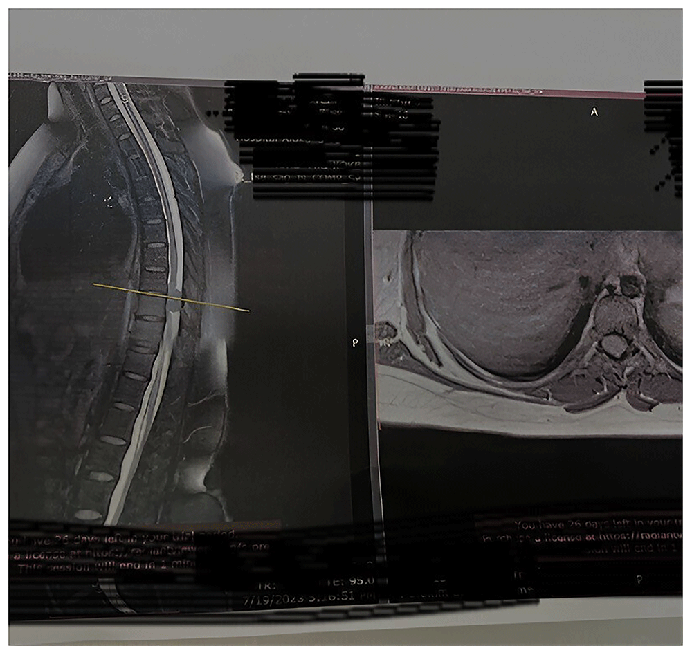
Figure 1. The image shows the magnetic resonance imaging (MRI) scan of the spinal axis, specifically highlighting an expansile intradural extramedullary process.
Treatment
The patient underwent urgent neurosurgical intervention to address the spinal meningioma. The surgical procedure involved total laminectomy at the TH6/7 vertebral level (Figure 2) to access and remove the tumour. Intraoperative neurosurgical procedure for the resection of a spinal meningioma is shown in Figure 2. Intraoperatively, meticulous care was taken to preserve neural structures while achieving complete tumour resection as shown in Figure 3. The excised mass was sent for histopathological examination to confirm the diagnosis and assess for any concerning features.
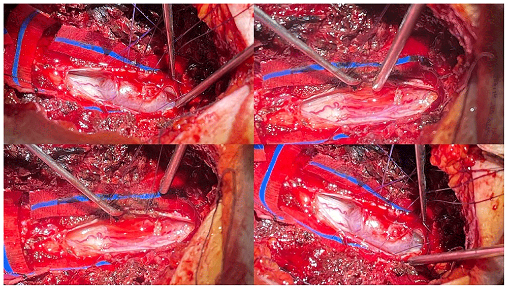
Figure 2. The image shows Intraoperative neurosurgical procedure for the resection of a spinal meningioma.
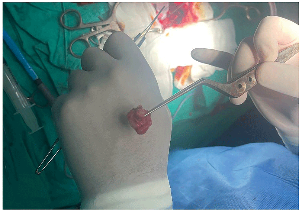
Figure 3. Complete tumour resection.
Follow-up and outcome
Following surgery, the patient was closely monitored in the postoperative period for any signs of neurological improvement or complications. Gradually, the patient exhibited notable improvement in muscle strength, with postoperative muscle strength reaching 4/5 compared to the preoperative level of 2/5. Regular follow-up appointments were scheduled to monitor the patient's progress, ensuring optimal recovery and functional outcomes.
Discussion
Although meningiomas are generally benign tumours, atypical and rare clinical presentations have been reported in the literature, emphasizing the need for individualized diagnostic and therapeutic approaches (Gone et al., 2024). The successful management of this case underscores the efficacy of prompt neurosurgical intervention coupled with comprehensive preoperative imaging and meticulous tumour resection in achieving favourable outcomes for spinal meningiomas. The utilization of magnetic resonance imaging (MRI) facilitated accurate preoperative planning, enabling the surgical team to approach the tumour with precision while minimizing damage to adjacent neural structures (Hohenberger et al., 2023; Serratrice et al., 2023). Total laminectomy at the TH6/7 vertebral level provided adequate exposure for complete tumour resection, with intraoperative vigilance ensuring preservation of neurological function. Previous studies have demonstrated that gross total resection is associated with lower recurrence rates and improved long-term neurological outcomes compared with subtotal resection (Materi et al., 2020). The observed postoperative improvement in muscle strength highlights the potential for significant neurological recovery following surgical intervention for spinal meningiomas. Thoracic tumours often present with progressive neurological deficits, and early surgical management can lead to significant functional recovery (Sun et al., 2020). Emerging evidence suggests that genomic subgroups of meningiomas may correlate with distinct clinical behaviours and outcomes, highlighting the potential role of molecular profiling in future risk stratification and management strategies (Youngblood et al., 2019). Regular follow-up appointments are essential for monitoring long-term outcomes and addressing any potential complications, emphasizing the importance of a multidisciplinary approach in optimizing patient care.
Conclusion
This case exemplifies the successful management of a spinal meningioma through prompt neurosurgical intervention guided by preoperative imaging. The observed neurological improvement underscores the importance of surgical resection in restoring function in affected patients. Long-term follow-up is crucial for monitoring outcomes and addressing any potential complications. Continued research efforts are needed to further elucidate optimal treatment strategies and improve outcomes for patients with spinal meningiomas.
Ethics and consent
Ethical approval not required. Written informed consent was obtained from the patient for participation in the study and for publication of this case report.
Data availability
All data underlying the findings of this case report are included within the article. Additional clinical data are archived at the University Hospital Centre and are not publicly available due to ethical and privacy considerations, as they contain potentially identifiable patient information. The CARE checklist for this case report is available in Zenodo under the title “CARE checklist for Rare Presentation of Spinal Meningioma in Kosova: A Case Report”, DOI: 10.5281/zenodo.17969909 licensed under CC0 (Hasimi & Ymeri, 2025).
Faculty Opinions recommendedReferences
- Gone J, Kennedy B, Fontaine T, et al.:
Creeping through the cranium: a rare and interesting presentation of a meningioma.
Radiol Case Rep.
2024; 19(9): 3928–3933. PubMed Abstract
| Publisher Full Text
| Free Full Text
- Hasimi F, Ymeri F:
Rare presentation of spinal meningioma in Kosova: case report. Zenodo. 2025. http://www.doi.org/10.5281/ZENODO.17969909
- Hohenberger C, Hau P, Schebesch KM, et al.:
Spinal meningiomas.
Neurooncol Adv.
2023; 5(Suppl 1): i112–i121. PubMed Abstract
| Publisher Full Text
| Free Full Text
- Materi J, Mampre D, Ehresman J, et al.:
Predictors of recurrence and high growth rate of residual meningiomas after subtotal resection.
J Neurosurg.
2020; 134(2): 410–416. PubMed Abstract
| Publisher Full Text
- Serratrice N, Lameche I, Attieh C, et al.:
Spinal meningiomas, from biology to management — A literature review.
Front Oncol.
2023; 12: 1084404. PubMed Abstract
| Publisher Full Text
| Free Full Text
- Sun C, Dou Z, Wu J, et al.:
The preferred locations of meningioma according to different biological characteristics based on voxel-wise analysis.
Front Oncol.
2020; 10: 1412. PubMed Abstract
| Publisher Full Text
| Free Full Text
- Youngblood MW, Duran D, Montejo JD, et al.:
Correlations between genomic subgroup and clinical features in a cohort of more than 3000 meningiomas.
J Neurosurg.
2019; 133(5): 1345–1354. PubMed Abstract
| Publisher Full Text