Introduction
Gambusia holbrooki, a member of the Poeciliidae family, is a freshwater fish native to eastern North America (Pyke, 2005). Along with its sister species Gambusia affinis, it has been introduced globally as a means of mosquito control, with often severe impacts on local biodiversity (Fryxell et al., 2022). In Europe, Gambusia holbrooki was introduced to Spain and Italy in 1921 and has since become established throughout the Southern part of the European continent (Fryxell et al., 2022). Mosquitofish (G. holbrooki and G. affinis) can have devastating impacts on local biodiversity and has been listed among the top 100 invasive alien species (Lowe et al., 2000). Being omnivorous and tolerant to a variety of environmental conditions, mosquitofish quickly spread with negative consequences for local biodiversity. In addition to its well-documented invasion history, the mosquitofish is a longstanding model system for life history evolution research and was key in the development of life history theory (Stearns, 1983). Gambusia holbrooki is listed as Least Concern (LC) in the IUCN Red List on Threatened Species (last assessed in 2012).
The generation of this reference resource was coordinated by the European Reference Genome Atlas (ERGA) initiative’s Biodiversity Genomics Europe (BGE) project, supporting ERGA’s aims of promoting transnational cooperation to promote advances in the application of genomics technologies to protect and restore biodiversity (Mazzoni et al., 2023).
Materials & methods
ERGA's sequencing strategy includes Oxford Nanopore Technology (ONT) and/or Pacific Biosciences (PacBio) for long-read sequencing, along with Hi-C sequencing for chromosomal architecture, Illumina Paired-End (PE) for polishing (i.e. recommended for ONT-only assemblies), and RNA sequencing for transcriptomic profiling, to facilitate genome assembly and annotation.
Sample and sampling information
On 24 July 2024, Clelia Gasparini and Francesco Santi sampled 8 male specimens of Gambusia holbrooki, which were identified based on morphology using the gonopodium structure (Lloyd & Tomasov, 1985) at the University of Padua, Italy. Specimens were sampled in the Piovego canal (which connects the Bacchiglione and Brenta rivers), Padua, Italy. Gambusia holbrooki is the only mosquitofish species that has so far been described in Italy. Sampling was performed under permission number 382 (16 May 2024) issued by the Regione del Veneto, Italy. Specimens were caught with a dip net and immediately placed into a collection container. Specimens were then euthanized with an overdose of MS222 and subsequently put in dry ice. Until DNA extraction, samples were preserved at -80 °C.
Vouchering information
Physical reference materials for the here sequenced specimen have been deposited in the Leibniz Institute for the Analysis of Biodiversity Change https://leibniz-lib.de/en/index.html under accession numbers ZFMK-TIS-97651 (male organism) and ZFMK-TIS-97652 (female organism).
Frozen somatic animal tissue is available from proxy individuals at the Biobank of the Leibniz Institute for the Analysis of Biodiversity Change https://leibniz-lib.de/en/index.html under the voucher IDs ZFMK-TIS-97677, ZFMK-TIS-97679, ZFMK-TIS-97680, ZFMK-TIS-97682 to ZFMK-TIS-97685, ZFMK-TIS-97688, ZFMK-TIS-97689, ZFMK-TIS-97692. Additionally, somatic cell cultures were established and frozen in liquid nitrogen under the ID ZFMK-TIS-98713.
Genetic information
The estimated genome size, based on ancestral taxa, is 0.83 Gb. This is a diploid genome with a haploid number of 24 chromosomes (2n=48) and XX/XY as sex determination mechanism (Kottler et al., 2020). All information for this species was retrieved from Genomes on a Tree (Challis et al., 2023).
DNA/RNA processing
Protocols for high molecular weight (HMW) DNA extraction developed at the Wellcome Sanger Institute (WSI) Tree of Life Core Laboratory are available on protocols.io (Denton et al., 2023b; Howard et al., 2025). The fGamHol4 sample was weighed and triaged (Jay et al., 2023) to determine the appropriate extraction protocol. Tissue from the mid-body was homogenised by powermashing using a PowerMasher II tissue disruptor (Denton et al., 2023a). HMW DNA was extracted using the Automated MagAttract v2 protocol (Oatley et al., 2025a). DNA was sheared into an average fragment size of 12–20 kb following the Megaruptor®3 for LI PacBio protocol (Bates et al., 2023). Sheared DNA was purified by automated SPRI (solid-phase reversible immobilisation) (Oatley et al., 2025b). The concentration of the sheared and purified DNA was assessed using a Nanodrop spectrophotometer and Qubit Fluorometer using the Qubit dsDNA High Sensitivity Assay kit. Fragment size distribution was evaluated by running the sample on the FemtoPulse system.
Library preparation and sequencing
Library preparation and sequencing were performed at the WSI Scientific Operations core. Libraries were prepared using the SMRTbell Prep Kit 3.0 (Pacific Biosciences, California, USA), according to the manufacturer’s instructions. The kit includes reagents for end repair/A-tailing, adapter ligation, post-ligation SMRTbell bead clean-up, and nuclease treatment. Size selection and clean-up were performed using diluted AMPure PB beads (Pacific Biosciences). DNA concentration was quantified using a Qubit Fluorometer v4.0 (ThermoFisher Scientific) and the Qubit 1X dsDNA HS assay kit. Final library fragment size was assessed with the Agilent Femto Pulse Automated Pulsed Field CE Instrument (Agilent Technologies) using the gDNA 55 kb BAC analysis kit.
The sample was sequenced on a Revio instrument (Pacific Biosciences). The prepared library was normalised to 2 nM, and 15 μL was used for making complexes. Primers were annealed and polymerases bound to generate circularised complexes, following the manufacturer’s instructions. Complexes were purified using 1.2X SMRTbell beads, then diluted to the Revio loading concentration (200–300 pM) and spiked with a Revio sequencing internal control. The sample was sequenced on a Revio 25M SMRT cell. The SMRT Link software (Pacific Biosciences), a web-based workflow manager, was used to configure and monitor the run and to carry out primary and secondary data analysis.
Biotinylated DNA constructs were fragmented using a Covaris E220 sonicator and size selected to 400–600 bp using SPRISelect beads. DNA was enriched with Arima-HiC v2 kit Enrichment beads. End repair, A-tailing, and adapter ligation were carried out with the NEBNext Ultra II DNA Library Prep Kit (New England Biolabs), following a modified protocol where library preparation occurs while DNA remains bound to the Enrichment beads. Library amplification was performed using KAPA HiFi HotStart mix and a custom Unique Dual Index (UDI) barcode set (Integrated DNA Technologies). Depending on sample concentration and biotinylation percentage determined at the crosslinking stage, libraries were amplified with 10–16 PCR cycles. Post-PCR clean-up was performed with SPRISelect beads. Libraries were quantified using the AccuClear Ultra High Sensitivity dsDNA Standards Assay Kit (Biotium) and a FLUOstar Omega plate reader (BMG Labtech). Prior to sequencing, libraries were normalised to 10 ng/μL. Normalised libraries were quantified again and equimolar and/or weighted 2.8 nM pools. Pool concentrations were checked using the Agilent 4200 TapeStation (Agilent) with High Sensitivity D500 reagents before sequencing. Sequencing was performed using paired-end 150 bp reads on the Illumina NovaSeq X. In total 56x HiFi and 147x HiC data were sequenced to generate the assembly.
Genome assembly methods
The HiFi reads were assembled using Hifiasm (Cheng et al., 2021) in Hi-C phasing mode, where data were separated into two haplotypes. These haplotypes were then curated to generate a final assembly. The Hi-C reads were aligned to the contigs using bwa-mem2 (Vasimuddin et al., 2019), and contigs were scaffolded with YaHS (Zhou et al., 2023), using the --break option for handling potential misassemblies. The resulting scaffolded assemblies were evaluated using Gfastats (Formenti et al., 2022), BUSCO (Manni et al., 2021), and MERQURY.FK (Rhie et al., 2020).
The mitochondrial genome was assembled using oatk (Zhou et al., 2024) as a single circular contig of 18,667 bp and it is included in the released assembly (GCA_965178065.1). Manual curation was primarily conducted using PretextView (Harry, 2022), with additional insights provided by JBrowse2 (Diesh et al., 2023) and HiGlass (Kerpedjiev et al., 2018). Scaffolds were visually inspected and corrected as described by (Howe et al., 2021). Any identified contamination, missed joins, and mis-joins were corrected, and duplicate sequences were tagged and removed. The curation process is documented at (Denton et al., 2023) (article in preparation). Summary analysis of the released assembly was performed using the ERGA-BGE Genome Report ASM Galaxy workflow (doi.org/10.48546/workflowhub.workflow.1104.1).
Genome annotation methods
A gene set was generated using the Ensembl Gene Annotation system (Aken et al., 2016), primarily by aligning publicly available short-read RNA-seq data from BioSample: SAMN14167175, SAMN14167173, SAMN11245730, SAMN11245729, SAMN11245733, SAMN11245732, SAMN14167176, SAMN14167159, SAMN14167158, SAMN11245734, SAMN11245731, SAMN14167178, SAMN14167177, SAMN14167174, SAMN14167160, SAMN14167157, SAMN14167156, and SAMN14167155 to the genome. Gaps in the annotation were filled via protein-to-genome alignments of a select set of vertebrate proteins from (UniProt Consortium, 2019), which had experimental evidence at the protein or transcript level. At each locus, data were aggregated and consolidated, prioritising models derived from RNA-seq data, resulting in a final set of gene models and associated non-redundant transcript sets. To distinguish true isoforms from fragments, the likelihood of each open reading frame (ORF) was evaluated against known vertebrate proteins. Low-quality transcript models, such as those showing evidence of fragmented ORFs, were removed. In cases where RNA-seq data were fragmented or absent, homology data were prioritised, favouring longer transcripts with strong intron support from short-read data. The resulting gene models were classified into three categories: protein-coding, pseudogene, and long non-coding. Models with hits to known proteins and few structural abnormalities were classified as protein-coding. Models with hits to known proteins but displaying abnormalities, such as the absence of a start codon, non-canonical splicing, unusually small intron structures (<75 bp), or excessive repeat coverage, were reclassified as pseudogenes. Single-exon models with a corresponding multi-exon copy elsewhere in the genome were classified as processed (retrotransposed) pseudogenes. Models that did not fit any of the previously described categories did not overlap protein-coding genes and were constructed from transcriptomic data were considered potential lncRNAs. Potential lncRNAs were further filtered to remove single-exon loci due to their unreliability. Putative miRNAs were predicted by performing a BLAST search of miRBase (Kozomara et al., 2019) against the genome, followed by RNAfold analysis (Gruber et al., 2008). Other small non-coding loci were identified by scanning the genome with Rfam (Kalvari et al., 2018) and passing the results through Infernal (Nawrocki & Eddy, 2013). Summary analysis of the released annotation was carried out using the ERGA-BGE Genome Report ANNOT Galaxy workflow (10.48546/workflowhub.workflow.1096.1).
Results
Genome assembly
The genome assembly has a total length of 674,622,098 bp in 318 scaffolds including the mitogenome (Figure 1 & Figure 2), with a GC content of 39%. The assembly has a contig N50 of 15,918,108 bp and L50 of 16 and a scaffold N50 of 29,628,722 bp and L50 of 11. The assembly has a total of 103 gaps, totalling 11 kb in cumulative size. The single-copy gene content analysis using the Cyprinodontiformes database (odb10) with BUSCO (Manni et al., 2021) resulted in 99.3% completeness (99.0% single and 0.3% duplicated). 93.8% of reads k-mers were present in the assembly and the assembly has a base accuracy Quality Value (QV) of 59.8 as calculated by Merqury (Rhie et al., 2020).
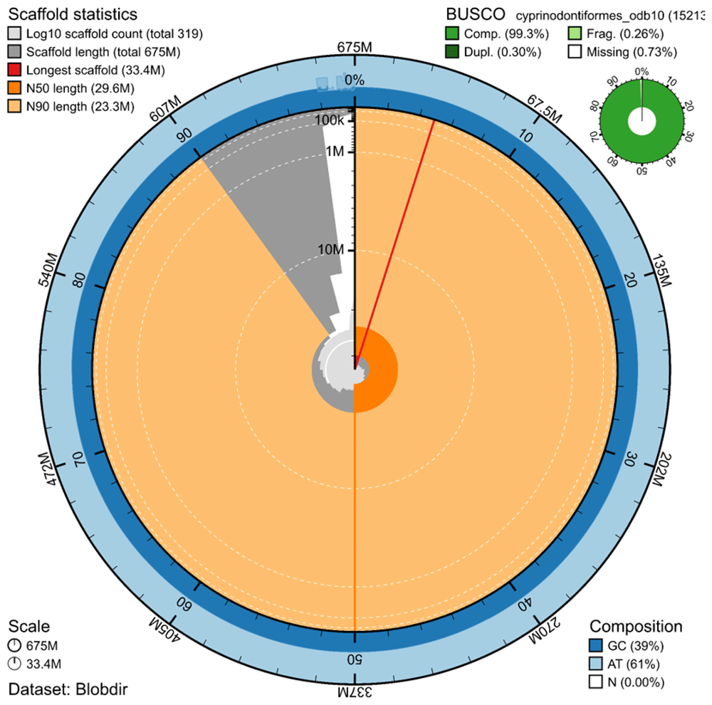
Figure 1. Snail plot summary of assembly statistics.
The main plot is divided into 1,000 size-ordered bins around the circumference, with each bin representing 0.1% of the 674,622,098 bp assembly including the mitochondrial genome. The distribution of sequence lengths is shown in dark grey, with the plot radius scaled to the longest sequence present in the assembly (33.4 Mp, shown in red). Orange and pale-orange arcs show the scaffold N50 and N90 sequence lengths (29,628,722 and 23,341,799 bp), respectively. The pale grey spiral shows the cumulative sequence count on a log-scale, with white scale lines showing successive orders of magnitude. The blue and pale-blue area around the outside of the plot shows the distribution of GC, AT, and N percentages in the same bins as the inner plot. A summary of complete, fragmented, duplicated, and missing BUSCO genes found in the assembled genome from the Cyprinodontiformes database (odb10) is shown in the top right.
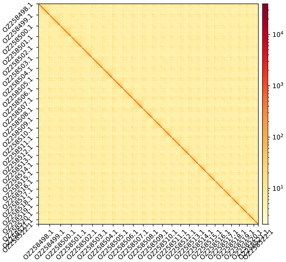
Figure 2. Hi-C contact map showing spatial interactions between regions of the genome.
The diagonal corresponds to intra-chromosomal contacts, depicting chromosome boundaries. The frequency of contacts is shown on a logarithmic heatmap scale. Hi-C matrix bins were merged into a 200 kb bin size for plotting.
Genome annotation
The genome annotation consists of 21,050 protein-coding genes with associated 56,794 transcripts, in addition to 3,687 non-coding genes (Table 1). Using the longest isoform per transcript, the single-copy gene content analysis using the Cyprinodontiformes database (odb10) database with BUSCO resulted in 88.5% completeness. Using the OMAmer Metazoa-v2.0.0.h5 database for OMArk (Nevers et al., 2025) resulted in 94.0% completeness and 98.5% consistency (Table 2).
Table 1. Statistics from assembled gene models.
| No.
genes | No.
transcripts | Mean gene
length (bp) | No. single-
exon genes | Mean exons
per transcript |
|---|
| mRNA | 21,050 | 56,794 | 18,920 | 517 | 14.3 |
| pseudogene | 228 | 228 | 22,143 | 1 | 24.5 |
| snoRNA | 158 | 158 | 114 | 158 | 1.0 |
| lncRNA | 2,085 | 2,520 | 4,158 | 1,283 | 2.6 |
| miRNA | 25 | 25 | 69 | 25 | 1.0 |
| snRNA | 118 | 118 | 153 | 118 | 1.0 |
| rRNA | 1,066 | 1,066 | 612 | 1,066 | 1.0 |
| scRNA | 7 | 7 | 223 | 7 | 1.0 |
| Other ncRNA | 13 | 13 | 98 - 275 | 13 | 1.0 - 1.0 |
Table 2. Annotation completeness and consistency scores calculated by BUSCO run in protein mode (cyprinodontiformes_odb10) and OMArk (Metazoa-v2.0.0.h5).
| Complete | Singular | Duplicated | Fragmented | Missing |
|---|
| BUSCO | 13,470 (88.5%) | 13,391 (88.0%) | 79 (0.5%) | 197 (1.3%) | 1,546 (10.2%) |
| OMArk | 17,336 (94.0%) | 17,053 (92.5%) | 283 (1.5%) | - | 1,105 (5.6%) |
| Consistent | Inconsistent | Contaminants | Unknown |
|---|
| OMArk | 20,764 (98.5%) | 142 (0.7%) | 0.0 (0.0%) | 166 (0.8%) |
Data availability
Gambusia holbrooki and the related genomic study were assigned to Tree of Life ID (ToLID) 'fGamHol4' and all sample, sequence, and assembly information are available under the umbrella BioProject PRJEB84145. The sample information is available at the following BioSample accessions: SAMEA115931355 and SAMEA115931357. The genome assembly is accessible from ENA under accession number GCA_965282415.1 and the annotated genome is available through the Ensembl website (https://projects.ensembl.org/erga-bge/). Sequencing data produced as part of this project are available from ENA at the following accessions: ERX13553727 and ERX14241339. Documentation related to the genome assembly and curation can be found in the ERGA Assembly Report (EAR) document available at https://github.com/ERGA-consortium/EARs/tree/main/Assembly_Reports/Gambusia_holbrooki/fGamHol4. Further details and data about the project are hosted on the ERGA portal at https://portal.erga-biodiversity.eu/data_portal/37273.
Author contributions
BF and CG Fraser coordinated the project; CG and FS collected the species; FS identified the species; FS and CG sampled and preserved biological material and provided metadata; AsB, RM, TM, RAO, and THS provided support in sampling, shipping of biological material, metadata collection, and management; DNA extraction, library preparation and sequencing were performed by AD, CH and the teams at the Wellcome Sanger Institute; assembly and curation was carried out by SMC and JDMW under the supervision of KH and MB; FM, SS, and LH performed genome annotation; CB generated the analysis and report. All authors contributed to the writing, review, and editing of this genome note and read and approved the final version.
Acknowledgements
We acknowledge the support of the Freiburg Galaxy Team: Saim Momin and Björn Grüning, Bioinformatics, University of Freiburg (Germany), funded by the German Federal Ministry of Education and Research BMBF grant 031 A538A de.NBI-RBC and the Ministry of Science, Research and the Arts Baden-Württemberg (MWK) within the framework of LIBIS/de.NBI Freiburg. We would like to acknowledge the assembly reviewer, Jessica Gomez Garrido from the Centro Nacional de Análisis Genómico.
Faculty Opinions recommendedReferences
- Aken BL, Ayling S, Barrell D, et al.:
The Ensembl gene annotation system.
Database (Oxford).
2016; 2016: baw093. PubMed Abstract
| Publisher Full Text
| Free Full Text
- Bates A, Clayton-Lucey I, Howard C:
Sanger Tree of Life HMW DNA fragmentation: diagenode megaruptor® 3 for LI pacBio. 2023. Reference Source
- Challis R, Kumar S, Sotero-Caio C, et al.:
Genomes on a Tree (GoaT): a versatile, scalable search engine for genomic and sequencing project metadata across the eukaryotic Tree of Life [version 1; peer review: 2 approved].
Wellcome Open Res.
2023; 8: 24. PubMed Abstract
| Publisher Full Text
| Free Full Text
- Cheng H, Concepcion GT, Feng X, et al.:
Haplotype-resolved de novo assembly using phased assembly graphs with hifiasm.
Nat Methods.
2021; 18(2): 170–175. PubMed Abstract
| Publisher Full Text
| Free Full Text
- Denton A, Oatley G, Cornwell C, et al.:
Sanger Tree of Life sample homogenisation: powerMash. 2023a. Publisher Full Text
- Denton A, Yatsenko H, Jay J, et al.:
Sanger Tree of Life wet laboratory protocol collection V.1. 2023b. Publisher Full Text
- Diesh C, Stevens GJ, Xie P, et al.:
JBrowse 2: a modular genome browser with views of synteny and structural variation.
Genome Biol.
2023; 24(1): 74. PubMed Abstract
| Publisher Full Text
| Free Full Text
- Formenti G, Abueg L, Brajuka A, et al.:
Gfastats: conversion, evaluation and manipulation of genome sequences using assembly graphs.
Bioinformatics.
2022; 38(17): 4214–4216. PubMed Abstract
| Publisher Full Text
| Free Full Text
- Fryxell DC, Moffett ER, Kinnison MT, et al.:
From southern swamps to cosmopolitan model: humanity’s unfinished history with mosquitofish.
Fish and Fisheries.
2022; 23(1): 143–161. Publisher Full Text
- Gruber AR, Lorenz R, Bernhart SH, et al.:
The vienna RNA websuite.
Nucleic Acids Res.
2008; 36(suppl_2): W70–W74. PubMed Abstract
| Publisher Full Text
| Free Full Text
- Harry E:
PretextView (Paired REad TEXTure Viewer): a desktop application for viewing pretext contact maps. 2022. Reference Source
- Howard C, Denton A, Jackson B, et al.:
On the path to reference genomes for all biodiversity: lessons learned and laboratory protocols created in the Sanger Tree of Life core laboratory over the first 2000 species.
bioRxiv.
2025; 2025-04. Publisher Full Text
- Howe K, Chow W, Collins J, et al.:
Significantly improving the quality of genome assemblies through curation.
GigaScience.
2021; 10(1): giaa153. PubMed Abstract
| Publisher Full Text
| Free Full Text
- Jay J, Yatsenko H, Narváez-Gómez JP, et al.:
Sanger Tree of Life sample preparation: triage and dissection. 2023. Reference Source
- Kalvari I, Nawrocki EP, Argasinska J, et al.:
Non-coding RNA analysis using the Rfam database.
Curr Protoc Bioinformatics.
2018; 62(1): e51. PubMed Abstract
| Publisher Full Text
| Free Full Text
- Kerpedjiev P, Abdennur N, Lekschas F, et al.:
HiGlass: Web-based visual exploration and analysis of genome interaction maps.
Genome Biol.
2018; 19(1): 125. PubMed Abstract
| Publisher Full Text
| Free Full Text
- Kottler VA, Feron R, Nanda I, et al.:
Independent origin of XY and ZW sex determination mechanisms in mosquitofish sister species.
Genetics.
2020; 214(1): 193–209. PubMed Abstract
| Publisher Full Text
| Free Full Text
- Kozomara A, Birgaoanu M, Griffiths-Jones S:
miRBase: from microRNA sequences to function.
Nucleic Acids Res.
2019; 47(D1): D155–D162. PubMed Abstract
| Publisher Full Text
| Free Full Text
- Lloyd LN, Tomasov JF:
Taxonomic status of the mosquitofish, Gambusia affinis (Poeciliidae), in Australia.
Mar Freshw Res.
1985; 36(3): 447–451. Publisher Full Text
- Lowe S, Browne M, Boudjelas S, et al.:
100 of the world’s worst invasive alien species: A selection from the global invasive species database. (Vol. 12). Invasive Species Specialist Group Auckland. 2000; 12. Reference Source
- Manni M, Berkeley MR, Seppey M, et al.:
BUSCO update: novel and streamlined workflows along with broader and deeper phylogenetic coverage for scoring of eukaryotic, prokaryotic, and viral genomes.
Mol Biol Evol.
2021; 38(10): 4647–4654. PubMed Abstract
| Publisher Full Text
| Free Full Text
- Mazzoni CJ, Ciofi C, Waterhouse RM:
Biodiversity: an atlas of European reference genomes.
Nature.
2023; 619(7969): 252. PubMed Abstract
| Publisher Full Text
- Nawrocki EP, Eddy SR:
Infernal 1.1: 100-fold faster RNA homology searches.
Bioinformatics.
2013; 29(22): 2933–2935. PubMed Abstract
| Publisher Full Text
| Free Full Text
- Nevers Y, Warwick Vesztrocy A, Rossier V, et al.:
Quality assessment of gene repertoire annotations with OMArk.
Nat Biotechnol.
2025; 43(1): 124–133. PubMed Abstract
| Publisher Full Text
| Free Full Text
- Oatley G, Denton A, Kovalevskaia A, et al.:
Sanger Tree of Life HMW DNA extraction: automated magAttract v.3. 2025a. Publisher Full Text
- Oatley G, Sampaio F, Denton A, et al.:
Sanger Tree of Life fragmented DNA clean up: automated SPRI. 2025b. Publisher Full Text
- Pyke GH:
A review of the biology of Gambusia affinis and G. holbrooki.
Rev Fish Biol Fisheries.
2005; 15(4): 339–365. Publisher Full Text
- Rhie A, Walenz BP, Koren S, et al.:
Merqury: reference-free quality, completeness, and phasing assessment for genome assemblies.
Genome Biol.
2020; 21(1): 245. PubMed Abstract
| Publisher Full Text
| Free Full Text
- Stearns SC:
The genetic basis of differences in life-history traits among six populations of mosquitofish (Gambusia affinis) that shared ancestors in 1905.
Evolution.
1983; 37(3): 618–627. PubMed Abstract
| Publisher Full Text
- UniProt Consortium:
UniProt: a worldwide hub of protein knowledge.
Nucleic Acids Res.
2019; 47(D1): D506–D515. PubMed Abstract
| Publisher Full Text
| Free Full Text
- Vasimuddin M, Misra S, Li H, et al.:
Efficient architecture-aware acceleration of BWA-MEM for multicore systems.
2019 IEEE International Parallel and Distributed Processing Symposium (IPDPS).
2019; 314–324. Reference Source
- Zhou C, Brown M, Blaxter M, et al.:
Oatk: a de novo assembly tool for complex plant organelle genomes.
bioRxiv.
2024; 2024–10. Reference Source
- Zhou C, McCarthy SA, Durbin R:
YaHS: Yet another Hi-C Scaffolding tool.
Bioinformatics.
2023; 39(1): btac808. PubMed Abstract
| Publisher Full Text
| Free Full Text







