Introduction
Iron is a fundamental nutrient for nearly all living organisms, supporting processes including replication, metabolism, and oxidative stress management (Andrews et al., 2003; Udipi et al., 2012; Cassat and Skaar, 2013). In pathogenic bacteria, competition for iron at the host–pathogen interface is a critical determinant of virulence. Bacteria therefore deploy a wide range of iron uptake systems, including siderophores and dedicated transporters that allow efficient acquisition under iron-limited conditions while preventing toxicity during iron abundance (Clarke et al., 2001; Raymond et al., 2015; Page, 2019; Marlovits et al., 2002; Hagelueken et al., 2016).
The Feo system is one such transport mechanism that enables ferrous iron (Fe2+) uptake under anaerobic or microaerobic conditions (Cartron et al., 2006; Brown et al., 2021). The central component of this system, FeoB, is an integral membrane protein comprising transmembrane helices and a cytosolic N-terminal GTP-binding domain (Kammler et al., 1993; Guilfoyle et al., 2009; Petermann et al., 2010). This G-domain contains conserved motifs and switch regions involved in nucleotide binding, hydrolysis, and conformational cycling (Eng et al., 2008; Marlovits et al., 2002; Ash et al., 2011a,b; Wittinghofer, & Vetter, 2011). GTP hydrolysis is believed to drive the transport of Fe2+ across the membrane (Lau et al., 2016; Seyedmohammad et al., 2016).
Although FeoB has been examined in bacterial cells, progress toward defining the mechanism is limited by the surrounding cellular milieu, uncharacterized cofactors, and variation in membrane composition. Reconstituting FeoB into synthetic lipid vesicles provides a means to examine intrinsic properties within a controlled environment. Here, we use cell-based and cell-free expression systems and synthetic membrane vesicles to produce, reconstitute, and functionally characterize full-length
FeoB.
Materials and methods
Plasmid construction
FeoB-YFP fusion constructs were assembled using Gibson assembly into pUC57 with an incorporated T7 promoter and pGEX-6p1 with a tac promoter. A His-tagged variant (FeoB-YFP-His) was prepared in pGEX-6p1. All constructs were verified by sequencing. Concentrations were measured with a NanoDrop spectrophotometer (Thermo Fisher NanoDrop 2000).
Bioinformatic analysis
Domain architecture was analyzed using SMART (Schultz et al., 2000). Disorder prediction and binding-region identification were obtained using IUPred2A (Mészáros et al., 2018). Functional annotations were retrieved from UniProt.
Recombinant protein expression and purification
FeoB-YFP-His was expressed and purified from Escherichia coli BL21(DE3). Protein production was induced at an OD660 of ~0.4–0.5 by adding 1.0 mM isopropyl β-D-thiogalactoside (IPTG) (Merck Life Sciences S.r.l. 367931), followed by overnight incubation at 18 °C. Cells were harvested by centrifugation at 5,000 rpm for 30 min at 4 °C and resuspended in cell-resuspension buffer (50 mM HEPES (Merck Life Sciences S.r.l. 40340), 300 mM NaCl (Merck Life Sciences S.r.l. S7653), 2 mM MgCl2 (Merck Life Sciences S.r.l. M2670), EDTA-free protease inhibitor tablet (Merck Life Sciences S.r.l. E5134), 0.1 mg/mL DNase I (Merck Life Sciences S.r.l. 10104159001), and lysozyme; pH 7.0 (Merck Life Sciences S.r.l. 10837059001). The suspension was sonicated and clarified by centrifugation at 10,000 rpm for 30 min at 4 °C.
The supernatant was transferred to ultracentrifuge tubes and centrifuged at 35,000 rpm for 50 min at 4 °C to pellet the membrane fraction. This pellet was resuspended in membrane-resuspension buffer (50 mM HEPES, 300 mM NaCl, 5% glycerol (Merck Life Sciences S.r.l., G5516), EDTA-free protease inhibitor; pH 7.0) and centrifuged again at 35,000 rpm for 50 min at 4 °C. The resulting membrane pellet was solubilized in the same buffer supplemented with 2% (w/v) n-dodecyl β-D-maltoside (DDM) (Merck Life Sciences S.r.l. 850520P) and incubated for 2 h at 4 °C with gentle shaking. Insoluble material was removed by ultracentrifugation at 35,000 rpm for 50 min at 4 °C.
Ni2+–nitrilotriacetate (Ni-NTA) resin (Merck Life Sciences S.r.l., 70666) was equilibrated with binding buffer (50 mM HEPES, 300 mM NaCl, 5% glycerol, 0.2% DDM; pH 7.0), and the solubilized protein was incubated with the resin overnight at 4 °C. The resin was transferred to a gravity-flow column and was subsequently washed with 10 column volumes of binding buffer. FeoB-YFP-His was eluted with 50 mM HEPES, 300 mM NaCl, 5% glycerol, 250 mM imidazole (Merck Life Sciences S.r.l., I2399), 0.2% DDM; pH 7.0. Protein yield and purity were assessed by 10% SDS-PAGE. Fractions containing the target protein were pooled and concentrated at 4 °C.
GTPase assay
GTP hydrolysis was quantified using a modified GTPase-Glo assay (Promega, Madison, WI, USA; cat. V7681) under anaerobic conditions in nitrogen-purged glovebox. Reactions contained 10 μM FeoB (recombinantly expressed and purified from E. coli BL21(DE3)) in the absence of vesicles, 400 μM GTP, and iron and potassium cofactors where indicated. After a 10 min incubation, luminescence was measured using an EnSight Multimode Plate Reader.
Cell-free extract preparation
E. coli Rosetta 2(DE3) was grown overnight in 2 × YTP medium (Merck Life Sciences S.r.l., Y1375) supplemented with 34 μg/mL chloramphenicol at 37 °C and 220 rpm. The culture was then diluted to an OD600 of approximately 0.01 and incubated for an additional 3.5 h under the same conditions. Cells were harvested by centrifugation at 6000 rpm for 10 min at 4 °C.
Extract preparation followed a protocol adapted from the Noireaux laboratory (Marshall & Noireaux, 2019), with the modification of using Snakeskin dialysis membranes (10 kDa MWCO, Thermo Fisher 68100). Freshly prepared extract was aliquoted, flash-frozen in liquid nitrogen, and stored at −80 °C.
The amino acid solution was prepared according to Caschera and Noireaux to achieve equimolar concentrations at pH 7.86 (Caschera & Noireaux, 2014). Cell-free reaction mixtures contained 10 nM plasmid DNA, 33% (v/v) extract, 7.14% (v/v) energy solution, 1.5 mM amino acid solution, and 2% (v/v) PEG-8000 (Sun et al., 2013). Reactions were supplemented with 12 mM maltose for ATP regeneration. Magnesium glutamate (Merck Life Sciences S.r.l., S595179) was added at 5 mM for reactions using T7 RNA polymerase or at 10 mM when relying on the endogenous E. coli RNA polymerase.
Cell-free transcription-translation
Unless stated otherwise, reactions employing the cell-free extract were carried out in 10 μL volumes containing 10 nM plasmid DNA. All components were assembled on ice, and incubation at 30 °C initiated the reaction. Fluorescence was recorded continuously for 5 h using a Rotor-Gene Q 6plex system (Qiagen). The yellow channel was selected for YFP detection (excitation: 513 nm; emission: 527 nm). Each reaction was performed in at least three biological replicates.
Cell-free protein synthesis was also examined with the PURE system (PUREfrex 2.0, Cosmo Bio). Reaction mixtures contained 5 μL Solution I, 0.5 μL Solution II, and 1 μL Solution III, prepared according to the manufacturer’s instructions. FluoroTect™ Green Lys (1.0 μL, Promega) was included, together with plasmid DNA (0.25 to 1 nM). Reaction mixtures were incubated for 2 h and 4 h, respectively, in a thermal cycler at 37 °C. Products were then resolved by PAGE in a dark room and visualized using an Amersham™ Typhoon™ Biomolecular Imager.
Liposome preparation and encapsulation
Liposomes were prepared using a water-in-oil emulsion–transfer method. POPC (1-palmitoyl-2-oleoyl-sn-glycero-3-phosphocholine) and POPG [1-palmitoyl-2-oleoyl-sn-glycero-3-(phospho-rac-(1-glycerol))], each supplied in chloroform at 100 mg/mL (Avanti Polar Lipids), were combined in a round-bottom flask in a 1:2 molar ratio. The lipid mixture was rotary evaporated at room temperature for 3 h to yield a dry thin film. Mineral oil (500 μL) was added to the film and vortexed at high speed for 20 s. The suspension was heated in a 70 °C water bath for 1 min, vortexed for 30 s at room temperature, incubated again at 70 °C for 1 min, and vortexed vigorously for an additional 2–3 min until the mixture returned to room temperature.
An inner solution (20 μL; 2.4 mg Ficoll in 0.2 M sucrose) was introduced into the lipid–oil suspension and vortexed for 30 s, producing a translucent white emulsion. In a separate tube, 300 μL of an outer solution (0.2 M glucose) was prepared, and the oil emulsion was layered on top. The two-phase system was centrifuged at 10,000 g for 5 min at 20 °C. The mineral-oil layer and residual white debris at the interface were removed carefully with a pipette. Giant unilamellar vesicles (GUVs), collected from the bottom aqueous layer, were transferred gently to a fresh tube for subsequent experiments.
Cell-free transcription-translation in GUVs
Following formation of the lipid thin film, mineral oil was added to resuspend the lipids as described earlier. The cell-free reaction mixture and plasmid DNA were then incorporated together with 2.4 mg Ficoll and 2 M sucrose to generate the inner solution. The outer solution consisted of a cell-free reaction mixture supplemented with spermidine and 2.5 M glucose. Liposome preparation proceeded as described above, and the resulting pellet was incubated at 37 °C for 4 h. Protein synthesis within the vesicles was subsequently visualized by confocal microscopy. For experiments using a GTP analogue, 20 nM BODIPY FL–labeled GTP was added to the inner solution, and all subsequent steps were performed in the dark.
Results
Expression and purification of FeoB-YFP
FeoB-YFP was expressed in E. coli using both T7- and tac-driven constructs. Lower expression was observed from the T7-driven FeoB-YFP construct compared to the tac-driven construct (
Figure 1). Expression improved markedly at reduced temperatures, consistent with known folding constraints for membrane proteins. Purification via Ni-NTA chromatography yielded FeoB at the expected molecular weight (~85 kDa) with Coomassie-stained polyacrylamide gels confirming successful enrichment (
Figure 2).
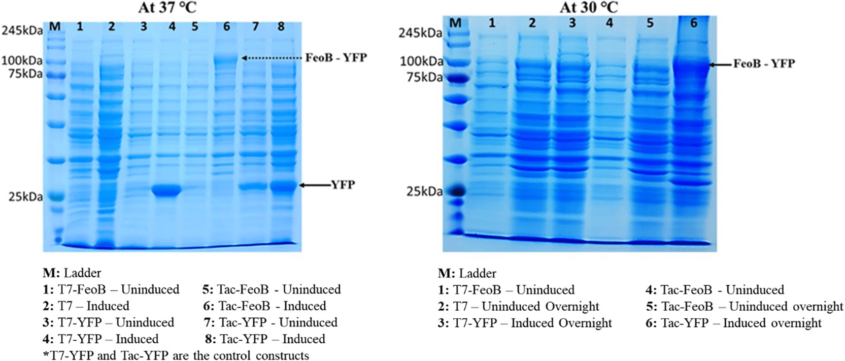
Figure 1. SDS–PAGE analysis of recombinant FeoB–YFP expressed in E. coli BL21 (DE3).
The left panel (37 °C) shows protein expression from T7- and Tac-driven constructs. Lanes 1 and 5: uninduced T7-FeoB–YFP and Tac-FeoB–YFP; lanes 2 and 6: induced T7-FeoB–YFP and Tac-FeoB–YFP; lanes 3 and 7: uninduced samples (replicates); lanes 4 and 8: induced samples (replicates). The band corresponding to FeoB–YFP is highlighted. Control constructs (T7-YFP and Tac-YFP) show strong YFP overexpression independent of promoter type. The right panel shows expression profiles of the same constructs at 30 °C. The PageRuler Prestained Protein Ladder (Thermo Scientific, USA) is indicated as M.
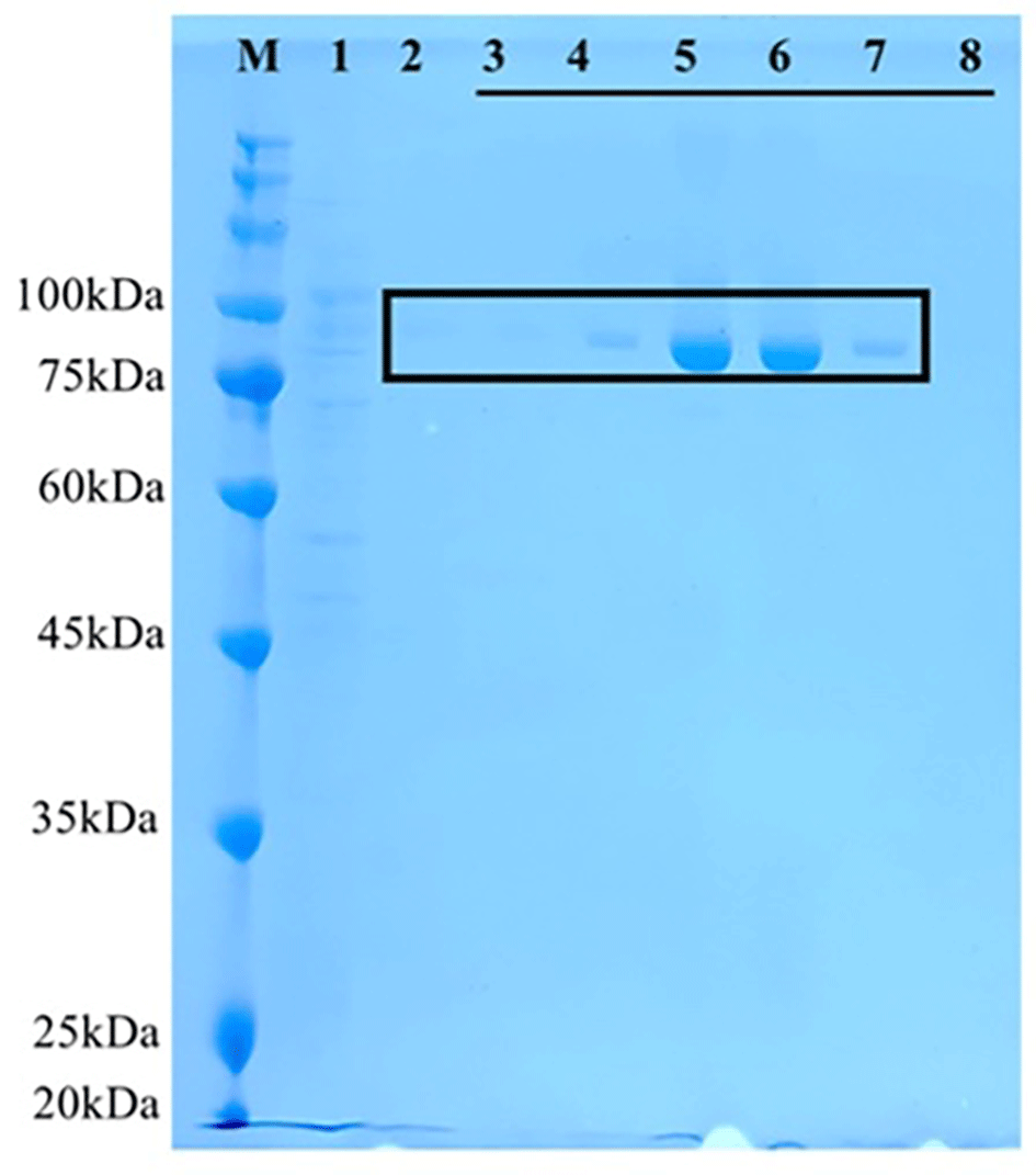
Figure 2. Purification of FeoB by Ni–NTA affinity chromatography.
The boxed region highlights elution fractions containing FeoB (~84.5 kDa). Elution fractions in lanes 5 and 6 were pooled for downstream analysis. Lane M: PageRuler Prestained Protein Ladder (Thermo Scientific™, USA); lane 1: lysate flowthrough; lane 2: binding/wash flowthrough; lanes 3–8: successive elution fractions containing FeoB.
Bioinformatic validation of FeoB architecture
SMART, IUPred2A, and UniProt analyses confirmed that the FeoB sequence contains canonical G-protein motifs, switch regions, ordered/disordered segments, and the predicted seven transmembrane helices (
Figure 3). These results validated the integrity of the FeoB constructs used for downstream assays.
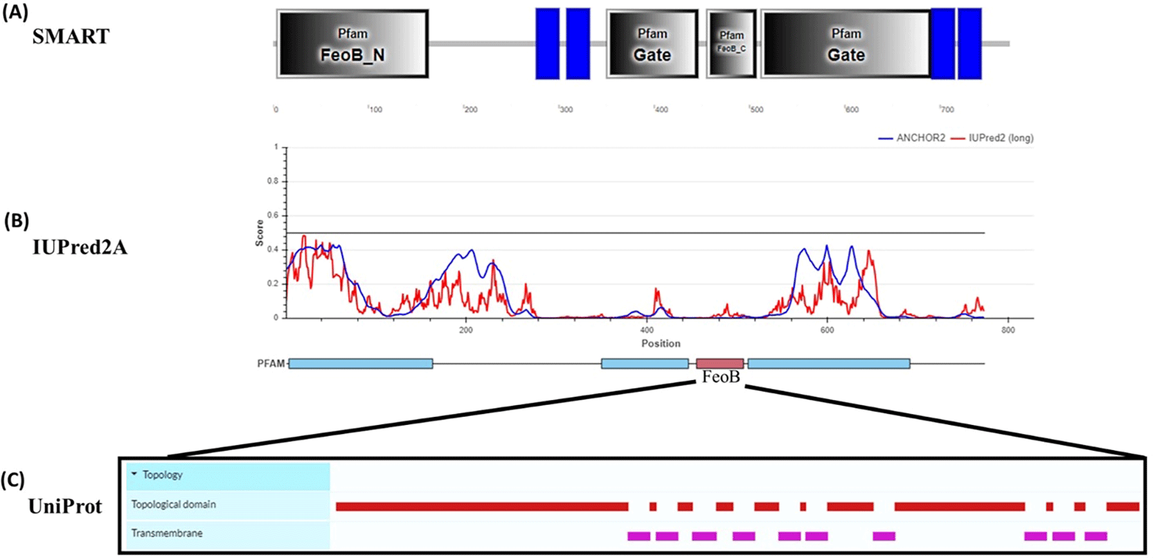
Figure 3. Bioinformatic analysis of the FeoB sequence.
(A) Visualization of predicted functional domains. (B) Amino acid sequence and homology predictions. (C) Functional annotations of the FeoB sequence.
GTPase activity is enhanced by iron and potassium
To test whether the GTPase domain retained activity, a luminescence-based GTPase assay was used to monitor time-dependent GTP hydrolysis by the purified protein. The activity of FeoB was significantly increased in the presence of Fe2+ and further enhanced upon addition of K+ under anaerobic conditions (
Figure 4). Control reactions lacking FeoB showed no activity. These findings align with earlier reports showing potassium-dependent activation of G-protein domains in FeoB. (Ash et al., 2010 (c)).
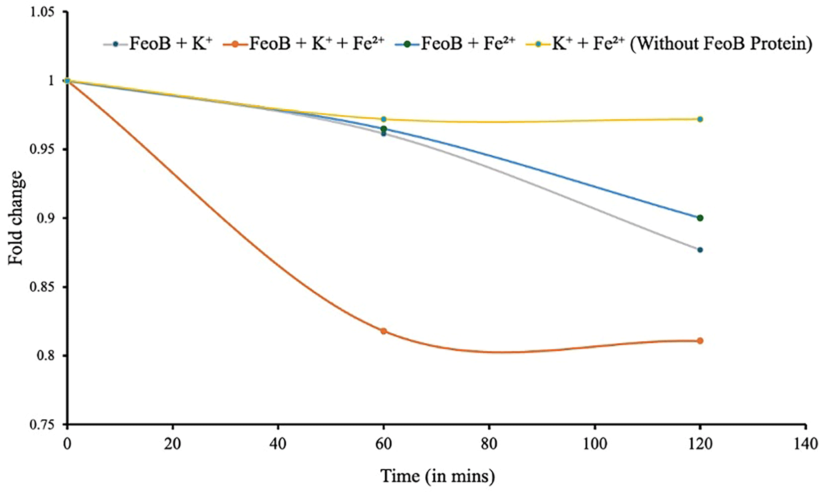
Figure 4. GTPase activity of FeoB measured using a luminescence-based assay.
A 10 μM FeoB sample exhibited measurable GTPase activity in the presence of 1 mM Fe2+, and this activity increased further upon addition of 200 mM K+. No detectable GTPase activity was observed in the absence of FeoB. Lower values indicate increased GTPase activity.
In vitro expression using cell-free systems
To facilitate reconstitution in lipid membranes, we probed whether FeoB-YFP could be expressed via in vitro transcription-translation. With E. coli Rosetta 2(DE3) cell extract, reactions expressing FeoB-YFP showed temperature-dependent profiles (
Figure 5). At 37 °C, expression was limited; however, robust YFP fluorescence emerged at 25–30 °C. The production of protein was confirmed by repeating the reactions with fluorescently labeled Lys residues with the PURE system and resolving the protein on an SDS-PAGE (
Figure 6).
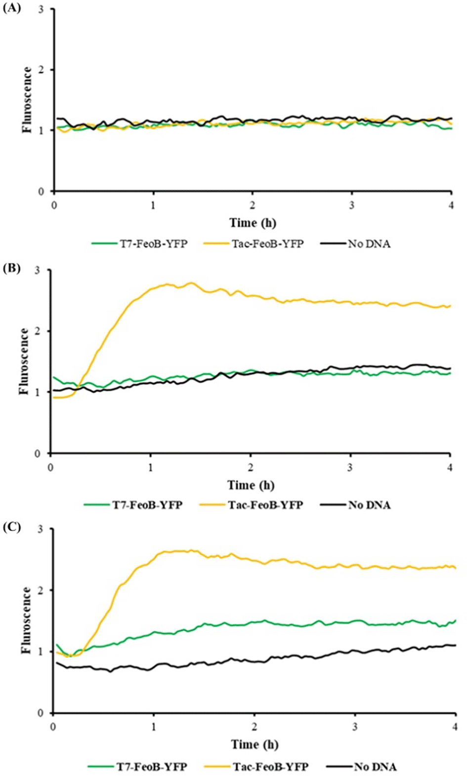
Figure 5. Fluorescence profiles of in vitro–expressed constructs T7-FeoB-YFP and Tac-FeoB-YFP at different temperatures using the E. coli Rosetta 2(DE3) cell-free system.
(A) Expression of the YFP-tagged FeoB driven by either the T7 or tac promoter was undetectable at 37 °C. (B) Lowering the temperature to 30 °C resulted in enhanced expression with a tac promoter. (C) At 25 °C, both constructs expressed protein, with a greater amount of detectable protein synthesized with the tac promoter. Yellow, T7 transcriptional promoter; green, tac transcriptional promoter; black, no template DNA.
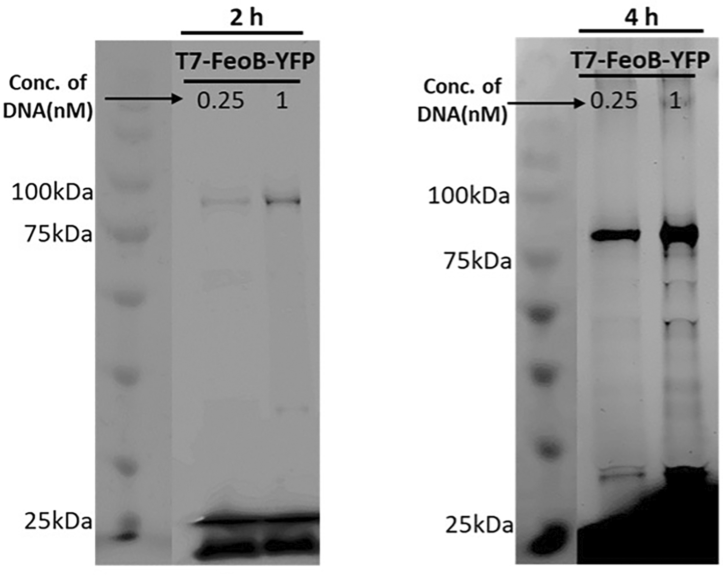
Figure 6. Detection of synthesized FeoB by fluorescent labeling.
Transcription-translation was with the PURE system, and the translated product was labeled with FluoroTect™ GreenLys. Reaction mixtures were resolved on a 12% SDS–PAGE gel, and fluorescence was detected with a laser scanner. FeoB synthesis was evident at both time points, with markedly stronger signal observed after 4 h. Aliquots were treated with RNaseA prior to loading on the gel.
Encapsulation of FeoB in lipid vesicles
In vitro transcription-translation in the presence of GUVs revealed the formation of GUVs containing FeoB-YFP integrated into the membrane, as determined by confocal fluorescence microscopy (
Figure 7). Fluorescent signal localized to vesicle membranes, indicating membrane integration and/or association. Osmotic balancing prevented vesicle rupture, improving reproducibility.
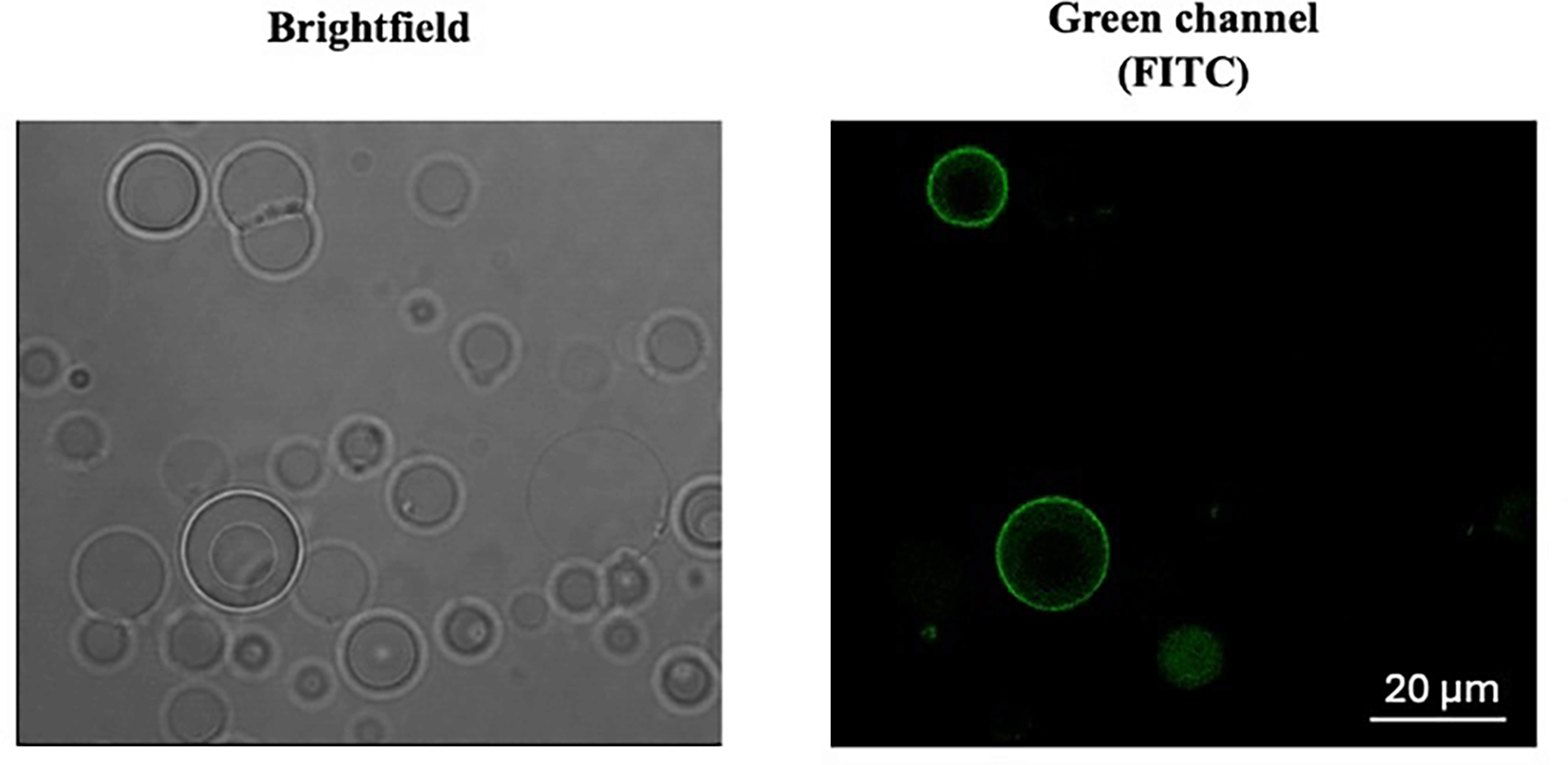
Figure 7. Confocal microscopy of 1:2 molar ratio POPC:POPG vesicles reveals a heterogeneous population of vesicle sizes containing FeoB–YFP after transcription–translation reaction with a tac-driven construct.
Encapsulated transcription-translation was with the E. coli Rosetta 2(DE3) cell extract. Reactions were run for 4 h at 37 °C. The left panel shows vesicles in brightfield phase contrast, enabling visualization of overall morphology and structural integrity. The right panel displays the corresponding green fluorescence, demonstrating the presence of FeoB–YFP within the vesicle lumen and confirming successful encapsulation of the fluorescent fusion protein.
Functional assessment using GTP analogs
Fluorescent BODIPY-GTP did not associate with protein-free vesicles but specifically bound to vesicles containing FeoB that had been encapsulated with BODIPY FL–labeled GTP (
Figure 8). This selective interaction indicates functional nucleotide binding by the G-domain within the reconstituted membrane environment.
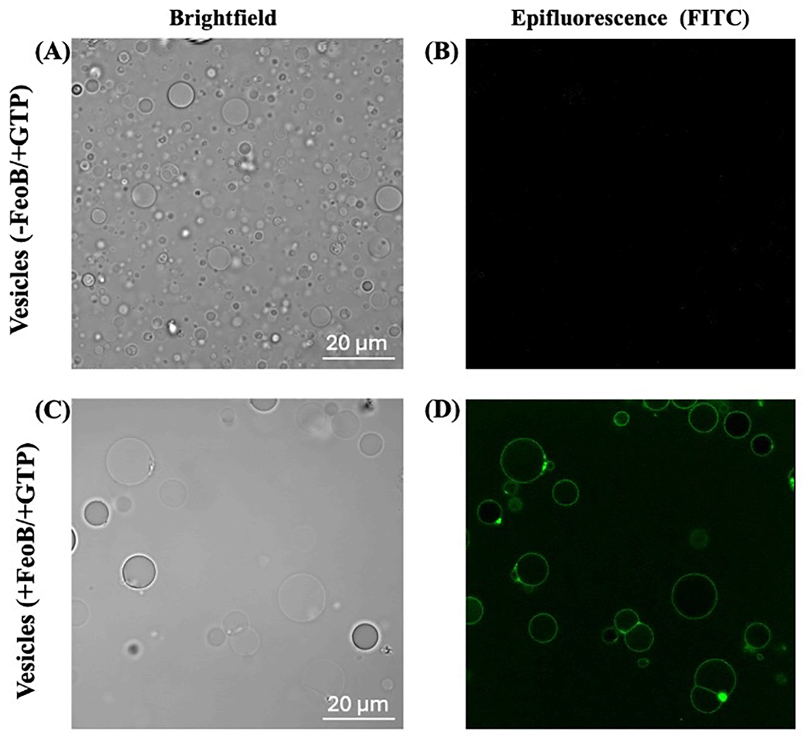
Figure 8. Functional assessment of FeoB using a fluorescent GTP analog.
In the upper panels (A, B), the brightfield image (left) shows the vesicle population, while the corresponding fluorescence image (right) demonstrates that green fluorescent BODIPY-labeled GTP (BODIPY FL GTP) does not associate with vesicles lacking protein. In contrast, the lower panels (C, D) reveal that vesicles reconstituted with FeoB exhibit clear binding of the fluorescent GTP analog, as indicated by the green BODIPY FL GTP signal.
Discussion
We established a platform to express, purify, and functionally characterize FeoB outside the native cellular context. Using complementary cell-based and cell-free approaches, we generated full-length FeoB and reconstituted the protein within synthetic 1:2 molar ratio POPC:POPG vesicles. This controlled environment enabled direct interrogation of FeoB’s biochemical properties, independent of cellular complexity.
Our observations confirm that the G-protein domain of FeoB remains active following reconstitution, binding fluorescent GTP analogs and catalyzing GTP hydrolysis. The enhancement of activity by Fe2+ and K+ is consistent with models proposing ion-dependent modulation of FeoB during ferrous iron transport (Yamanaka et al., 2000; Scrima, & Wittinghofer, 2006). These results support the view that FeoB employs GTP hydrolysis in a tightly regulated manner, potentially involving additional cofactors such as FeoA in vivo (Cartron et al., 2006).
The artificial membrane system developed here can serve as a modular platform to dissect mechanistic features of FeoB, including pore formation, nucleotide cycling, and ion coordination. Moreover, such systems provide a highly controllable environment for screening small-molecule inhibitors targeting FeoB-mediated iron acquisition—an attractive strategy for anti-virulence therapies.
Conclusions
We demonstrate that full-length FeoB can be successfully expressed, purified, and functionally reconstituted within synthetic lipid vesicles using cell-free systems. The resulting proteoliposomes retain GTP-binding and GTPase activity, modulated by iron and potassium ions. This work highlights the potential of minimal, cell-free membrane systems to uncover mechanistic details of complex membrane proteins and to support the rational development of FeoB-targeted inhibitors.
Ethics and consent
Ethical approval and consent were not required.
Data availability
All data supporting the findings of this study will be made available in an open repository upon publication (https://doi.org/10.5281/zenodo.20503086) (Rajanya, B. 2026). This includes raw fluorescence data, gel images, GTPase measurements, and plasmid sequences.
Acknowledgements
We would like to extend our gratitude to the staff and colleagues of Department of Cellular Computational and Integrative Biology for their contribution. This research was funded in part from the European Union’s Horizon 2020 research and innovation programme under grant agreement No. 824060 (project ACDC).
References
- Andrews SC, Robinson AK, Rodríguez-Quiñones F:
Bacterial iron homeostasis.
FEMS Microbiol. Rev.
2003; 27(2–3): 215–237. Publisher Full Text
- Ash MR, Guilfoyle A, Clarke RJ, et al.:
Potassium-activated GTPase reaction in the G Protein-coupled ferrous iron transporter B.
J. Biol. Chem.
2010; 285(19): 14594–14602. PubMed Abstract
| Publisher Full Text
| Free Full Text
- Ash MR, Maher MJ, Guss JM, et al.:
The initiation of GTP hydrolysis by the G-domain of FeoB: insights from a transition-state complex structure.
PLoS One.
2011b; 6(8): e23355. Publisher Full Text
- Ash MR, Maher MJ, Guss JM, et al.:
A suite of Switch I and Switch II mutant structures from the G-protein domain of FeoB.
Acta Crystallogr. D Biol. Crystallogr.
2011a; 67(11): 973–980. Publisher Full Text
- Brown R, Naujoks S, Richardson DJ, et al.:
Ins and outs: recent advancements in membrane protein-mediated prokaryotic ferrous iron transport.
Biochemist.
2021; 60(44): 3277–3291. Publisher Full Text
- Cartron ML, Maddocks S, Gillingham P, et al.:
Feo–transport of ferrous iron into bacteria.
Biometals.
2006; 19(2): 143–157. Publisher Full Text
- Caschera F, Noireaux V:
Synthesis of 2.3 mg/ml of protein with an all Escherichia coli cell-free transcription–translation system.
Biochimie.
2014; 99: 162–168. PubMed Abstract
| Publisher Full Text
- Cassat JE, Skaar EP:
Iron in infection and immunity.
Cell Host Microbe.
2013; 13(5): 509–519. PubMed Abstract
| Publisher Full Text
| Free Full Text
- Clarke TE, Tari LW, Vogel HJ:
Structural biology of bacterial iron uptake systems.
Curr. Top. Med. Chem.
2001; 1(1): 7–30. Publisher Full Text
- Eng ET, Jalilian AR, Spasov KA, et al.:
Characterization of a novel prokaryotic GDP dissociation inhibitor domain from the G protein coupled membrane protein FeoB.
J. Mol. Biol.
2008; 375(4): 1086–1097. PubMed Abstract
| Publisher Full Text
| Free Full Text
- Guilfoyle AP, Papp-Wallace KM, McGuire ML, et al.:
Structural basis of GDP release and gating in G protein coupled Fe2+ transport.
EMBO J.
2009; 28(17): 2677–2685. PubMed Abstract
| Publisher Full Text
| Free Full Text
- Hagelueken G, Huang H, Mainprize IL, et al.:
Studies on the X-ray and solution structure of FeoB from Escherichia coli BL21.
Biophys. J.
2016; 110(12): 2642–2650. PubMed Abstract
| Publisher Full Text
| Free Full Text
- Kammler M, Schön C, Hantke K:
Characterization of the ferrous iron uptake system of Escherichia coli.
J. Bacteriol.
1993; 175(19): 6212–6219. PubMed Abstract
| Publisher Full Text
| Free Full Text
- Lau CKY, Krewulak KD, Vogel HJ:
Bacterial ferrous iron transport: the Feo system.
FEMS Microbiol. Rev.
2016; 40(2): 273–298. Publisher Full Text
- Marlovits TC, Haase W, Herrmann C, et al.:
The membrane protein FeoB contains an intramolecular G protein essential for Fe (II) uptake in bacteria.
Proc. Natl. Acad. Sci. USA.
2002; 99(25): 16243–16248. PubMed Abstract
| Publisher Full Text
| Free Full Text
- Marshall R, Noireaux V:
Quantitative modeling of transcription and translation of an all-E. coli cell-free system.
Sci. Rep.
2019; 9(1): 11980. PubMed Abstract
| Publisher Full Text
| Free Full Text
- Mészáros B, Erdos G, Dosztányi Z, et al.:
IUPred2A: contextdependent prediction of protein disorder as a function of redox state and protein binding.
Nucleic Acids Res.
2018; 46(W1): W329–337. Publisher Full Text
- Page MGP:
The role of iron and siderophores in infection, and the development of siderophore antibiotics.
Clin. Infect. Dis.
2019; 69(Suppl 7): S529–S537. PubMed Abstract
| Publisher Full Text
| Free Full Text
- Petermann N, Hansen G, Schmidt CL, et al.:
Structure of the GTPase and GDI domains of FeoB, the ferrous iron transporter of Legionella pneumophila.
FEBS Lett.
2010; 584(4): 733–738. PubMed Abstract
| Publisher Full Text
- Rajanya B:
TX-TL Reaction, Microscopy. [Data set].
Zenodo.
2026. Publisher Full Text
- Raymond KN, Dertz EA, Kim SS:
Coordination chemistry of microbial iron transport.
Acc. Chem. Res.
2015; 48(9): 2496–2505. PubMed Abstract
| Publisher Full Text
| Free Full Text
- Scrima A, Wittinghofer A:
Dimerisation-dependent GTPase reaction of MnmE: how potassium acts as a GTPase-activating element.
EMBO J.
2006; 25(13): 2940–2951. Publisher Full Text
- Schultz J, Copley RR, Doerks T, et al.:
SMART: a web-based tool for the study of genetically mobile domains.
Nucleic Acids Res.
2000; 28(1): 231–4. Publisher Full Text
- Seyedmohammad S, Fuentealba NA, Marriott RA, et al.:
Structural and functional characterization of the bacterial ferrous iron transporter FeoB.
Biochem. J.
2016; 473(23): 3863–3874. Publisher Full Text
- Sun ZZ, Hayes CA, Shin J, et al.:
Protocols for implementing an Escherichia coli based TX-TL cell-free expression system for synthetic biology.
J. Vis. Exp.
2013; 79: e50762. PubMed Abstract
| Publisher Full Text
| Free Full Text
- Udipi SA, Ghugre PS, Gokhale CV:
Iron, oxidative stress and health.
Oxidative Stress – Molecular Mechanisms and Biological Effects.
InTech;
2012; 73–108. Publisher Full Text
- Wittinghofer A, Vetter IR:
Structure–function relationships of the G domain, a canonical switch motif.
Annu. Rev. Biochem.
2011; 80: 943–971. Publisher Full Text
- Yamanaka K, Hwang J, Inouye M:
Characterization of GTPase activity of TrmE, a member of a novel GTPase superfamily, from Thermotoga maritima.
J. Bacteriol.
2000; 182(24): 7078–7082. PubMed Abstract
| Publisher Full Text
| Free Full Text








