Plain language summary
Mushroom-forming fungi are an important sustainable food source. Furthermore, they make important medicinal substances and enzymes for biofuel production. To alter the characteristics of mushrooms, it is interesting to look at the effect of the integration of new genes that produce proteins. However, currently this often does not succeed or results in a sick mushroom, because the gene integrates in a random spot in the genome. Here, we developed a method to very precisely place a gene into a selected area with molecular scissors to reduce the variability of protein production. We test this method by inserting red-fluorescent protein and compare targeted and random integration. With targeted integrations, all obtained strains were very similar in the production of the red-fluorescent protein, while random integrations sometimes resulted in no, or very low production. This method can help us to get a better understanding of how mushrooms work and improve their production.
Introduction
Mushrooms are primarily used as a sustainable food source, but they also produce many pharmacological compounds and carbohydrate-active enzymes (de Mattos-Shipley et al., 2016). Schizophyllum commune is a mushroom-forming basidiomycete in the order Agaricales and serves as a model for fruiting body development due to its short life cycle and genetic accessibility (Ohm et al., 2010b). Unlike most mushroom-forming fungi, high transformation efficiency can be achieved in S. commune with PEG-mediated transformation of protoplasts (De Jong et al., 2010; Ohm et al., 2010a; Vonk et al., 2019). Additionally, an efficient gene-deletion (knock-out) method is available (Vonk et al., 2019). These methods provide quick and efficient genome editing tools to study the function of genes S. commune.
The study of gene function often requires the complementation of gene deletions, over-expression of genes, the expression of recombinant genes, or the heterologous expression of genes encoding fluorescent markers (Ohm et al., 2011; Ohm et al., 2013; Pelkmans et al., 2017; Vonk & Ohm, 2021a). The current gene-integration method relies on random integration of a plasmid that comprises the gene of interest and a selectable marker (Schuren & Wessels, 1994). This method reliably creates transformant strains, but expression of the integrated gene of interest can vary considerably between these strains and some do not show any expression of the integrated gene (Van Peer et al., 2009). This can likely be attributed to variations in copy number, to partial integration of the plasmid into the genome (which may result in an incomplete gene), and to the epigenetic state of the locus of integration. Moreover, some strains show phenotypes that are not caused by the integrated gene but by the interruption of a gene at the integration site. It is often difficult to distinguish between these two, which complicates the study of gene function. Fungal genomes generally have a high gene density. The 38.67 Mbp genome assembly of S. commune encodes 16,204 predicted genes, resulting in 419.03 genes per Mbp and a coding content of 52.89% (Ohm et al., 2010b). Including regulatory elements of the genome, such as promoters, enhancers, terminators and non-coding RNA transcripts, this results in a very dense genome where random integrations are likely to affect genetic elements at the site of integration.
This high variation of phenotypes between the transformants complicates the study of gene function, especially since the observed phenotype may not even be caused by the gene of interest. For this reason, generally several transformants are phenotyped in an effort to identify the actual phenotype of the gene integration. Instead, it would be beneficial to predictably target the gene of interest to a predetermined locus, leading to consistent expression dependent only on the selected promotor. This approach has already been applied in other fungal model species, including Saccharomyces cerevisiae and Neurospora crassa (Matsu-ura et al., 2015; Ronda et al., 2015), but not in any mushroom-forming fungus. Previously, homologous recombination (which is required for targeted integration) was not efficient enough in S. commune to make this a feasible strategy. Furthermore, transformation in S. commune always requires a selection marker, making the method used in S. cerevisiae (which does not require a selection marker) unfeasible. However, we recently developed a CRISPR-Cas9-based method that considerably increases the occurrence of homologous recombination (Vonk et al., 2019). Here, we modified this method to efficiently target a gene of interest and a nourseothricin selection marker to a non-coding locus by homologous recombination. We show that this greatly enhances the reproducibility of integration and gene expression compared with ectopic integrations.
Methods
Culture conditions and strains
S. commune was grown from a small agar inoculum on solid S. commune minimal medium (SCMM) supplemented with 1.5% agar at 30°C (Van Peer et al., 2009). All strains are derived from S. commune H4–8 (matA43matB41; FGSC 9210) (Ohm et al., 2010b). For targeted integration, a previously published Δku80 strain in a H4–8 background was used (De Jong et al., 2010). For selection on nourseothricin (Bio-Connect, Netherlands) or phleomycin (Bio-Connect, Netherlands), SCMM was supplemented with 15 μg/mL and 25 μg/mL antibiotic, respectively (Alves et al., 2004).
Plasmid construction
The locus selected for targeted integration was on scaffold 6: 2369122–2369196. The flanks upstream and downstream of this region were amplified with spi_up_fw + spi_up_NcoI_rv and spi_down_fw + spi_down_rv respectively (Table 2), resulting in 1106 bp and 1107 bp fragments. The primer spi_up_NcoI_rv introduced an NcoI site into the 3′ end of the upstream fragment for future cloning. The targeted integration plasmid was constructed as previously described for gene deletion plasmids (Vonk & Ohm, 2021a), resulting in plasmid pPV033 (available from Addgene under plasmid #178359). To construct the dTomato targeted integration plasmid, the tubulin promoter and dTomato were amplified from pPV040 with primers ptub_dtom_fw + ptub_dTom_rv resulting in a 1475 fragment (Table 2). The hom2 terminator was amplified from the S. commune H4–8 genome with primers hom2_term_fw and hom2_term_rv, resulting in a 1021 bp fragment (Table 2). Primers ptub_dtom_fw and hom2_term_rv contained 20 bp overhangs with pPV033 at the NcoI site. The tubulin promotor with dTomato and hom2 terminator were integrated in pPV033 digested with NcoI using NEB HiFi DNA Assembly Master Mix (New England Biolabs, MA, USA) resulting in plasmid pPV048.
Transformation of S. commune
Ectopic integration was performed as previously described (Van Peer et al., 2009). Targeted integration (knock-in) was performed using a similar approach as previously described for gene deletions (knock-out) (Vonk & Ohm, 2021a). A sgRNA was designed on the 75 bp and produced with the primers spi_sgRNA_fw + spi_sgRNA_rv (Table 2) with the GeneArt Precision sgRNA Synthesis Kit (ThermoFisher Scientific, MA, USA) according to manufacturer’s specifications (Vonk et al., 2019). The plasmid pPV048 was linearized with Anza NdeI (ThermoFisher Scientific, MA, USA). The candidate transformants were verified by PCR with two primers (spi_chk_fw + spi_chk_rv, Table 2), binding outside the upstream and downstream flanks site of integration. In the case of successful integration, this results in the insertion of 3780 bp at the 75 bp region inside the selected upstream and downstream flanks. For the check PCR this results in a change of band size from 2473 bp in the wild type to 6177 bp in transformants with a successful targeted integration.
Fluorescent imaging and analysis
The transformants and wild type H4–8 were grown for five days in triplicate at 25°C with a 16-hour light, 8-hour dark cycle. Imaging was done with an Euromex sCMED-20 camera on a Leica MZ16FA with a Leica Planapo 0.63× with fluorescent light from a Leica EL6000 at 1 second exposure and 10× digital gain. Pictures were converted to 8-bit grey-scale and the average fluorescence of pixels with an intensity >14 in a range from 0 to 255 was determined in ImageJ version 1.53c (RRID:SCR_003070) and used as an arbitrary unit of measurement of dTomato fluorescence (AU). The fluorescence intensity was normalized by subtracting the measured autofluorescence of the wild type H4–8. Both the raw and processed fluorescence data are available in the extended data (Vonk & Ohm, 2021).
Results
We identified a suitable locus for targeted integration using the following criteria: the neighboring genes (i) should be convergently transcribed and (ii) should be spaced at least 2 kbp apart, to minimize the odds of disrupting regulatory elements in a promoter. The neighboring genes (iii) should have stable expression during development and (iv) active histone 3 lysine 4 dimethylation (H3K4me2) during development (which indicates that the chromatin structure facilitates transcription), to increase the odds of stable expression solely under control of the selected promoter. Based on these criteria, a 2723-bp area on scaffold 6 from 2367903–2370626 was selected (Figure 1a). This region is flanked by a putative vacuolar transporter chaperone (protein ID Schco3|2627341, 57% amino acid identity match to S. cerevisiae VTC4) and a fungal-specific transcription factor (protein ID Schco3|2543274) that both have stable expression at approximately 40 FPKM during development (Almási et al., 2019). Furthermore, we previously determined that this region has high H3K4 dimethylation that does not change during development (Vonk & Ohm, 2021a).
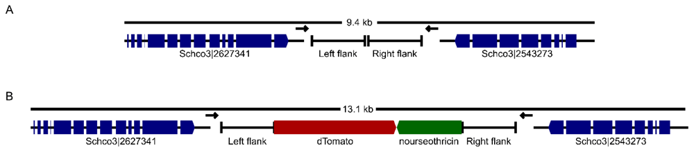
Figure 1.
Genomic region of scaffold 6 (2,364,297–2,373,735 bp) before (A) and after (B) integration of the targeted insertion. Arrows indicate the location of PCR primers to verify successful targeted knock-in transformants.
We created a plasmid to facilitate targeted integration, comprising a nourseothricin resistance cassette flanked by 1.1 kbp upstream and downstream homology flanks. Any promoter, gene of interest and terminator can be subcloned into an NcoI restriction site between the upstream flank and nourseothricin resistance cassette. Moreover, a phleomycin resistance cassette is located outside the homology flanks, allowing for counter-selection of transformants that did not originate from a double cross-over. Upon successful integration into the genome, this would result in the deletion of 75 bp at the integration site, replacing it with the nourseothricin resistance cassette and the gene of interest (Figure 1). Homologous recombination during the transformation is further induced by a CRISPR-Cas9 ribonucleoprotein programmed to cut in the integration site.
We determined the efficacy of our method by comparing transformants generated by the traditional ectopic integration approach with transformants generated with our novel targeted integration approach. As a marker gene, we used dTomato under control of the tubulin promoter (Figure 1b), which allowed us to efficiently determine the variation between the transformants by measuring the colony-wide intensity of red fluorescence as well as its distribution pattern across the colony. We first transformed wild-type S. commune with our plasmid but without Cas9-RNPs, which resulted in random integration, as verified by PCR (Figure 2a). 12 nourseothricin-resistant transformants, named E1–E12 were screened for dTomato fluorescence (Figure 2b). The fluorescence intensity varied greatly between the transformants with a spread of 115.4 arbitrary units of fluorescence (AU) between the strain with the lowest and highest fluorescence (0.6 AU and 116 AU, respectively) (Table 1). Strain E11 showed very little fluorescence, while strain E1 had the highest fluorescence (Figure 2d).
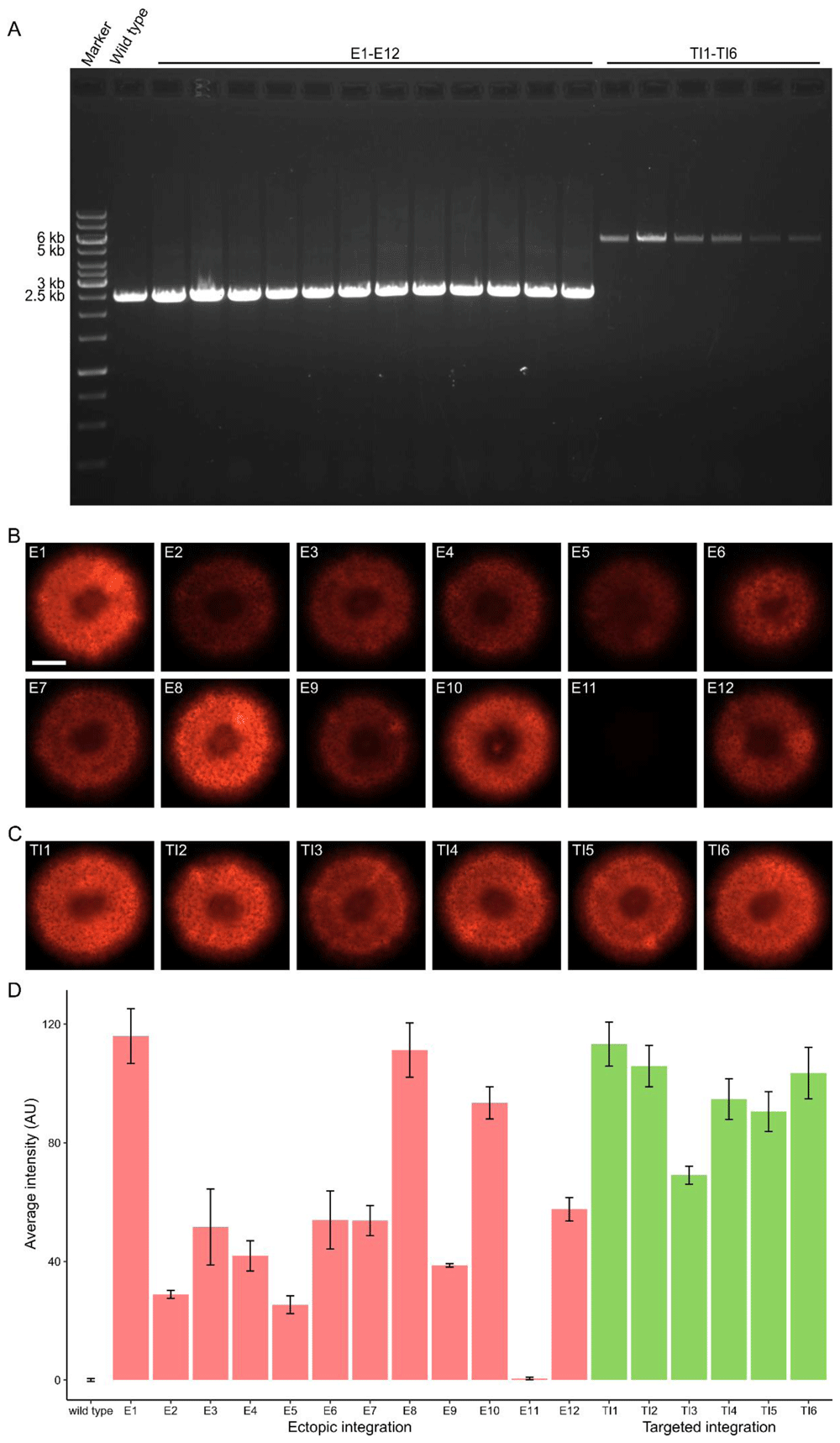
Figure 2. Genotyping and phenotyping the strains transformed with the dTomato targeted insertion plasmid.
A: PCR on the targeted integration locus. Integration of the dTomato expression cassette and nourseothricin cassette results in an increase in band size from 2473 bp to 6178 bp. All ectopic integration strains show the wild type band, while all targeted knock-in strains show the larger band that indicates successful integration. B: Strains with ectopic integration of dTomato. The white bar indicates 1 cm. C: Strains with targeted integration of dTomato. D: Quantification of fluorescence intensity in arbitrary units. On average, fluorescence is more consistent in strains generated by targeted integration, compared to ectopic integration. The fluorescence was measured in three biological replicates. The raw fluorescence photos and quantifications can be found in the supplementary data.
Table 1. Statistics on dTomato fluorescence after ectopic and targeted integration.
| Strain | Average
fluorescence
intensity | Minimal
fluorescence
intensity | Maximum
fluorescence
intensity | Spread | Variance |
|---|
| Ectopic integration | 56.1 | 0.6 | 116 | 115.4 | 1110.96 |
| Targeted integration | 96.2 | 69.1 | 113.3 | 44.2 | 201.2 |
Table 2. Primers used in this study.
Underlined sequences indicate homologous sequences for Gibson assembly.
| Primer name | Sequence |
|---|
| spi_up_fw | CTATGACCATGATTACGCCAGCCAACAATCAGATCCGCTAGC |
| spi_up_NcoI_rv | TCCCAGACCACCATGCCGGGCCATGGAGCAACCTCGGTGCTTATGACT |
| spi_down_fw | GTCCCCCTCGAGGCGCGCCGGGGTCCTTTCTGACGATGACCA |
| spi_down_rv | GATAACCTTCAGCAGAACGACGTGACTTTTCGCCGTCTGTAC |
| ptub_dtom_fw | CATAAGCACCGAGGTTGCTCAAGCTTTGGAACCGGGCCTCAA |
| ptub_dtom_rv | CTTGTACAGCTCGTCCATGC |
| hom2_term_fw | GCATGGACGAGCTGTACAAGTAGCCCCTGGCAATCATTTGC |
| hom2_term_rv | CCCAGACCACCATGCCGGGCTGACCTTGGGGAAAATCAAG |
| spi_sgRNA_fw | TAATACGACTCACTATAGGTCCTCCAGAGCTCCGCGCCA |
| spi_sgRNA_rv | TTCTAGCTCTAAAACTGGCGCGGAGCTCTGGAGGA |
| spi_chk_fw | GCAGATGCTGGATAGGCTTC |
| spi_chk_rv | GGGACATGGTGGTAGAGAGC |
Next, we created targeted knock-in transformants by introducing the plasmid in a Δku80 strain (which is deficient in non-homologous end-joining) assisted by Cas9-RNPs. This resulted in six nourseothricin-resistant, phleomycin-sensitive strains, named TI1–TI6, that were all red fluorescent (Figure 2c). With PCR, it was confirmed that all targeted knock-in transformants integrated the dTomato gene and nourseothricin resistance cassette at the targeted locus (Figure 2a). Compared to the ectopic integration transformants, the spread in fluorescence intensity between the lowest and highest fluorescent strain was much lower at 44.2 AU (69.3 AU and 113.2 AU, respectively) (Table 1). Furthermore, the variance was markedly lower. Additionally, the average intensity was 40.1 AU higher in the targeted integration transformants. While no strain showed more intense fluorescence than ectopic integration strain E1 (possibly due to several integrations of the plasmid), only three of the twelve ectopic integration strains had a higher fluorescence than the lowest fluorescence intensity measured in the targeted knock-in transformants (Figure 2d).
Discussion
We developed a method for targeted gene integration (knock-in) in S. commune. This method greatly reduced the variation in fluorescence intensity between the transformants from 115.4 AU to 44.2 AU (in transformants resulting from ectopic integration and targeted integration, respectively). Average dTomato fluorescence was also stronger during targeted integration, indicating that the selected region for targeted integration is suitable for expression of genes. Unlike a similar method applied in S. cerevisiae, our method does rely on the integration of a selection marker (Ronda et al., 2015). It was previously shown that homologous recombination and transformation efficiency are not sufficient for transformation without a selection marker in S. commune and therefore a selection marker is still required (Vonk et al., 2019). Although the transformation efficiency with targeted integration is still markedly lower than with ectopic integrations, it is comparable to the efficiency of Cas9-RNP-mediated gene deletion and therefore sufficient for gene integration. Although the size of the integration was considerably larger (3.8 kbp) than in the case of a gene deletion (1.3 kbp), the transformation efficiency did not appear to be negatively affected.
Together, this method expands on the toolkit available for functional genomics in S. commune with reliable and consistent expression of integrated genes. This can be used for the complementation of previously deleted genes, but also the integration of genes modified in their active sites. An example of this is the mutation of the phosphorylation sites of the homeodomain transcription factor hom2 (Pelkmans et al., 2017). Furthermore, combined with our previously developed ChIP-Seq protocol, it enables efficient integration and expression of epitope-tagged transcription factors to identify their binding sites (Vonk & Ohm, 2021).
Data and software availability