Background
Malaria remains a major public health problem with an estimated 282 million malaria cases in 2024.1 Most of the cases were caused by Plasmodium falciparum, the deadliest parasite species that is dominant in sub-Saharan Africa. P. vivax is the second leading cause of malaria worldwide and is mostly associated with malaria-related disease burden in South and Southeast Asia, and South America.2 However, P. vivax also contributes substantially to malaria cases on the African continent, where it was long considered absent due to the high prevalence of Duffy negativity.3 This is supported by a recent study from Ethiopia that demonstrates P. vivax can infect Duffy-negative individuals.4 Recognizing this substantial disease burden, the World Health Organization has prioritized P. vivax research in its Malaria Vaccine Technology Roadmap.5
P. vivax biology differs markedly from P. falciparum and poses unique challenges.6 One of its most distinctive features is the ability to form hypnozoites, dormant liver stage parasites that can reactivate weeks to months after the initial infection and are responsible for most recurrent episodes.7 The study of P. vivax and their hypnozoite formation would benefit from a reliable source of viable sporozoites. However, the reproducible establishment of long-term continuous in vitro culture of P. vivax has so far proven to be an unattainable goal.8 This is largely due to P. vivax exclusively invading immature red blood cells, so-called reticulocytes. As a result, studying sporozoites and hypnozoites continues to be dependent on mosquitoes infected by natural gametocyte carriers, typically involving patients at remote field sites where experimental conditions can be challenging. Although natural infections can reach high parasite densities, and P. vivax gametocytes appear early in infection, mosquito infection success remains variable and unpredictable.9 These challenges pose a relevant bottleneck for research on pre-erythrocytic vaccines as well as for studies on hypnozoites. A method to achieve higher and more reproducible mosquito infection would be highly valuable for downstream applications.
Two gametocyte enrichment approaches have proven to be effective for P. falciparum: magnetic-activated cell sorting (MACS), exploiting the paramagnetism of mature gametocytes,10–12 and Percoll density gradient centrifugation (DGC), separating infected erythrocytes by density.13 While both methods have been repeatedly used for P. falciparum, information on their suitability for P. vivax is sparser.14–16 Here, we utilized gametocyte-positive blood samples from a P. vivax PvW1 controlled human malaria infection (CHMI) to systematically compare both techniques and establish a protocol that achieves the highest proportion of infected mosquitoes and the highest infection burden (oocyst density) in these mosquitoes. We also confirm completion of sporogonic development in mosquitoes by detecting sporozoites in mosquito salivary glands.
Methods
Plasmodium falciparum and Plasmodium vivax gametocytes
Concentration methods were first tested using P. falciparum gametocytes (NF54) that were cultured in a tipper system,17 as previously described.18 Mosquito feeding experiments with P. falciparum were performed on day 14 post seeding, by spiking pelleted cultured gametocytes at a concentration of 0.75 million/mL into 10 mL fresh heparinized blood (ref 367526, BD). P. vivax gametocytes were the primary focus of this study and obtained during the RaViCHMI1 clinical trial (ID NL-OMON57011) that will be published separately. Briefly, study participants were inoculated intravenously with the blood-stage P. vivax W1 challenge inoculum (PvW1), provided by the University of Oxford.19 PvW1 is a clonal parasite line from Thailand that has been cryopreserved in 2021.20 P. vivax mosquito feeding experiments were conducted using blood samples from eight participants with a positive thick blood smear at a parasitemia between 2–38 parasites/μL between day 18 to 24 post infection (ID NL-OMON57011). Heparinized blood was obtained from study participants by venipuncture and immediately transferred to a water bath set at 37 °C until sample processing. All additional blood products required were supplied by the national blood bank (Sanquin Nijmegen, The Netherlands).
Mosquito husbandry and Plasmodium infection
Anopheles stephensi mosquitoes of the Sind-Kasur strain were reared at 30 +/− 1 °C and ~ 80% relative humidity, in a room with a reversed light period (12 h shifts).21 P. vivax infected mosquitoes received an extra uninfected blood-meal on day 6 post-infection to harmonize oocyst and sporozoite development22 and were housed at 26 +/− 1 °C with ~80% relative humidity. For all membrane feeding assays An. stephensi mosquitoes were allowed to feed for 10 min in the dark on blood in water-jacketed membrane feeders. For the membrane feeding assay with un-enriched gametocytes, about 100 female mosquitoes between 1–3 days old were collected in cages and fed using midi glass feeders (approximately 1.2 mL).23 For gametocyte enriched feeds, about 50 female mosquitoes (1–3 days old) were collected in cups and fed using mini glass feeders that contained approximately ~300 μL material.23 Unfed and partially fed mosquitoes were removed. Fully fed mosquitoes were maintained on 5% glucose (D+ Glucose Monohydrate, ref 49159, Sigma) and dissected 5 to 8 days post feeding to count the number of developed oocysts on midguts by microscopy after 1% mercurochrome staining.
Membrane feeding assays
All steps until mosquito feeding, including centrifugation, were performed at 37 °C to prevent early activation of gametocytes and thereby a potential loss of infectivity. Blood meals were prepared with lithium heparinized blood (ref 367526, BD) for P. falciparum cultured gametocytes, or with blood obtained by venipuncture (again in lithium heparin tubes) from participants enrolled in a P. vivax CHMI. The non-enriched blood meal (control) for P. falciparum was prepared as previously described.24 For P. vivax-infected blood, plasma for the non-enriched feed was replaced with malaria-naive serum (group AB; Sanquin blood bank, The Netherlands).
Prior to gametocyte-enrichment, both P. falciparum cultured gametocytes and P. vivax gametocyte-infected in whole blood were leukodepleted by Plasmodipur (Europroxima) filtration. After flushing the filter with 2 mL pre-warmed RPMI medium, 10 mL blood material was gently filtered into a 15 mL tube. Tubes were spun for 5 min at 760 g after which plasma was removed without disturbing the top layer of erythrocytes. Blood was then brought to a hematocrit of 50% by resuspension with pre-warmed RPMI medium.18
The MACS procedure was performed as previously described,12 with minor modifications. Briefly, columns were attached to the QuadroMACS™ separator (Miltenyi Biotech) in a 37 °C incubator, a hypodermic needle (25G) was attached, and the column was equilibrated with 1 mL pre-warmed RPMI medium. Subsequently, 3 mL of blood diluted in RPMI (50% hematocrit) was loaded onto the column; this was repeated on the same column until a total volume of 10 mL blood was processed. Columns were subsequently washed two times with 1 mL RPMI medium to remove unbound erythrocytes (referred to as wash 1 and wash 2). Columns were then detached from the magnet and placed in 15 mL tubes. Twice 1 mL RPMI medium was added to collect the bound fraction; the plunger that was provided with the column was used after the second 1 mL medium addition to expel all the retained liquid. Collection tubes were spun at 760 g for 1 min. The supernatant was carefully removed and approximately 1 mL was left on top of the pellet.
Percoll density gradient centrifugation was performed as previously described,13 with minor modifications. A 90% isotonic Percoll (SIP) solution (Cytiva, 17089101) was prepared with PBS (10x) on the day of use. The SIP solution was subsequently used to prepare 65% and 70% Percoll working solutions (v/v) in PBS (1x). 6 mL aliquots were prepared in 15 mL tubes and pre-warmed at 37 °C. The 10 mL blood sample was divided over 3 Percoll tubes as follows: a maximum of 3.3 mL blood (50% hematocrit) was gently layered via the wall of the tube onto the Percoll, using a 3 mL syringe with blunt needle. Tubes were spun at 760 g or 1500 g for 15 min at 37 °C without break. A 3 mL syringe with blunt needle was used to collect and transfer the layer on top of the gradient to a new 15 mL tube. Samples were topped up to 12 mL with pre-warmed RPMI and spun at 760 g for 5 min at 37 °C. The supernatant was carefully removed without disturbing the pellet, and the washing procedure was repeated two times. After the final wash, 0.5 mL of supernatant was left on top of the pellet. Subsequently, pellets from three tubes (from one study participant) were resuspended and combined.
The resuspended pellet obtained by MACS or DGC was transferred to a 1.5 mL Eppendorf tube in a heating block set at 37 °C. 180 μL of pelleted erythrocytes from a universal donor (for P. falciparum) or from the same clinical trial participant were added. Tubes were spun in a benchtop centrifuge for 20 s at 10,000 rpm. The supernatant was very carefully removed, and the pellet was resuspended in 150 μL serum (group AB). The complete protocols can be found at protocol.io (Extended data).
Plasmodium vivax sporozoite quantification by microscopy
A subset of mosquitoes was dissected between day 11 and 20 post-blood feeding to collect the salivary glands, collecting 2 glands with 3 lobes each, into a glass pestle grinder containing 1 mL PBS (1x). After tissue homogenization, 10 μL from the suspension was loaded into a Bürker-Türk counting chamber that was left for 20 min at RT to allow sporozoites to settle. Sporozoites were counted using a phase contrast light microscope with a final magnification of 400x.
Plasmodium vivax quantification by q (RT)PCR
To quantify P. vivax parasites in material collected during MACS processing, 100 μL sample was collected in a 1.5 mL Eppendorf tube and mixed with 400 μL of RNA protect Cell Reagent (Qiagen). Samples were stored at −20 °C and extracted within 10 weeks post-collection. Total nucleic acid (NA) was extracted with the automated MagNAPure 96 instrument (Roche) using the MagNA Pure 96 DNA and Viral NA Large Volume Kit (Roche, Basel, Switzerland) and eluted in 50 μL. The number of P. vivax parasites was quantified by 18S rRNA quantitative PCR (qPCR) as described previously,25 using the forward- 5′-GCTTTGTAATTGGAATGATGGGAAT-3′ and reverse primer 5′- ATGCGCACAAAGTCGATACGAAG −3′. A probe was used for amplicon detection 6FAM-AGCAACGCTTCTAGCTTA-MGBNFQ. P. vivax gametocytes were quantified by qRT-PCR, amplifying pvs25 as described previously,25 using the forward- 5′- ACACTTGTGTGCTTGATGTATGTC -3′ and reverse primer 5′- ACTTTGCCAATAGCACATGAGCAA −3′ set. A probe was used for amplicon detection 6FAM-TGCATTGTTGAGTACCTCTCGGAA-BHQ1. The number of parasite copies was calculated with a standard curve, obtained using 10-fold dilutions of plasmids as previously described by Tadesse et al.26 The MACS-column binding efficiency (%) was calculated by dividing copies in the bound fraction against the the total number of copies in the sample (total copies in bound + flow-through + wash 1 and 2 fractions).
Statistical analysis
Statistical analyses were performed using R (version 4.3.2). For oocyst density, values were analysed on the log10 scale. We fitted a linear model with condition (direct vs each enrichment method) as a categorical predictor and tested each enrichment method against the direct control using planned pairwise contrasts, reporting estimated mean differences with 95% confidence intervals and two-sided p-values. For ease of interpretation, these estimated differences on the log10 scale were back-transformed to mean fold changes (ratios) by exponentiation, yielding the enrichment-to-control oocyst density ratio. We analyzed the percentage infected mosquitoes using a linear model with condition as predictor, again testing each enrichment method versus the direct non-enriched control. Data are presented as violin plots with 95% confidence intervals. Correlations in proportion of infected mosquitoes and oocyst density between control and enrichment methods were visualized as scatter plots with dashed lines indicating a 1:1 reference. The relationship between oocyst prevalence and oocyst density was assessed using a logistic regression model, and 95% confidence intervals were calculated for the model fit. The significance level was set at a two-tailed p < 0.05.
Results
Optimizing P. falciparum gametocyte enrichment to enhance mosquito infection
The enrichment protocols were first examined for P. falciparum gametocytes. Methods tested were MACS with LS columns and two DGC gradients with two centrifugation speeds: Percoll 65%14 (P65) and 70%15 (P70) spun at a centrifugation speed of 760 g11 or 1500 g14 (Figure 1). All gametocyte enrichment methods increased the oocyst density and the proportion of infected mosquitoes (Figure 2A, B) relative to the non-enriched sample (control). Mean oocyst densities were as follows: control, 3.0 (range 0.6–6.2); MACS, 11.0 (range 4.9–21.2); DGC65-760 g 16.1 (7.9–29.4); DGC65–1500 g,
10.0 (6.3–16.1); DGC70–760 g,
17.4 (11.2–23.6); DGC70–1500 g,
21.2 (18.6–23.8) (Extended data). Mean infection rates were control, 66% (76/116); MACS, 88% (105/119); DGC65-760 g,
98% (55/56); DGC65-1500 g,
85% (51/60); DGC70-760 g,
90% (36/40); DGC70-1500 g,
97% (36/37) (Extended data). DGC70 enrichment resulted in the highest oocyst densities (Figure 2B) compared with control. DGC70-760 g and 1500 g increased oocyst density 7.5-fold (95% CI 2.0–29.1; p = 0.007) and 9.8-fold (95% CI 2.5–37.6; p = 0.004), respectively. The proportion of mosquitoes that became infected similarly increased with absolute increases of 24.8% (95% CI −1.8 to 51.4; p = 0.06) and 31.9% (95% CI 5.2–58.5; p = 0.02) for DGC-760 g and 1500 g, respectively (Figure 2A). DGC65–760 g achieved a 6.3-fold increase in oocyst density compared with control (95% CI 1.9–21.1; p = 0.006) (Figure 2B). This was accompanied by an absolute increase in the proportion of infected mosquitoes of 33.1% (95% CI 9.3–57.0; p = 0.01) (Figure 2A). In comparison, DGC65–1500 g and MACS resulted in smaller increases in oocyst density of 4.3-fold (95% CI 1.3–14.2; p = 0.02) and 4.4-fold (95% CI 1.3–14.8; p = 0.02), respectively. Their effects on the proportion of infected mosquitoes were comparable to those observed with DGC70–760 g, with absolute increases of 19.8% (95% CI 4.0–43.6; p = 0.09) for DGC65–1500 g and 23.1% (95% CI −0.7 to 46.9; p = 0.06) for MACS.
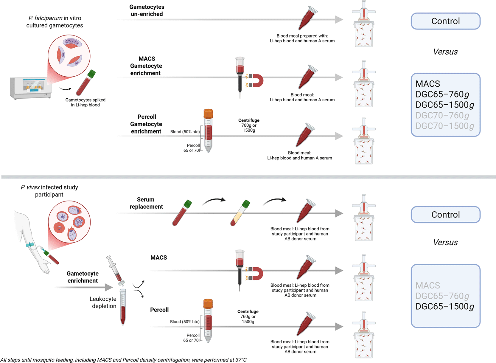
Figure 1. Membrane feeding assays performed with P. falciparum and P. vivax gametocytes.
Plasmodium falciparum (in vitro cultured) and P. vivax (ex vivo) gametocytes were MACS and Percoll enriched, and fed to Anopheles stephensi mosquitoes to compare the proportion of infected mosquitoes and oocyst density against non-enriched blood (control). The boxes on the right of the figure indicate the tested control or enrichment conditions; enrichment conditions presented in grey were tested but found to be inferior to those presented in black.
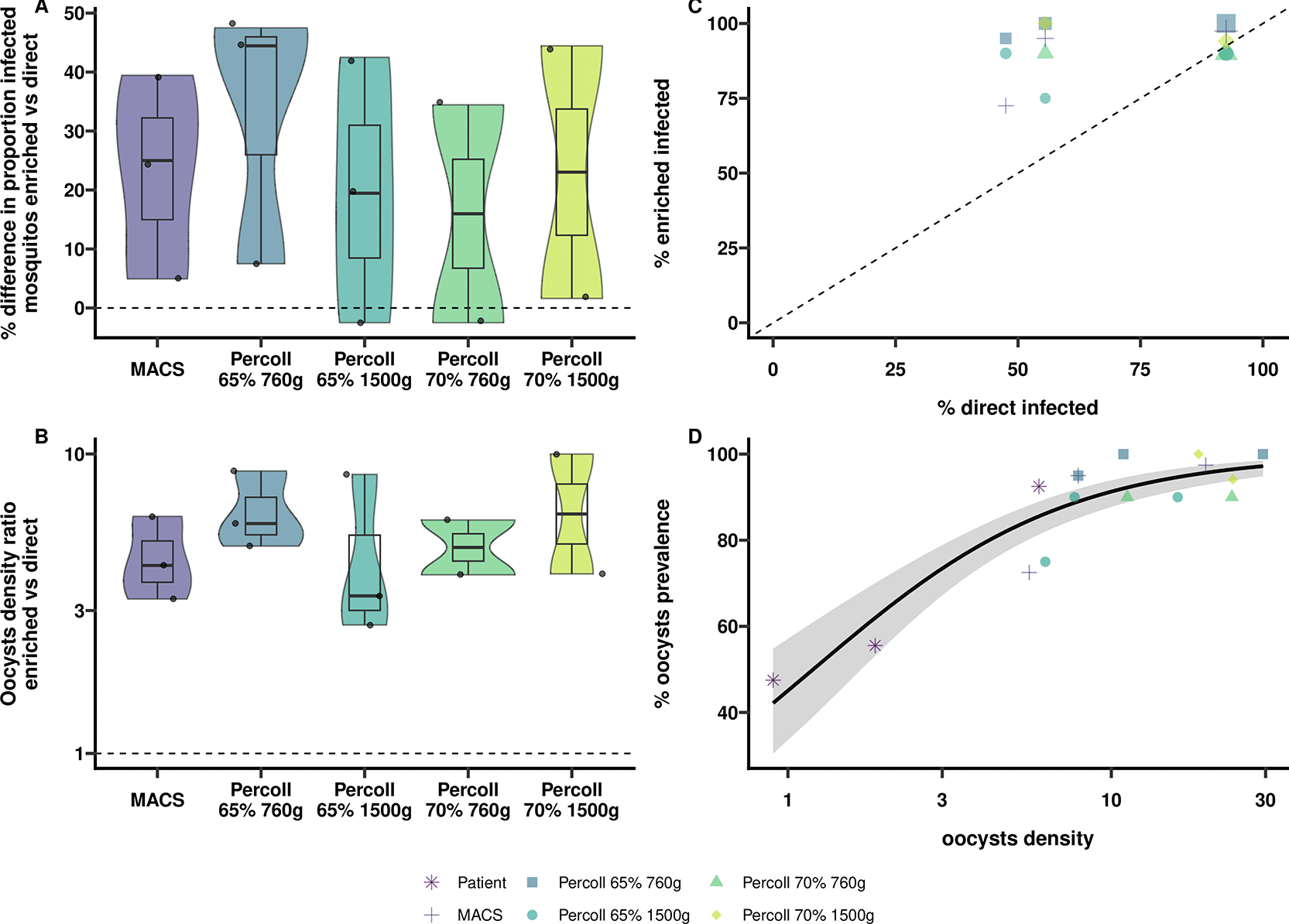
Figure 2. Comparing enrichment methods for P. falciparum cultured gametocytes.
Blood from healthy malaria-naive blood donors was supplemented with mature cultured P. falciparum gametocytes. Non-enriched blood (control) was compared to gametocyte enriched blood (volume 2.5 mL) in the membrane feeding assay, using Anopheles stephensi mosquitoes and mini-feeders (300 μL). (A) Violin plots with 95% CI comparing the difference in proportion of infected mosquitoes (%) between control and gametocyte enriched blood feeding, using MACS and Percoll density gradient centrifugation (at two concentrations and spinning speeds). The dashed horizontal line shows a difference of 0 (i.e. equality). (B) Violin plots with 95% CI comparing mosquito oocyst density ratio of control feeding versus feeding after gametocyte enrichment method. Dashed horizontal line shows a ratio of 1 (i.e. equality). (C) Association of mosquito infection prevalence (%) between control and enriched-feeds, the size of the geometric figures depict the oocyst intensity. The dashed line shows a 1:1 association. (D) Trend for oocyst prevalence amongst positives versus oocyst density found by fitting a logistic regression model. The grey shaded area reflects the pointwise 95% confidence interval.
Although enrichment of P. falciparum gametocytes with DGC70 was achieved in two experiments, blood occasionally failed to penetrate the gradient after centrifugation (data not shown). Because this was observed repeatedly, independent of centrifugation speed, this condition was not considered in further evaluations. Both DGC65 and MACS were further assessed for their applicability in enriching P. vivax gametocytes.
Comparing P. vivax gametocyte enrichment methods to enhance mosquito infection
Next, we evaluated P. vivax gametocyte enrichment by DGC65 (spun at 760 g or 1500 g) or MACS (LS columns); both were examined relative to the non-enriched control (Figure 1). Blood was drawn from eight study participants in a P. vivax blood-stage CHMI. Paired observations were collected for un-enriched versus enriched gametocytes with a total of 27 observations for DGC65-760 g, 28 for DGC65-1500 g, and 8 for MACS. For each condition, 11 mosquitoes were dissected to determine oocyst counts. DGC65-1500 g achieved the highest oocyst density and proportion of infected mosquitoes (Figure 3A, B). Compared with control, DGC65-1500 g increased the oocyst density 86-fold (95% CI 14–525; p < 0.0001) and increased the proportion of infected mosquitoes by 58.3% (95% CI 42.6–74.0; p < 0.0001). More modest effects were observed with DGC65-760 g that increased oocyst density 11-fold (95% CI 2.9–42.6; p < 0.001) and the proportion of infected mosquitoes by 28.0% (95% CI 12.1–43.9; p < 0.001) (Figure 3A, B). In contrast to all DGC conditions, MACS enrichment did not improve P. vivax mosquito infection, showing no increase in oocyst density (density ratio 0.68; 95% CI 0.09–5.28; p = 0.71) or in the proportion of infected mosquitoes (−5.4%; 95% CI −29.6 to 18.9; p = 0.66) compared to control (Figure 3A, B).
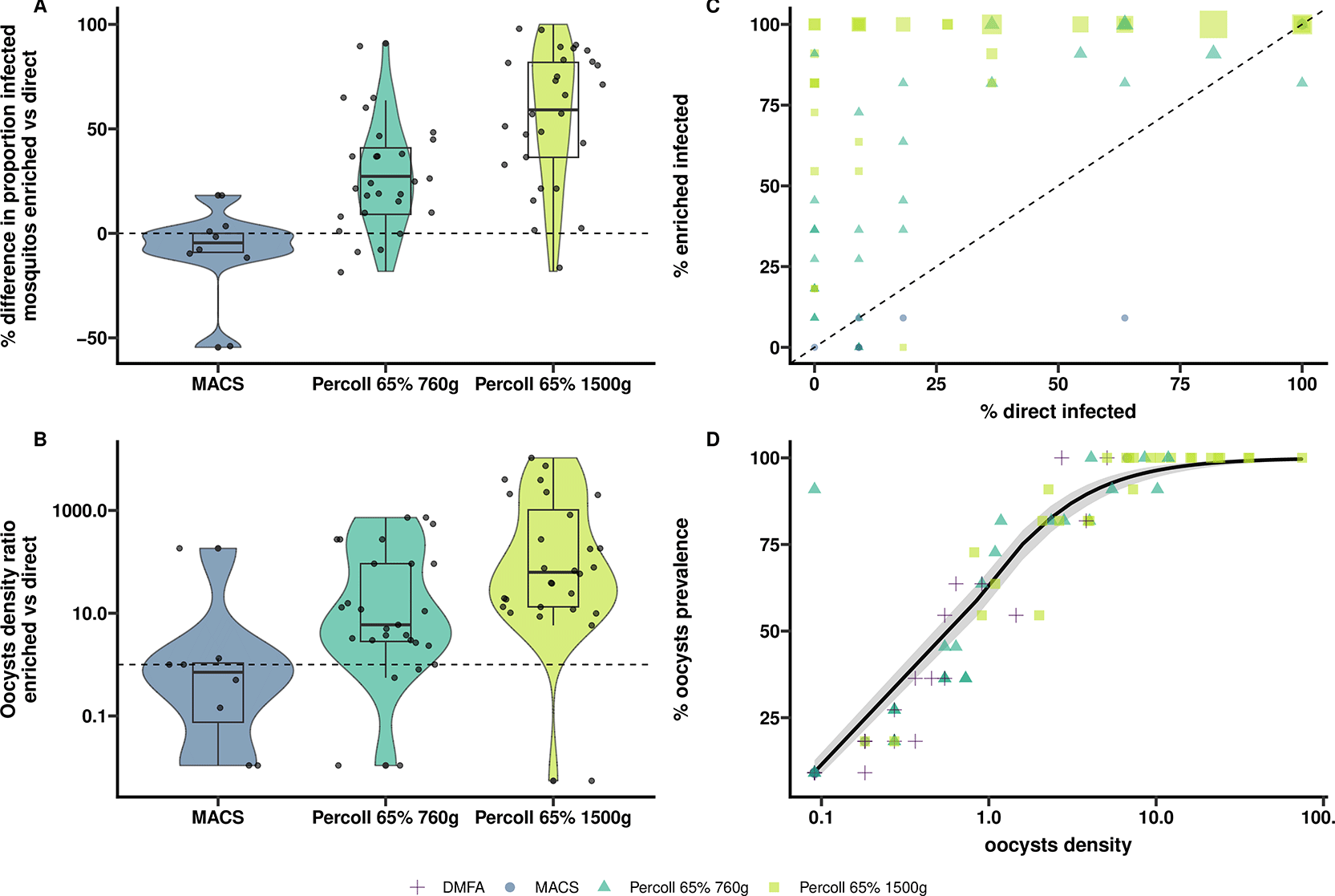
Figure 3. Comparing enrichment methods for P. vivax gametocytes.
Gametocyte infected blood drawn from P. vivax CHMI study participants was fed non-enriched (control) and enriched (start volume 10 mL) to Anopheles stephensi mosquitoes in membrane feeders (300 μL). (A) Violin plots with 95% CI comparing the difference in proportion (%) of infected mosquitoes between non-enriched and gametocyte enriched blood feeding utilizing MACS and Percoll density gradient centrifugation (65%) using two different spinning speeds. The dashed horizontal line shows a difference of 0 (i.e. equality). (B) Violin plots with 95% CI comparing mosquito oocyst density ratio of control versus gametocyte enriched blood. Dashed horizontal line shows a ratio of 1 (i.e. equality). (C) Association of mosquito infection prevalence (%) between non-enriched and enriched-feeds, the size of the geometric figures depict the oocyst intensity. Dashed line shows a 1:1 association. (D) Trend for oocyst prevalence amongst positives versus oocyst density found by fitting a logistic regression model. The grey shaded area represents the pointwise 95% confidence interval.
Gametocyte enrichment using DGC65 improved transmission significantly (Figure 3C). This was most apparent when mosquitoes were infected with blood containing low parasite densities, with 567 parasites/mL being the lowest parasitemia at which we observed transmission. DGC at a speed of 1500 g was superior and resulted in a higher oocyst intensity compared to 760 g (Figure 3D).
A subset of mosquitoes that fed on DGC65-1500 g gametocyte enriched blood were salivary gland dissected between day 11 and 20 post-blood feeding for quantification of sporozoites. Mean sporozoite load increased approximately proportionally with oocyst density in a weighted log–log regression model, adjusting for days post-infection relative to day 14 (β = 0.91, 95% CI [0.51, 1.30], p = 0.0003). The model-predicted relationship between oocyst density and sporozoite load shows that oocyst burden and sporozoite load were strongly associated (Figure 4).
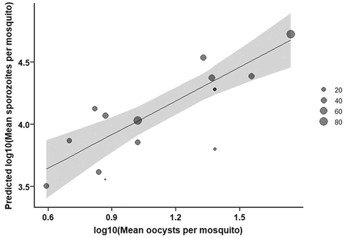
Figure 4. Model-predicted relationship between oocyst density and sporozoite load.
Plot shows the predicted log10-transformed mean sporozoite load based on log10-transformed oocyst density from a weighted log-log regression model, adjusting for days post-infection relative to day 14. The shaded area represents the 95% confidence interval around the predicted relationship; point size reflects the number of dissected mosquitoes that ranged from 7–77.
Effect of leukocyte depletion on P. vivax mosquito infection
A subset of 7 Percoll-enriched gametocyte samples was processed with and without filtration to explore whether leukocyte depletion by Plasmodipur filtration is an essential component to increase P. vivax transmission. For the proportion of infected mosquitoes, absolute differences between filtered and unfiltered conditions were small and not statistically significant: 9.09% (95% CI: −47.30 to 65.49; p > 0.99) for 1500 g, and − 0.00% (95% CI: −89.17 to 89.17; p > 0.99) for 760 g. Differences in oocyst intensity were similarly negligible: 0.29 (95% CI: −1.00 to 1.57; p = 0.96) for 1500 g, and 0.16 (95% CI: −1.87 to 2.19; p = 0.96) for 760 g.
Quantifying binding of P. vivax gametocytes to MACS LS and LD columns
As detailed above, P. vivax gametocyte enrichment by MACS did not result in increased mosquito infection prevalence and intensity compared to the control conditions. To explore whether this was due to inefficient column binding, we molecularly quantified enriched parasites. Blood from two study participants was processed in parallel on LS and LD columns and the following fractions were collected: starting, flow-through, wash 1, wash 2, and bound (as described elsewhere11). Parasites were quantified by qPCR targeting 18S (total parasites) and Pv25 (gametocytes) transcripts.24 The proportion of P. vivax gametocytes in the bound fraction was 0 and 0.1% when using LS columns and 17.7 and 33.6% when using LD columns (Extended data).
Discussion
This study aimed to determine the best method to enrich P. vivax gametocytes out of whole blood samples to enhance mosquito infection. We demonstrated that Percoll density gradient centrifugation at a concentration of 65%, when centrifuged at a speed of 1500 g, results in the highest mosquito infection and oocyst density. While MACS can successfully enhance mosquito infectivity for P. falciparum gametocytes, we observed no increase when the method was applied on P. vivax gametocytes which was plausibly due to inefficient column-binding of gametocytes. Our optimized P. vivax enrichment protocol allows for reproducible mosquito infections even at low-density gametocyte infections.
Two previous studies evaluated MACS and Percoll enrichment for P. vivax gametocytes.15,16 A study of Vera et al. used naturally infected malaria patients, with parasitemia ranging from 297.5 to 30,600 parasites/μL (of which 13.3% ± 16.7% gametocytes), to develop a method to purify and concentrate viable gametocytes. This study compared enrichment using DGC45, DGC60 and MACS, with DGC60 resulting in a significantly higher percentage of gametocytes. However, gametocyte infectivity was only accessed for DGC45, with a limited number of mosquito membrane feedings (n = 8) and without parallel control feeds of unenriched material. We can therefore not directly compare our findings to their work. A study of Ramos et al. used P. vivax gametocytes from naturally infected participants, with parasitemia ranging from 501–10,000 parasites/μL, to evaluate viability of enriched gametocytes after short-term culture. Gametocytes were enriched using DGC70 and MACS and fed to mosquitoes at different time intervals post enrichment. Their independent assays allowed them to study gametocyte viability over time for each condition, but comparison between enrichment methods or to a control was not possible. Both studies reported gametocytemia as the proportion of gametocytes relative to total parasites (gametocytes/total parasites). This measure reflects purity (i.e. the depletion of asexual parasites) rather than yield (the absolute number of gametocytes recovered). Consequently, enrichment may appear successful even if substantial numbers of gametocytes are lost during processing— this would only become evident when gametocyte numbers are quantified, or infectivity is assessed in mosquito feeding assays as we did in the current study. We therefore consider our study of value to formally compare the effect of different methods of gametocyte enrichment on transmission.
To our surprise, MACS was successful in enhancing mosquito infection for P. falciparum gametocytes, which was in line with previous studies,11,12,27–29 but not for P. vivax. To explore why MACS LS columns did not result in a measurable increase in mosquito infection rates, we determined the fraction of gametocytes in the bound and flow-through fractions for both LS- and LD columns in a small set of experiments. In these experiments MACS LS columns failed to retain P. vivax gametocytes effectively (Extended data). An increase in enrichment was observed when using MACS LD columns, which are specifically designed to capture weaker magnetic cells. One possible explanation for this observation is that P. vivax gametocytes have a different hemozoin content or intracellular distribution, resulting in reduced paramagnetic properties. Even though no direct comparative data on hemozoin levels between P. vivax and P. falciparum is available, the distribution of hemozoin in gametocytes is morphologically different: the hemozoin in the cresent shaped P. falciparum gametocyte is more condensed compared to P. vivax, which is oval shaped with a more dispersed pattern. This may result in different paramagnetic properties and could explain the difference in MACS enrichment between the two parasite species. Dedicated studies using methods such as quantitative electron microscopy or magneto-optical assays to study absolute hemozoin concentration in both P. vivax and P. falciparum gametocytes could help support this hypothesis further. Of note, even the better performing LD columns did not achieve binding efficiencies in the range of those observed for P. falciparum.12
Beyond poor retention, impaired gametocyte quality caused by the MACS process may have further contributed to the poor transmission results we observed. The MACS magnetic field could induce oxidative or mechanical stress. Such stress-related damage may continue to accumulate even after removal from the magnetic field, ultimately impairing infectivity even if gametocytes are more efficiently enriched by LD columns. This damage may be more pronounced in P. vivax gametocytes because they preferentially invade reticulocytes, which are considered fragile.8 This hypothesis is supported by data from the Ramos et al. study, where they show a more rapid decline in infectivity of MACS-enriched gametocytes compared to Percoll — infectivity dropped after 6 hours of culture for MACS versus up to 12 hours for Percoll.15 Because we did not perform mosquito feeding assays following enrichment with MACS LD columns, the effect of the MACS magnetic field on P. vivax gametocytes remains uncertain.
This study has some limitations. First, we did not quantify gametocyte densities prior to enrichment or mosquito feeding. We are therefore unable to quantify and compare the gametocyte concentration efficacy of each enrichment method. Second, a subset of experiments was performed for protocol optimization and did not have appropriate power to detect statistically significant differences. Of particular interest were the experiments omitting leukodepletion. Even though we were unable to show statistical significance, we have shown that, in this small sample size, omitting the Plasmodipur filter was not detrimental to mosquito infectivity. A protocol without the need for leukodepletion would lower costs significantly (Plasmodipur filter costs ~25 €) and reduce handling time, making the methodology more accessible. For large scale experiments, the reduction in handling time may become particularly important since all procedures must take place at 37 °C.
In conclusion, we established the optimal methodology to reliably generate highly infected P. vivax mosquitoes through optimized gametocyte enrichment, even at low parasitemia. The resulting infected mosquitoes also completed sporozoite development, demonstrating the suitability of the protocol for downstream applications such as evaluating pre-erythrocytic interventions and studies on hypnozoite biology.
Ethics and consent
Written informed consent was obtained from CHMI volunteers who participated in the RaViCHMI1 clinical trial (ID NL-OMON57011), this study was approved by the Central Committee for Research Involving Human Subjects in The Netherlands (CCMO; NL-005553).
Acknowledgements
The authors are grateful to Jolanda Klaassen, Laura Pelser-Posthumus and Astrid Pouwelsen for their role in mosquito husbandry and extrapolating data from the P. vivax infected mosquitoes. We thank Weronika Machnik and Eyob Addise Workneh for technical assistance during sample processing; the BIO-006 study team (University of Oxford) and the OptiViVax Project Management team for assistance; and all the study participants of the RaViCHMI1 trial.
References
- 1.
Organization, W.H:
World malaria report.2024.
- 2.
Battle KE, Baird JK:
The global burden of Plasmodium vivax malaria is obscure and insidious.
PLoS Med.
2021; 18(10): e1003799. PubMed Abstract
| Publisher Full Text
| Free Full Text
- 3.
Twohig KA, Pfeffer DA, Baird JK, et al.:
Growing evidence of Plasmodium vivax across malaria-endemic Africa.
PLoS Negl. Trop. Dis.
2019; 13(1): e0007140. PubMed Abstract
| Publisher Full Text
| Free Full Text
- 4.
Bradley L, Yewhalaw D, Hemming-Schroeder E, et al.:
Epidemiology of Plasmodium vivax in Duffy negatives and Duffy positives from community and health centre collections in Ethiopia.
Malar. J.
2024; 23(1): 76. PubMed Abstract
| Publisher Full Text
| Free Full Text
- 5.
(WHO), W.H.O:
Malaria Vaccine Technology Roadmap.2013.
Reference Source
- 6.
Escalante AA, Cepeda AS, Pacheco MA:
Why Plasmodium vivax and Plasmodium falciparum are so different? A tale of two clades and their species diversities.
Malar. J.
2022; 21(1): 139. PubMed Abstract
| Publisher Full Text
| Free Full Text
- 7.
Commons RJ, Simpson JA, Watson J, et al.:
Estimating the Proportion of Plasmodium vivax Recurrences Caused by Relapse: A Systematic Review and Meta-Analysis.
Am J Trop Med Hyg.
2020; 103(3): 1094–1099. PubMed Abstract
| Publisher Full Text
| Free Full Text
- 8.
Bermudez M, et al.:
Plasmodium vivax in vitro continuous culture: the spoke in the wheel.
Malar. J.
2018; 17(1): 301. PubMed Abstract
| Publisher Full Text
| Free Full Text
- 9.
Miura K, Swihart BJ, Fay MP, et al.:
Evaluation and modeling of direct membrane-feeding assay with Plasmodium vivax to support development of transmission blocking vaccines.
Sci. Rep.
2020; 10(1): 12569. PubMed Abstract
| Publisher Full Text
| Free Full Text
- 10.
Karl S, Davis TM, St Pierre TG:
Short report: Quantification of Plasmodium falciparum gametocytes by magnetic fractionation.
Am J Trop Med Hyg.
2011; 84(1): 158–160. PubMed Abstract
| Publisher Full Text
| Free Full Text
- 11.
Reuling IJ, Stone WJR, van de Vegte-Bolmer M, et al.:
Concentration of Plasmodium falciparum gametocytes in whole blood samples by magnetic cell sorting enhances parasite infection rates in mosquito feeding assays.
Malar. J.
2017; 16(1): 315. Publisher Full Text
- 12.
Graumans W, Andolina C, Awandu SS, et al.:
Plasmodium falciparum Gametocyte Enrichment in Peripheral Blood Samples by Magnetic Fractionation: Gametocyte Yields and Possibilities to Reuse Columns.
Am J Trop Med Hyg.
2019; 100(3): 572–577. Publisher Full Text
- 13.
Collins KA, Wang CYT, Adams M, et al.:
A controlled human malaria infection model enabling evaluation of transmission-blocking interventions.
J. Clin. Invest.
2018; 128(4): 1551–1562. PubMed Abstract
| Publisher Full Text
| Free Full Text
- 14.
Collins KA, Wang CYT, Adams M, et al.:
A Plasmodium vivax experimental human infection model for evaluating efficacy of interventions.
J. Clin. Invest.
2020; 130(6): 2920–2927. PubMed Abstract
| Publisher Full Text
| Free Full Text
- 15.
Ramos GQ, Baia-da-Silva DC, Lacerda MVG, et al.:
Viability and Infectivity of Plasmodium vivax Gametocytes in Short-Term Culture.
Front. Cell. Infect. Microbiol.
2021; 11: 676276. PubMed Abstract
| Publisher Full Text
| Free Full Text
- 16.
Vera O, Brelas de Brito P, Albrecht L, et al.:
Purification Methodology for Viable and Infective Plasmodium vivax Gametocytes That Is Compatible with Transmission-Blocking Assays.
Antimicrob. Agents Chemother.
2015; 59(10): 6638–6641. PubMed Abstract
| Publisher Full Text
| Free Full Text
- 17.
Ponnudurai T, Lensen AHW, Leeuwenberg ADEM, et al.:
Cultivation of fertile Plasmodium falciparum gametocytes in semi-automated systems. 1. Static cultures.
Trans. R. Soc. Trop. Med. Hyg.
1982; 76(6): 812–818. PubMed Abstract
| Publisher Full Text
- 18.
van de Vegte-Bolmer M , Graumans W, Stoter R, et al.:
A portfolio of geographically distinct laboratory-adapted Plasmodium falciparum clones with consistent infection rates in Anopheles mosquitoes.
Malar. J.
2021; 20(1): 381. PubMed Abstract
| Publisher Full Text
| Free Full Text
- 19.
Minassian AM, Themistocleous Y, Silk SE, et al.:
Controlled human malaria infection with a clone of Plasmodium vivax with high-quality genome assembly.
JCI Insight.
2021; 6(23). Publisher Full Text
- 20.
Salkeld J, Themistocleous Y, Barrett JR, et al.:
Repeat controlled human malaria infection of healthy UK adults with blood-stage Plasmodium falciparum: Safety and parasite growth dynamics.
Front. Immunol.
2022; 13: 984323. PubMed Abstract
| Publisher Full Text
| Free Full Text
- 21.
Graumans W, et al.:
Comparative assessment of mosquito size and receptiveness to P. falciparum infection of two rearing procedures for Anopheles mosquitoes.
bioRxiv.
2023; 2023.03.09.531859.
- 22.
Shaw WR, Holmdahl IE, Itoe MA, et al.:
Multiple blood feeding in mosquitoes shortens the Plasmodium falciparum incubation period and increases malaria transmission potential.
PLoS Pathog.
2020; 16(12): e1009131. PubMed Abstract
| Publisher Full Text
| Free Full Text
- 23.
Graumans W, Heutink R, van Gemert GJ, et al.:
A mosquito feeding assay to examine Plasmodium transmission to mosquitoes using small blood volumes in 3D printed nano-feeders.
Parasit. Vectors.
2020; 13(1): 401. PubMed Abstract
| Publisher Full Text
| Free Full Text
- 24.
Ponnudurai T, Lensen AHW, van Gemert GJA, et al.:
Infectivity of cultured Plasmodium falciparum gametocytes to mosquitoes.
Parasitology.
1989; 98(Pt 2): 165–173. Publisher Full Text
- 25.
Tadesse FG, van den Hoogen L, Lanke K, et al.:
The shape of the iceberg: quantification of submicroscopic Plasmodium falciparum and Plasmodium vivax parasitaemia and gametocytaemia in five low endemic settings in Ethiopia.
Malar. J.
2017; 16(1): 99. PubMed Abstract
| Publisher Full Text
| Free Full Text
- 26.
Tadesse FG, Slater HC, Chali W, et al.:
The Relative Contribution of Symptomatic and Asymptomatic Plasmodium vivax and Plasmodium falciparum Infections to the Infectious Reservoir in a Low-Endemic Setting in Ethiopia.
Clin. Infect. Dis.
2018; 66(12): 1883–1891. PubMed Abstract
| Publisher Full Text
- 27.
Karl S, David M, Moore L, et al.:
Enhanced detection of gametocytes by magnetic deposition microscopy predicts higher potential for Plasmodium falciparum transmission.
Malar. J.
2008; 7: 66. PubMed Abstract
| Publisher Full Text
| Free Full Text
- 28.
Ribaut C, Berry A, Chevalley S, et al.:
Concentration and purification by magnetic separation of the erythrocytic stages of all human Plasmodium species.
Malar. J.
2008; 7: 45. PubMed Abstract
| Publisher Full Text
| Free Full Text
- 29.
Ahn S:
Magnetic separation: a highly effective method for synchronization of cultured erythrocytic Plasmodium falciparum.
Parasitol. Res.
2008; 102: 1195–1200. PubMed Abstract
| Publisher Full Text
- 30.
Geraedts T, et al.:
P. vivax gametocyte enrichment. [dataset].
Radboud University;
2026. Version 1. Publisher Full Text







