Introduction
Disclaimer
The views expressed in this article are those of the author(s). Publication in Open Research Europe does not imply endorsement by the European Commission.
Background and aims of STANDUP
Diabetes is one of the most common diseases in the world, and is correlated to a high mortality index. Uncontrolled diabetes can affect several organs, namely feet, eyes, heart, kidneys, nervous system and blood vessels1,2. The European Horizon 2020 STANDUP #777661 project is interested in health problems that arise from diabetic foot (DF), which can be infection, ulceration or destruction of deep tissues of the foot3. The severity of DF can lead to hospitalization or even to lower limb amputation, resulting in substantial costs and reduced quality of life. In the majority of cases, a proper risk assessment of diabetic patients associated with diabetic foot care can significantly prevent the development of diabetic foot disorders. Currently, risk assessment for DF is based solely on neuropathy and vascular analysis. However, according to diabetes experts, further improvements are needed.
In the literature, several studies have proven that the use of temperature can be of great relevance in the early diagnosis of DF problems4,5. Foot temperature varies from patient to patient and depends on the surrounding temperature and the activity level. Usually, the average temperature difference between the contralateral areas of the left and right feet does not exceed 1°C. A point-to-point temperature difference greater than 2.2°C is considered abnormal and is called hyperthermia6. Due to neuropathic sensory loss, diabetic patients rarely feel pain and injury in the feet, especially in the early stages of the disease. Therefore, temperature elevation can be a useful indicator of foot ulceration risk. A strategy including information on hyperthermia has been shown to reduce foot ulceration by 70%5,7.
According to 8, the temperature distribution in the thermogram of plantar foot of a healthy person is a bilateral butterfly pattern, whereas in diabetic patients, this temperature varies and shows no typical pattern as in healthy people. The cold stress test9 is another effective method to asses thermoregulatory problems. The temperature difference before and after a cold stress is an important indicator for the early detection of diabetic neuropathy problems10.
Within the framework of the European Horizon 2020 program, the main objective of the STANDUP #777661 project is to propose thermal solutions and technologies that could help doctors and specialists improve DF diagnosis, to detect hyperthermia at an early stage, and to develop better DF ulcer treatments using thermal information. STANDUP is a collaboration between clinicians, researchers, and industry partners. The specific research objectives of the project are:
Developing a smartphone thermal-based system to detect hyperthermia of the plantar foot surface and to study thermal variations with time after a cold stress test in targeted regions of interest (ROI).
Following-up on temperature, color, and 3D shape of DF ulcer along time.
Conducting clinical trials to assess thermoregulation quality of the plantar foot surface by analyzing temperature variation in targeted ROI, and to monitor DF ulcer temperature, color, and 3D shape over time.
Taking into account thermal factors to design new foot insoles, which will be tested in clinical trials on patients with DF ulcer.
Associating all results produced during STANDUP to converge into advanced smartphone-based prototype and foot insoles.
Project structure
STANDUP has five interrelated research work packages (WP, Figure 1) and two additional WPs (WPs 0, and 6) which focus on management, training, and ensuring impact. WP1 aims to develop a smartphone application (A1) to detect hyperthermia and to analyze thermal variation of the plantar foot surface through time.WP2 aims to create another smartphone application (A2) to assess variation of DF ulcers along time. WP3 focus on the improvement of insoles using thermal information.WP4 is dedicated to clinical trials for DF ulcer prevention and treatment using the applications (A1, A2) developed in the two previous work packages (WP1, WP2). In the last WP5, the smartphone applications will be tested in real clinical situation, and improved taking into accounts remarks from the users (medical staff, university members and patients), to result in advanced prototypes.
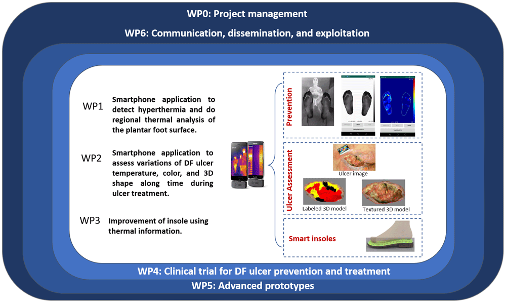
Figure 1. schematic overview showing the work packages included in the STANDUP project.
In this paper we focus specifically on a crucial step of the STANDUP project, which consists in creating a database of thermal and color images of the foot sole for healthy people and diabetic patients. Thermal and color images were acquired using the STANDUP protocol, i.e., freehandedly with a smartphone equipped with a dedicated thermal camera that allows the simultaneous recording of a thermal image and a red-green-blue (RGB) one. To our knowledge, this database is the first multispectral database (containing thermal and color images) of diabetic patients' plantar feet surfaces.
Materials and methods
Choice of thermal camera
Camera resolution
The largest foot that we considered was 30 cm long. The vertical field of view we chose was therefore 40 cm: 30 cm to include the foot sole plus a 5 cm margin at the top and bottom of the foot. In the other direction, 40 cm was enough to contain both feet along their width (about 10 cm), including the same 10 cm margin for each foot. The field of view was then 40×40 cm² (see Figure 2).
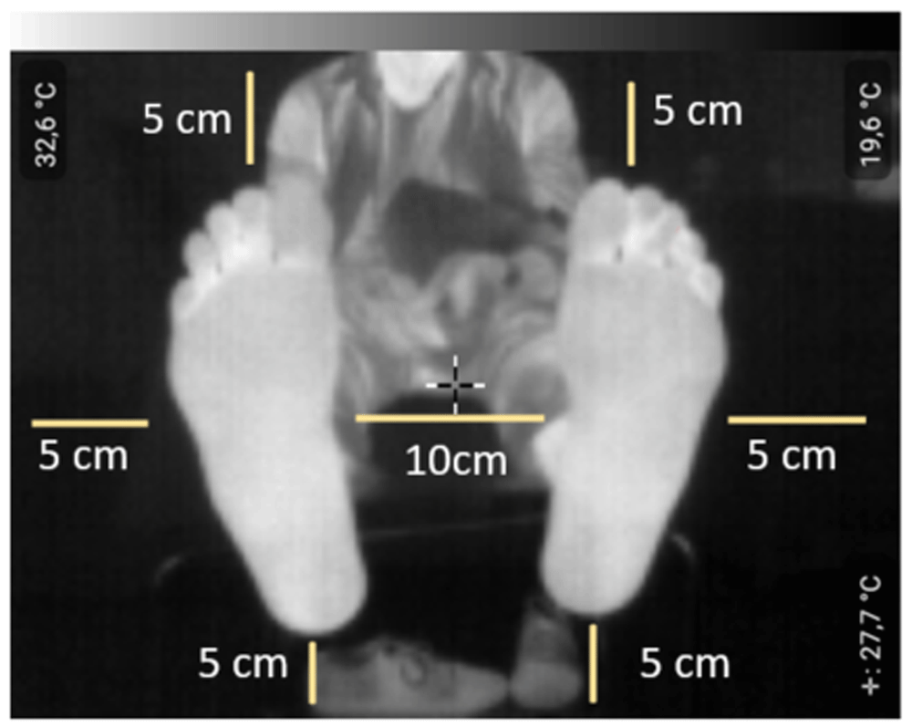
Figure 2. Margins to be respected during the acquisition of thermal images of plantar foot.
Hyperthermia occurs one week before an ulcer. The smallest hyperthermia area is a circle of approximately 1 cm in diameter. According to Shannon's first theorem and the values used in image processing, two pixels are required to detect the smallest risk area. This means that any camera with a resolution larger than 80×80 pixels can be chosen.
Camera sensitivity
Hyperthermia corresponds to a point-to-point temperature difference between contralateral feet higher than 2.2°C. The differential accuracy must therefore be better than 0.2°C (about 10% of the value to be evaluated).
Spectral range
For a healthy person, the mean skin temperature under normal conditions is between 33.5 and 36.9 °C11. Wien's law shows that the infrared radiation wavelength is around 9.5 µm. Therefore, the spectral range of the chosen camera must contain this wavelength.
In this study, we chose the FLIR ONE Pro camera. It consists of two sensors. The first is a thermal sensor that measures heat by infrared emission, characterized by a thermal image resolution of 160×120 pixels. The spectral range of the thermal sensor is 8–14 µm. FLIR ONE Pro can detect temperature differences of 0.1°C. The other sensor is a conventional 1440×1080 pixels RGB camera, designed to work in parallel with the thermal core. FLIR ONE Pro is designed to be plugged into a smartphone12.
The FLIR ONE Pro camera software includes an alignment control technology called Multi-Spectral Dynamic Imaging (MSX)13, allowing it to provide spatially registered RGB and thermal images. MSX increases the clarity of acquired images by integrating edge and contour details of visible light into thermal readings in real-time. This technology does not dilute the thermal image or reduce its transparency. A thermal image and an RGB image of the plantar foot surface are acquired simultaneously, as shown in Figure 3.
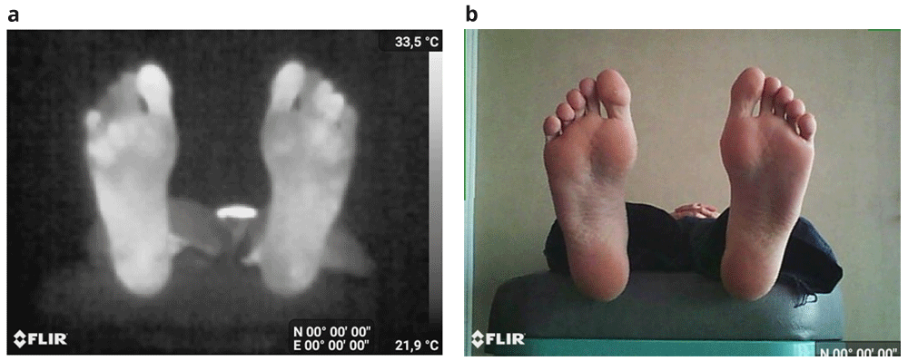
Figure 3.
An acquisition example: (a) the thermal image and (b) the corresponding RGB image.
Recruitment and acquisition protocols
Ethical considerations
The ethical approval for the project was obtained from the Biomedical Research Ethics Committee of Hospital Nacional Dos De Mayo (No. 075-2021-CEIB-HNDM) on 10/01/2019.
The study was conducted from January to March 2019 in Dos De Mayo hospital in Peru, under the supervision of Dr. H. Arbanil, responsible at the endocrinology service in HNDM, the former president of the Peruvian Society of Endocrinology, and member of biomedical research ethics committee of HNDM.
The main objective of this study was to use thermal images of the plantar foot in the prevention of diabetic ulcers at early stages. For this purpose, it was necessary to collect thermal images of the plantar foot as well as medical information measured by medical doctors.
Each volunteer participating in our clinical trial was informed of the ethical issues of the STANDUP project by a clear and informed consent form concerning the data collection and processing, the validation of the prototype which he/she read, understood, and signed.
This consent relating to the specific purpose of STANDUP project is informative, unambiguous, and freely given. It clearly defines the acquisition process, the purpose of the data collection, who will use it, and for what goal. Appropriate languages were used for each country.
Informed consent and patient details
The authors declare that this article does not contain any personal information that could lead to patients’ identification. The authors declare that they have obtained a written informed consent from patients and volunteers included in the article. The authors also confirm that the personal details of the patients and volunteers have been removed.
Healthy subjects
The first acquisition campaign was conducted in the PRISME laboratory of the University of Orléans (France) in January 2017. The participants were students of the Polytech Orléans engineering school on an internship at PRISME, Ph.D. students, and PRISME staff. A total of 22 people participated in this first acquisition campaign. This group was composed of 10 women and 12 men with an average age of 25 years. It corresponds to a total of 22 multispectral images.
From December 2018 to March 2019, we organized a second acquisition campaign at the Ibn Zohr University in Agadir, Morocco. A total of 17 students and members of the IRF-SIC laboratory participated. In addition, a cold stress test acquisition campaign detailed in the second section (Figure 6) was carried out for 43 people at two different times. This gives a total of 60 people, including 25 women and 35 men with an average age of 30 years. This corresponds to a total of 103 multispectral images.
The total number of healthy multispectral images (thermal and RGB) is 125.
Recruitment protocol of healthy subjects
The criteria for recruitment were as follows: any adult volunteer without diabetes and without foot problems.
Acquisition protocol for healthy subjects
To ensure high image quality, the room conditions had to be adapted to thermal image acquisition. We conducted the acquisition campaigns in rooms that were large enough to place the equipment and provide freedom of movement for the technician and volunteers. The lighting was controlled during acquisitions. Windows were partially covered or shielded to prevent outside infrared radiation from entering the room. Temperature was controlled at 20 ± 1°C. The protocol was as follows:
The person removes shoes and socks, and the acquisition takes place ten minutes later.
The person reads and signs the consent agreement form.
The data sheet is filled with the person's information.
The person lies down on a medical bed or sits in a chair, feet at the end, upright, and 10 cm apart.
An acquisition is made at a sufficient distance from both feet to meet the margins shown in Figure 2.
Two typical multispectral images (thermal + RGB) acquired respectively in PRISME are shown in Figure 4, and in IRF-SIC in Figure 5.
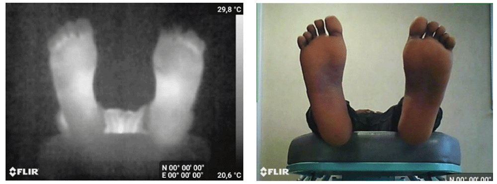
Figure 4. An example of images acquired at PRISME.
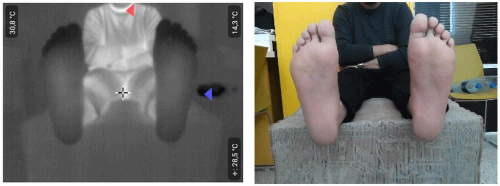
Figure 5. An example of images acquired at IRF-SIC.
Diabetic subjects
The image database of people with diabetes was acquired at the Hospital Nacional Dos de Mayo (HNDM) in Lima, Peru from 14 January, 2019, to 9 March 9. The approval (Number 075-2021-CEIB-HNDM) of this study by the ethics committee of the HNDM occurred on January 10, 2019. A total of 145 diabetic patients participated in this acquisition campaign. It was performed under the supervision of nurses and medical doctors. The thermal stress test was applied. For each subject, we obtained a pair of multispectral images (thermal + RGB). Thus, a total of 290 pair images for diabetic subjects, 91 women and 54 men with an average age of 63 years was collected.
Recruitment protocol of diabetic subjects
The subjects in HNDM were diabetic patients who have a regular examination in the diabetes department. We excluded patients with ulcers, partial or total amputations from this campaign.
Acquisition protocol for diabetic subjects
The objective of this campaign conducted in HNDM was to conduct a cold stress test for all subjects in order to perform a correlation study between thermal and medical into. This test consists in immersing the patient's feet in cold water at 15°C for one minute9,14. The campaign was performed in a north-facing room with small windows and controlled luminosity. The average temperature in the room was 20°C with a variation of less than 1°C. The acquisition protocol was as follows:
The person removes shoes and socks, and the acquisition takes place ten minutes later.
The person reads and signs the consent agreement form (Figure 6a).
Complete the data sheet with the person's information.
The person lies down on a medical bed and places his/her feet on the end of the medical bed, in an upright position and 10 cm apart. The distance between the camera and the feet is chosen so that the feet are completely visible in the image, as shown in Figure 2. The first acquisition (thermal and color images) at time T0 is performed (Figure 6b).
The patient is asked to sit in a chair. Each foot is inserted into a plastic bag. The feet are then immersed in cold water at 15°C for one minute (Figure 6c).
The patient lifts his feet, the plastic bags are removed and a waiting time of 10 min is respected before a second thermal and color acquisition corresponding to the time T10 is performed (Figure 6d).
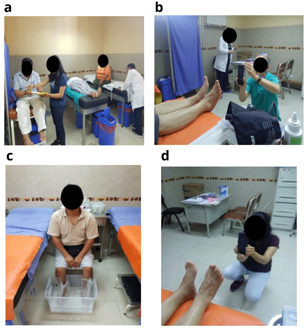
Figure 6. Cold stress test protocol.
a) Signature of the consent form (the two persons on the left side). b) First acquisition at time T0. c) Immerse both feet one minute in water at 15°C. (d) Second acquisition at time T10 10 min later.
Images at times T0 and T10 are shown in Figure 7.
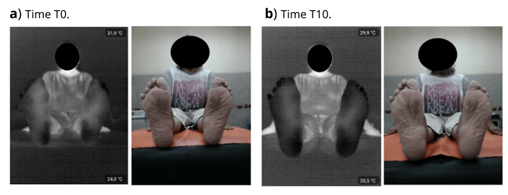
Figure 7. Example of a diabetic patient without ulcer or amputation (Dos De Mayo Hospital).
First (thermal and RGB) image acquisition at T0 (a) and second (thermal and RGB) acquisition at T10 (b). We can see that feet are colder at time T10 than time T0.
Scientific papers that used this database
Data availability
Due to the sensitive nature of the data, images are currently only available internally to our partners of the STANDUP #777661 project. It is also worth mentioning that the database will be publicly available on https://www.standupproject.eu/home/fr once the research project is achieved on June 30, 2023.
The project website (https://www.standupproject.eu/home/fr) contains the coordinator's contact; however, interested parties are advised to contact the project’s coordinator or the corresponding author (doha.bouallal@uiz.ac.ma) for further information to apply for data access.
Acknowledgements
We would like to thank all our colleagues from both universities of Orleans and Ibn Zohr who participated in our acquisition campaigns. We would also like to thank all the medical staff from the HNDM in Peru who accompanied us during the whole study and took care of the patients during the acquisition of the images. A special thanks to patients.
Faculty Opinions recommendedReferences
- 1.
Deshpande AD, Harris-Hayes M, Schootman M:
Epidemiology of Diabetes and Diabetes-Related Complications.
Phys Ther.
2008; 88(11): 1254–1264. PubMed Abstract
| Publisher Full Text
| Free Full Text
- 2.
Harding JL, Pavkov ME, Magliano DJ, et al.:
Global trends in diabetes complications: a review of current evidence.
Diabetologia.
2019; 62(1): 3–16. PubMed Abstract
| Publisher Full Text
- 3.
van Netten JJ, Bus SA, Apelqvist J, et al.:
Definitions and criteria for diabetic foot disease.
Diabetes Metab Res Rev.
2020; 36(Suppl 1): e3268. PubMed Abstract
| Publisher Full Text
- 4.
Bagavathiappan S, Philip J, Jayakumar T, et al.:
Correlation between Plantar Foot Temperature and Diabetic Neuropathy: A Case Study by Using an Infrared Thermal Imaging Technique.
J Diabetes Sci Technol.
2010; 4(6): 1386–1392. PubMed Abstract
| Publisher Full Text
| Free Full Text
- 5.
Armstrong DG, Holtz-Neiderer K, Wendel C, et al.:
Skin Temperature Monitoring Reduces the Risk for Diabetic Foot Ulceration in High-risk Patients.
Am J Med.
2007; 120(12): 1042–1046. PubMed Abstract
| Publisher Full Text
- 6.
Vilcahuaman L, Harba R, Canals R, et al.:
Automatic Analysis of Plantar Foot Thermal Images in at-Risk Type II Diabetes by Using an Infrared Camera.
World Congress on Medical Physics and Biomedical Engineering.
In D. A. Jaffray, Éd. Cham: Springer International Publishing. 2015; 51: 228–231. Publisher Full Text
- 7.
Sun PC, Lin HD, Jao SHE, et al.:
Relationship of skin temperature to sympathetic dysfunction in diabetic at-risk feet.
Diabetes Res Clin Pract.
2006; 73(1): 41–6. PubMed Abstract
| Publisher Full Text
- 8.
Chan AW, MacFarlane IA, Bowsher DR:
Contact Thermography of Painful Diabetic Neuropathic Foot.
Diabetes Care.
1991; 14(10): 918–22. PubMed Abstract
| Publisher Full Text
- 9.
Bharara M, Viswanathan V, Cobb JE:
Cold immersion recovery responses in the diabetic foot with neuropathy.
Int Wound J.
2008; 5(4): 562–9. PubMed Abstract
| Publisher Full Text
| Free Full Text
- 10.
Balbinot LF, Canani LH, Robinson CC, et al.:
Plantar thermography is useful in the early diagnosis of diabetic neuropathy.
Clinics (Sao Paulo).
2012; 67(12): 1419–25. PubMed Abstract
| Publisher Full Text
| Free Full Text
- 11.
BIERMAN W:
THE TEMPERATURE OF THE SKIN SURFACE.
JAMA.
1936; 106(14): 1158–1162. Publisher Full Text
- 12.
FLIR ONE Pro Thermal Imaging Camera for Smartphones FLIR Systems.
2021. Reference Source
- 13.
What is MSX®? 2021. Reference Source
- 14.
Bouallal D, Bougrine A, Douzi H, et al.:
Segmentation of plantar foot thermal images: application to diabetic foot diagnosis. In
2020 International Conference on Systems, Signals and Image Processing (IWSSIP).
Niterói, Brazil, juill. 2020; 116–121. Publisher Full Text