Plain language summary
This study investigates the effects of cigarette smoke on the visual system of zebrafish larvae. We exposed the larvae to cigarette smoke extract for 4, 24, or 48 hours and assessed their eye movements, retina morphology and oxidative stress gene expression. Exposure to cigarette smoke extract for 48 hours reduced eye movements behaviour in the zebrafish larvae and led to changes in the morphology of their hyaloid vasculature present in the lens and the number of phagosomes in their retinal pigment epithelium. When exposure was shortened to 4 or 24 hours, eye movements were still reduced and oxidative stress was affected. These results suggest that zebrafish larvae can be used as a valuable model to investigate ocular disorders related to cigarette smoke.
Abbreviations
CSE: Cigarette Smoke Extract
Dpf: Days post-fertilization
OKR: Optokinetic Response
VMR: Visual Motor Response
PCR: Polymerase Chain Reaction
RPE: Retinal Pigment Epithelium
TPM: Total Particulate Matter
PaHs: Polycyclic Aromatic Hydrocarbons
IARC: International Agency for Research on Cancer
AMD: Age-related Macular Degeneration
Hpf: Hours post-fertilization
AREC: Animal Research Ethics Committee
Wt: wild type
EGPF: Enhanced Green Fluorescent Protein
FTC: Federal Trade Commission Smoke
DMSO: Dimethyl sulfoxide
MTD: Maximum Tolerate Dose
VA: Visual Acuity
CS: Contrast Sensitivity
RGB: Red Green Blue
Cpd: Cycles per degree
Ms/s: milliseconds per second
PFA: paraformaldehyde
TEM: Transmission electron microscopy
Cat: Catalase
Gpx1a: Glutathione Peroxidase 1
Casp3a: Caspase 3
Nfe2l2a: nuclear factor erythroid 2–related factor 2
ROS: Reactive Oxygen Species
SD: Standard Deviation
FP: Front Plane
OA: Optic axis
AL: Axial Length
Introduction
Tobacco and smoking are global concerns due to their health, societal and economic impact1,2. Second-hand smoke from cigarettes is a complex mixture of mainstream smoke (smoke inhaled and exhaled by smokers) and sidestream smoke (smoke directly from the burning cigarette) for which even brief exposure can be harmful to health3–5. Sidestream smoke contains a mixture of more than 4000 chemicals6. This mixture is constituted by total particulate matter (TPM) which includes solid chemicals (e.g. alkaloids, polycyclic aromatic hydrocarbons also known as PaHs and N-nitrosamines) as well as gaseous components (e.g. carbon dioxide, carbon monoxide, nicotine, acetaldehyde, hydrocarbons, nitrogen, ammonia, nitrosamines and hydrogen cyanide)7. Second-hand smoke is a Group 1 carcinogen, the highest carcinogenic level of the International Agency for Research on Cancer (IARC)4,8. In 2019, smoking and second-hand smoke were directly related to 7.69 and 1.2 million deaths worldwide, respectively9,10.
Smoking is well-known to promote carcinogenicity, induce developmental toxicity5,11 and cardiovascular effects11,12. Second-hand smoke is a strong risk factor for developing dry or neovascular types of age-related macular degeneration (AMD)3,13,14; which is the leading cause of severe visual impairment in Europe6,15. Smokers and passive smokers are also at higher risk of nuclear cataracts and dry eye disease13.
The use of zebrafish as an animal model to identify environmental substances impairing vision has increased considerably in recent years16,17. Advantages of zebrafish include the similarity in eye structure with the human eye, accessibility of the model to handling and manipulation, the ability to efficiently perform systemic drug administration and rapid advances in visual behaviour assays16,18–20.
Recent studies investigating toxic effects of CSE in zebrafish identified lethality, abnormal swimming behaviour21, hyperactivity22,23 and cardiovascular anomalies24. Ellis et al. reported eye malformation in zebrafish exposed to CSE from 6 to 72 hours post-fertilization (hpf)21. However, there are no studies investigating the impact of CSE on zebrafish visual behaviour, despite previous studies showing CSE exerts oculotoxic effects in mice25–27 and RPE cells28. Putative research models of AMD were reported based on exposing mice to an experimental chamber for six months where cigarettes were artificially smoked27. This study showed CSE to trigger oxidative damage, cell degeneration and cell apoptosis in the RPE, as well as signs of drusen formation27. In another study, in which mice were exposed to CSE under similar conditions but for a shorter period of time (∼3.7 months), sub-RPE deposits and thickening of Bruch's membrane were observed26. Mice exposed to CSE for 12 weeks were reported to also show apoptosis of both corneal and conjunctival epithelium cells leading to dry eye, potentially induced by inflammation25. In summary, CSE exposure can adversely impact the eye and visual system, outcomes that warrant further investigation. Here we apply zebrafish as a vertebrate animal model to explore the effects of cigarette smoke on visual behaviour and retinal biology.
Methods
Zebrafish maintenance
All experiments using animals were carried out at University College Dublin and approved by the University College Dublin Animal Research Ethics Committee (AREC). All possible efforts were undertaken to minimise the suffering of animals. Adult zebrafish (Danio Rerio) WT-Tü (wt) and transgenic line Tg(fli1:eGFP) were maintained on a recirculating water system at 28°C and pH 7.1 on a 14:10 h light-dark cycle. Water conductivity averaged 1347 µS. Adult zebrafish were fed shrimp and dry pellet food twice daily. Following afternoon feeds, male and female adults were placed in breeding tanks. Embryos were obtained by natural spawning, collected the next morning and raised in embryo medium (0.137 M NaCl, 5.4 mM KCl, 5.5 mM Na2HPO4, 0.44 mM KH2PO4, 1.3 mM CaCl2, 1.0 mM MgSO4 and 4.2 mM NaHCO3 with 1 mL methylene blue) until 5 dpf29.
Cigarette smoke extract (CSE)
CSE was sourced from Murty Pharmaceuticals (US) where 3R4F Standard Research Cigarettes were smoked using a Federal Trade Commission (FTC) smoke machine under an ISO smoke regime, which takes one puff every two seconds and 35 mm of volume4,30. The resulting smoke extracted directly from the cigarette was collected by a Cambridge pad filter, into total particulate matter (TPM), and dissolved in DMSO at 40 µg/mL TPM per vial. Concentrated CSE stocks were diluted before each experiment to achieve desired concentrations.
Exposure of zebrafish larvae to CSE
CSE exposure was assessed for durations of 4, 24 or 48 h in larvae. For 48 hours exposure, CSE was added at 72 hpf, when embryos were placed in 6 cm Petri dishes. CSE doses were added directly into the embryo medium and then refreshed at 96 hpf. Medium was replaced with fresh embryo medium at 120 hpf to stop the exposure. Dose response assays identified the maximum tolerated dose (MTD) that lead to lethality, defined as larvae lacking a heartbeat and unresponsive to touch at <131 hpf. For CSE exposure of 4 and 24 h, 72 hpf embryos were placed in 6 cm Petri dishes and a CSE dose of 20 µg/mL was added directly into the embryo medium at 96 hpf. CSE exposure was stopped at 100 hpf and 120 hpf for 4 and 24 h treatments respectively. Wild-type zebrafish larvae treated with 0.05% DMSO were used as negative controls.
Visual behaviour, visual acuity and contrast sensitivity assays
Optokinetic response (OKR), visual acuity (VA) and contrast sensitivity (CS) assays were performed as described previously19. Briefly, ≤131 hpf larvae were immobilized in 9% methylcellulose and placed in the centre of a rotating drum. The drum was rotated at 18 rpm for 30 seconds clockwise and 30 seconds anti-clockwise. Saccades per minute were counted manually. For standard OKR, 0.02 cycles per degree (cpd) was used. For VA, 0.02 cpd, 0.06 cpd and 0.2 cpd 3D-printed drums were used. For CS, 0.02 cpd 2D-printed drums with a 100% and 20% black contrast were used. VA and CS assays were done separately (using different biological replicates). For VA assays, each larva from each group was tested with the 0.02, 0.06 and 0.2 cpd drums sequentially. For CS assays, each larva from each group was tested with the 100% and then 20% black-white contrast drums sequentially19.
Visual behaviour assays with coloured OKR drums
Coloured 0.02 cpd drums were designed with alternating Black-Red, Black-Green or Black-Blue stripes (named here RGB OKR drums) (Figure 3A). 3D-printed drums were manufactured by Materialise UK and printed with 3D printing technology in polylactic acid. The RGB codes used to 3D print the drums were as follows: RGB (255, 0, 0) for the black-red drum; RGB (0, 255, 0) for the black-green drum and RGB (0, 0, 255) for the black-blue drum. To reduce the number of larvae used for experiments and to profile responses from each larvae to all drums, we followed previously described Protocol I19, i.e. each larva from each group was subjected sequentially to standard Black-White, then the Black-Red, Black-Green and finally to the Black-Blue drums. Groups were assessed in the following order: control, 0.05% DMSO, 10, 15 and 20 µg/mL. Biological replicate experiments were run on separate days.
Locomotor and cardiovascular physiological assays
By gently touching the caudal fin with a pipette tip, the escape or startle response of 16 larvae in a 96 well plate was examined and manually recorded using an Olympus SZX16 fluorescent microscope. This locomotor response was classified into normal, reduced or absent16. For heartbeat analysis, larvae were placed into methylcellulose followed by a 10-second desensitization period. Heart rate was observed using a Nikon SMZ800 microscope and manually counted for 30 seconds. The number of beats was multiplied by 2 to calculate the heart rate per minute.
Visual motor response assays
To measure the visual motor response (VMR), we followed the protocol previously described by Deeti et al.16. Individual larvae were placed in a 96-well plate and immersed in 600 μL of embryo medium. A total of 12 larvae were used per treatment group. The plate was placed in the Zebrabox recording chamber (Viewpoint Life Sciences, France) to record larval locomotor activity in response to changes in light intensity for a period of 1 h and 40 min. After 30 min of adaptation to the new environment, the light intensity changed from ON to OFF (and from OFF to ON) in 20-min intervals. Locomotor activity data were exported to MS Excel and analysed using GraphPad Prism. In particular, the average overall activity over 1 h and 40 min and the ON and OFF peaks (activity recorded during the first 5 sec following the light change) were quantified for each treatment group. The activity of individual larvae was measured in milliseconds per second (ms/s).
RT-PCR
Eye dissection was carried out after 4 and 24 hours of 20 µg/mL CSE exposure. Before the dissection, 12 drug-treated larvae were washed three times with fresh embryo medium and stored in RNAlater (Merck) overnight at 4°C. Dissected eyes were homogenized through a 26-gauge needle/syringe and total RNA was extracted using the mirVana miRNA Isolation Kit (Thermo Fisher Scientific) according to the manufacturer’s instructions. RNA samples were precipitated using 100% ethanol and 3 M sodium acetate solution overnight at −20 °C, then washed with 80% ethanol. Pellets were dissolved in nuclease-free water. The concentration was measured using a Denovix DS-11 spectrophotometer. cDNA was synthesized using a PrimeScript RT reagent Kit (Perfect Real Time) (TaKaRa Bio Inc.) according to the manufacturer’s protocol. Quantitative real-time PCRs were carried out using a QuantStudio 7 Flex Real-Time PCR System (Applied Biosystems). Targets were detected using PowerSYBR Green PCR Master Mix (Applied Biosystems) under the following conditions: 50 °C for 2 min, 95 °C for 10 min, then 40 cycles at 95 °C for 15 s and 60 °C for 60 s. Primers used are: cat_Fwd: TGAGGCTGGGTCATCAGATA; cat_Rev: AAAGACGGAAACAGAAGCGT; gpx1a_Fwd: AGGCACAACAGTCAGGGATT; gpx1a_Rev: CAGGAACGCAAACAGAGGG; casp3a_Fwd: TAGTGTGTGTGTTGCTCAGTC; casp3a_Rev: CTCGACAAGCCTGAATAAAG; nfe2l2a _Fwd: GAGCGGGAGAAATCACACAGAATG; nfe2l2a _Rev: CAGGAGCTGCATGCACTCATCG; actb1 _Fwd: ACATCCGTAAGGACCTG and actb1_Rev: GGTCGTTCGTTTGAATCTC. All reactions were performed in technical triplicates. Relative expression of targets was assessed by the 2-ΔΔCT method using β-actin as the housekeeping gene.
Hyaloid vessels assay
Tg(fli1:EGFP) larvae treated with CSE from 72 to 120 hpf (48 h CSE exposure) were culled using 4% paraformaldehyde (PFA) fixative, gently shaken overnight at 4 °C and washed three times the next day in 1X PBS-0.1% Tween 20 PBST. Whole larvae were screened for overall defects before dissecting the lens using an Olympus SZX16 fluorescent microscope. For analysis, lenses were transferred to 9% methylcellulose on microscope depression slides and reoriented with tweezers and tungsten needles for optimal visualisation of hyaloid vessels as described previously31. The number of main branches radiating from the optic disc area was quantified and their diameter measured using arbitrary lines generated with OLYMPUS cellSens Standard software. Three replicates were run on separate days. Each replicate consisted of eight larvae each in the following groups: 0.05% DMSO, 15 µg/mL CSE or 20 µg/mL CSE.
Histological analysis
For light microscopy, larvae were fixed in glass vials with 2.5% glutaraldehyde, 2% paraformaldehyde (PFA) and 0.1% Sorenson’s phosphate buffer (pH 7.3) and placed at 4°C overnight. Samples were transferred to 1% osmium tetroxide before an ethanol gradient dehydration. Larvae were embedded in agar epoxy resin and sectioned using a glass knife and a Leica EM UC6 microtome. Retinal sections were placed on glass slides and stained with Toluidine blue (Sigma Aldrich, UK) and imaged using a Leica DMLB bright field illumination microscope with a Leica DFC 480 camera. For transmission electron microscopy (TEM), sections from light microscopy were stained with uranyl acetate and lead citrate and imaged with FEI Tecnai 120 transmission electron microscope (Thermo Fisher Scientific, Waltham, MA, United States). Morphological analysis was performed on retinal sections for light microscopy and using arbitrary lines from OLYMPUS cellSens Standard software with SZX2-ILLT Olympus stereo microscope. First, a straight arbitrary line connecting both marginal zones named here front plane was traced. Axial length was measured using a perpendicular line (named optic axis here) to front plane from anterior lens surface to RPE. Retinal layers were all measured at the optic axis. Measurements were done using an Olympus SDF PLAPO 1.6XPF objective and 11.5X magnification.
Transmission electronic microscopy (TEM) and phagosomes quantification
Samples preparation, imaging for TEM and phagosomes analysis were performed as reported previously32. Briefly, zebrafish samples were prepared for TEM using the same protocol for light microscopy. 80 nm sections were cut on a Leica EM UC6 microtome and mounted on copper grids and post-stained with 2% uranyl acetate and 3% lead citrate. The optic nerve was used as a reference point for sectioning. Imaging was performed on an FEI Tecnai 120 electron microscope. By TEM, phagosomes were manually counted, and the density was calculated as phagosomes per micron of RPE. A minimum of 350 μm of RPE surface was analysed per larva.
Data analysis
Statistical analysis was completed using GraphPad Prism 7.00 software (GraphPad, San Diego, CA). Statistical analyses of 48 hours-OKR, VA, CS, RGB OKR and Biometry analyses were evaluated using a one-way ANOVA and a Bonferroni’s multiple comparison test, comparing CSE-treated larvae to vehicle controls. 24 hours-OKR and phagosomes quantification were analysed by unpaired t-test. Visualmotor response assay data was analysed by unpaired t-test and a non-parametric Mann-Whitney test. RT-PCR analysis was evaluated with one-way ANOVA and a Dunnett’s multiple comparison test. Hyaloid vessels statistical analysis was carried out using ordinary one-way ANOVA and a Tukey’s multiple comparisons. Significance levels were set at p<0.05.
Results
Systemic cigarette smoke extract attenuates zebrafish visual function
To identify doses of CSE that selectively induce effects on vision, a dose-response analysis was undertaken exposing 72 hpf wild-type zebrafish larvae to CSE for 48 hours, before examining gross macroscopical morphology, visual behaviour, motility and cardiac function (Figure 1). CSE doses higher than 20 µg/mL were toxic (Data of zebrafish larvae treated with lethal doses of CSE are not included due to the severe state of decomposition, which rendered them unsuitable for presentation and analysis) with larvae displaying abnormal development, extensive oedema or lack of mobility. More specifically, concentrations of 40 and 50 µg/mL CSE were lethal, and 30 µg/mL CSE invoked developmental delay (as above, data not shown). Based on macroscopic assessment, larvae treated with 10, 15 or 20 µg/mL CSE displayed grossly normal morphology (Figure 1B). However, 41% of larvae treated with 15 or 20 µg/mL CSE did not display properly inflated swim bladders (Figure 1B–C). Swim bladders are inflated by zebrafish swimming-up to the water-air interface and an uninflated swim bladders are often observed in zebrafish with impaired vision33,34.
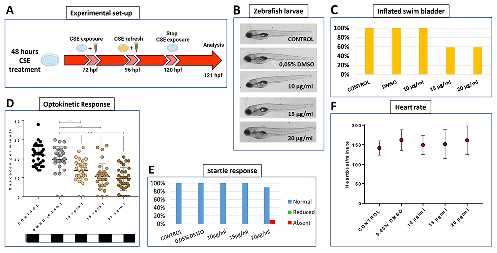
Figure 1. Cigarette Smoke Extract Impairs Visual Behaviour.
A. Schematic illustrating the timeline and experimental design of 48 hours cigarette smoke extract (CSE) treatment of zebrafish larvae and physiological analysis. B. Gross morphology of 5 dpf larvae treated with CSE showing no macroscopic abnormalities except for the incomplete inflation of swim bladder. C. The percentage of larvae with inflated swim bladders is only reduced at 15 or 20 µg/mL CSE, n=24 larvae per group, experiment repeated 3 times. D. Standard optokinetic response assay (0.02 cpd) at 5 dpf shows visual behaviour is significantly affected at 10, 15 and 20 µg/mL CSE; n=32 larvae per group, experiment repeated 4 times. Data were analysed by one-way ANOVA and Bonferroni´s multiple comparison test where **=p<0.01, ***=p<0.001 and ****=p<0.0001. Error bars indicate standard deviation (SD). E. Normal swimming response when caudal fin was touched at 5 dpf. N=16 larvae per group, experiment repeated 2 times. F. Heartbeat per minute average reveals no significant effects of CSE on cardiac rhythm at 5 dpf. N=8 larvae per group, experiment repeated 2 times.
The OKR is an established visual behaviour assay widely used for assessment of vision in zebrafish16,35. Larvae treated with 10, 15 or 20 µg/mL were assessed using a 0.02 cpd drum, with 100% contrast, considered the standard optokinetic response (Figure 1D). A dose-dependent reduction in visual behaviour was observed with CSE. Larvae treated with 10, 15 or 20 µg/mL CSE showed an average of 13.7, 10.7 or 9.4 saccades per minute, respectively, which were significantly lower compared with the 0.05% DMSO vehicle control group (p=0.0093, p=0.0004 and p<0.0001, respectively) that exhibited 19.7 saccades per minute. (Figure 1D).
To assess the selectivity of the CSE effect on visual behaviour, other more general, systemic readouts of physiology were analysed. The touch or startle response is a locomotor test assessing general motility16,36. Larvae treated with 10 or 15 µg/mL CSE showed a normal touch response, and only one of 16 larvae treated with 20 µg/mL CSE showed an absent touch response (Figure 1E). Cardiac physiology assessed by heart rate was not altered in any of the 10, 15 or 20 µg/mL CSE-treated groups (149.63, 151.75 or 161.38 bpm, respectively) when compared with the 0.05% DMSO and untreated control groups (141.75 or 162 bpm, respectively) (Figure 1F). In conclusion, specific concentrations of CSE appear to induce a selective visual behaviour deficit in zebrafish larvae.
Cigarette smoke extract attenuates visual acuity and contrast sensitivity
To assess effects of CSE on visual behaviour in more depth, visual acuity (VA) and contrast sensitivity (CS) were assessed (Figure 2). To examine VA, each larva was assessed consecutively with a 0.02, then a 0.06, and finally a 0.2 cpd drum, all with 100% contrast19. The responses for the 0.02 cpd drum show CSE-treated larvae with reduced saccadic eye movements. Responses of 13.5 and 12.3 saccades per minute, respectively with the 15 and 20 µg/mL CSE-treated larvae were significantly lower (p=0.0015 and p=0.0001, respectively) than the 0.05% DMSO control group which evoked 24.2 saccades per minute (Figure 2A). Surprisingly, although the average OKR to the mid-spatial frequency drum of 0.06 cpd, was lower for the 15 and 20 µg/mL CSE-treated larvae than the DMSO vehicle control group, this was not statistically significant (Figure 2A). However, the higher doses, 15 or 20 µg/mL CSE, also significantly impaired (p<0.0001) the ability of zebrafish larvae to respond to the highest spatial frequency tested with the 0.2 cpd 3D-printed drum (Figure 2A), lowering the number of saccades per minute to 1.6 and 0.9, respectively, versus the DMSO vehicle control group which responded with 6.5 saccades per minute.
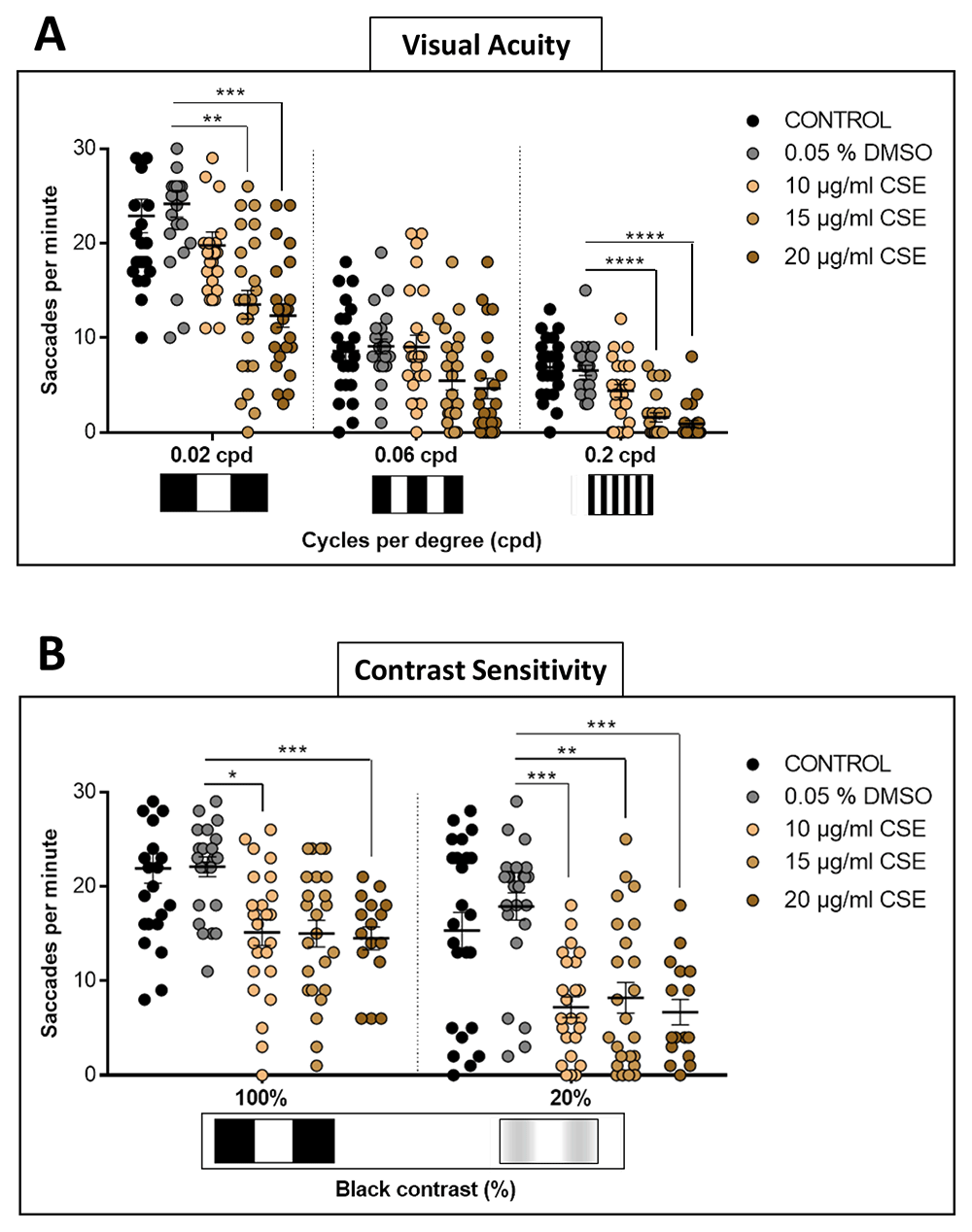
Figure 2. Visual Acuity and Contrast Sensitivity are affected by Cigarette Smoke Extract Treatment.
A. Larvae treated with 15 or 20 µg/mL CSE have a decreased visual acuity response to 0.2 cpd, 100% contrast drum, compared to DMSO treated larvae. B. For contrast sensitivity, all CSE concentrations tested result in a significantly reduced number of saccadic eye responses with the 20% black contrast drum, compared to DMSO controls. Data were analysed by repeated measures one-way ANOVA and Bonferroni’s multiple comparison test, where *=p>0.01, **=p<0.01, ***=p<0.001 and ****=p<0.0001. Error bars indicate standard deviation (SD). Midline of error bars represents the group average. Protocol I used, three replicates of eight larvae, n=24 measurements per pattern per group.
CS was tested comparing 0.02 cpd drums but with 100% or 20% contrast19. With the 20% black-white contrast drum, all zebrafish groups treated with CSE displayed significantly decreased visual responses (Figure 2B). CSE-treated groups with 10, 15 and 20 µg/mL showed 7.2, 8.2 and 7.2 saccades per minute, respectively, which were significantly lower (p=0.0006, p=0.0043 and p=0.0002 respectively) than the 0.05% DMSO vehicle control-treated larvae which respond with 17.9 saccades per minute. In summary, these bespoke visual behaviour assays showed that CSE impacts visual acuity and contrast sensitivity in zebrafish.
Cigarette smoke extract-treated larvae show diminished saccadic responses to coloured drums
To investigate if CSE treatment selectively affects optokinetic responses to coloured patterns, visual behaviour responses to drums with coloured stripes contrasted with alternating black stripes were quantified (Figure 3A). Thus, larvae treated with 10, 15 or 20 µg/mL CSE were confronted with rotating black-white (standard), then black-red, black-green or black-blue 0.02 3D-printed drums (Figure 3). As shown earlier, larvae treated with CSE display reduced saccades with the standard OKR black-white 0.02 drum (Figure 1D, Figure 3B). When testing the black-red drum, 10, 15 or 20 µg/mL CSE-treated larvae generated significantly impaired visual responses; reduced by 82% (3.9 saccades per minute, p=0.0126), 94.5% (1.2 saccades per minute, p<0.0001) and 98.3% (0.4 saccades per minute, p<0.0001), respectively compared to the 0.05% DMSO vehicle control larvae (Figure 3B–C). With the black-green drum, 15 µg/mL CSE significantly reduced the number of saccades by 85.6% (3.1 saccades per minute, p<0.0001) and 20 µg/mL CSE by 92.6% (1.6 saccades per minute, p<0.0001). With the black-blue drum, 15 and 20 µg/mL CSE-treated larvae significantly reduced their saccades per minute by 98.9% and 99.4%, respectively (p<0.0001 and p<0.0001) (Figure 3B–C). Overall, the biggest absolute reduction in visual behaviour was observed with the black-white drum wherein 20 µg/mL CSE displayed an average reduction of 13.5 saccades per minute, followed by black-green, black-blue and black-red with saccade per minute reductions of 12.5, 9 and 5.5, respectively (Figure 3C). Notably, the rank order of the largest percentage reduction in OKR was with the black-blue, black-red, black-green and black-white drum when exposed to 20 µg/mL CSE (Figure 3C).
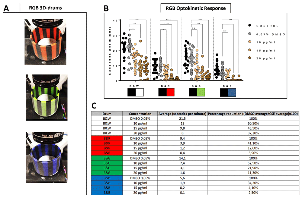
Figure 3. Cigarette Smoke Extract decreases the ability to discriminate colour patterns.
A. Larvae treated with 10, 15 and 20 µg/mL have a poorer optokinetic response to customised RGB 0.02 cpd drums. B. Average of number of saccades and reduction between 0.05% DMSO control group and CSE-treated larvae. C. Table showing OKR response from CSE-treated larvae to Red-Black, Green-Black and Blue-Black G response is lower than standard OKR (black-white stripes). Data were analysed by repeated measures one-way ANOVA and Bonferroni’s multiple comparison test where ****=p<0.0001 and **=p<0.01. Error bars indicate standard deviation. Midline of error bars represents the group average. Protocol I (19 used, three replicates of eight larvae, n=24 measurements per pattern per group.
Short cigarette smoke extract exposure induces abnormal visual behaviour
To analyse shorter-term effects of CSE, wild-type zebrafish larvae were exposed at 96 hpf to CSE for only 24 hours and the standard optokinetic response (0.02 cpd, 100% contrast) performed at 120 hpf (Figure 4A–B). A 20 µg/mL CSE concentration was chosen as it produced the most adverse effects on visual behaviour in the earlier assays with 48 hours CSE treatment. A 24-hour exposure to CSE resulted in larvae exhibiting a significantly lower number of eye movements (15.53 saccades per minute, p<0.0001) compared with vehicle controls (25.41 saccades per minute). Following the same exposure time (Figure 4A), the VMR was assessed in larvae treated with 20 µg/mL CSE-treated or 0.05% DMSO vehicle control (Figure 4C). The VMR is a visual behaviour assay that analyses how zebrafish acutely respond to lights turned ON and OFF16,37,38. Here, we analysed overall locomotor activity and peak locomotor activity when lights go ON and OFF (Figure 4C). In the overall activity, 20 µg/mL CSE results in significantly lower (52.5% reduced, p<0.0001) larval activity than the 0.05% DMSO vehicle group. (Figure 4C.1). CSE treated larvae showed 0.01±0.008 milliseconds per second (ms/s) average activity, versus the 0.05% DMSO vehicle group, which showed 0.021±0.009 ms/s average activity. In relation to changes in locomotor activity when lights were switched ON/OFF, although CSE-treated larvae exhibited markedly reduced activity before either change in light (Figure 4C.2–3), they could sense the lighting status and did increase their activity, albeit not to the same level as the DMSO controls (Figure 4C.2–3). In relation to peak activity at the light changes, there was no significant change in the average maximum peak OFF activity between 20 µg/mL CSE-treated larvae (0.1±0.07 ms/s) compared to vehicle controls (0.12±0.058 ms/s) (Figure 4C.3). However, CSE treatment resulted in a significant reduction in the average maximum peak ON activity (0.05±0.06 ms/s activity, p=0.0342) compared to 0.05% DMSO controls (0.09±0.07 ms/s) (Figure 4C.2).
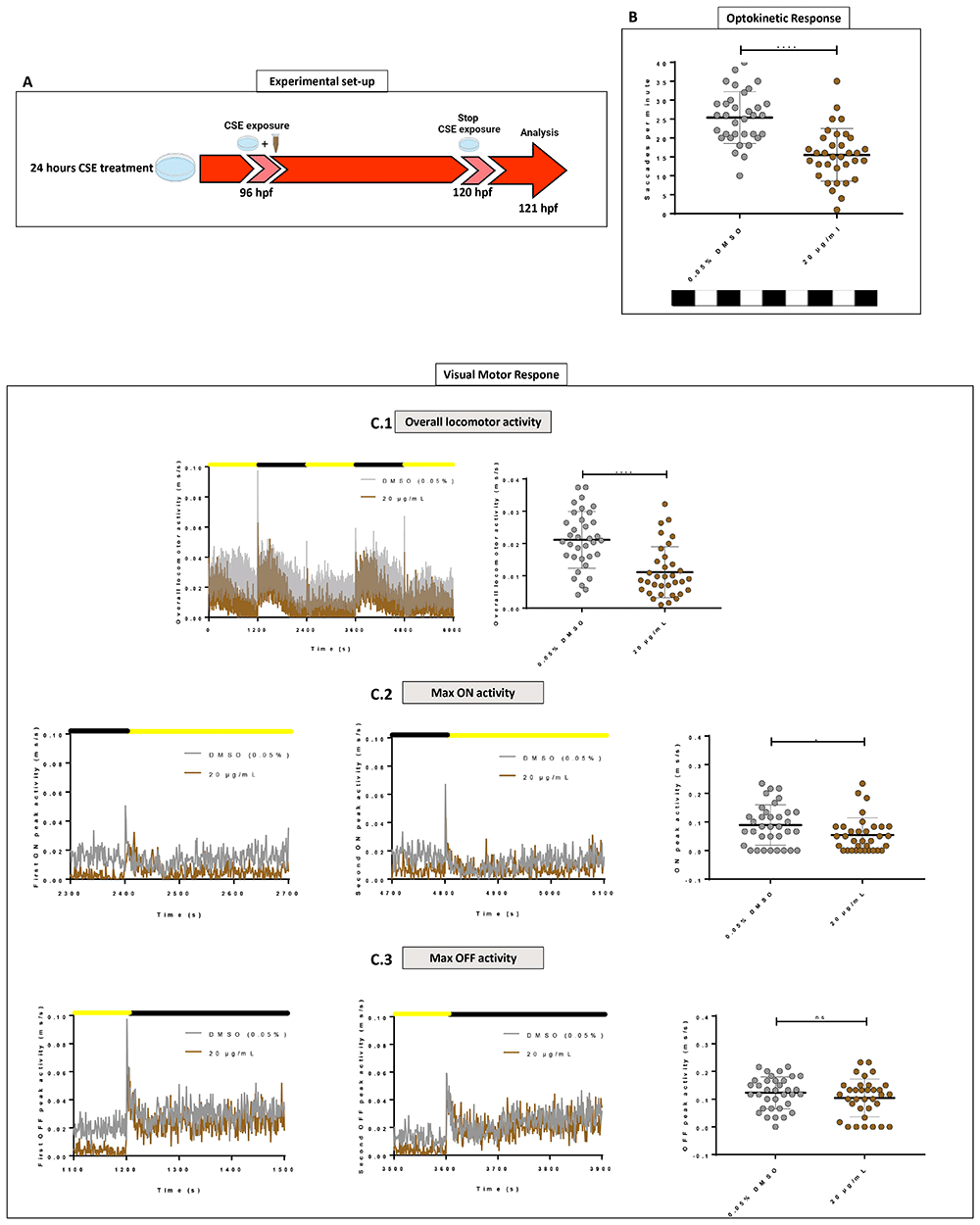
Figure 4. Standard Optokinetic Response and Visual Motor Response are affected when CSE is applied for 24 hours.
A. Schematic illustrating the timeline and experimental design of 24 hours cigarette smoke extract (CSE) treatment of zebrafish larvae. B. Standard optokinetic response (0.02 cpd, 100% contrast) at 121 hpf after 24 hours CSE-treatment. Larvae treated with 20 µg/mL show a lower optokinetic response than 0.05% DMSO vehicle control group. Statistical analysis carried out by student t-test between groups where ****=p<0.0001. Midline of error bars represents the group average. Three replicates, n=36 measurements per treatment group. C. Visualmotor response traces showing overall locomotor activity (C.1) and max ON (C.2) and max OFF (C.3) peak activities revealed 20 µg/mL CSE-treated larvae display less overall locomotor movements (C.1) but still react to light changes (C2–C3). Statistical analysis was carried out by unpaired t-test and a non-parametric Mann-Whitney test where *=p>0.01 and ns=no significant. Error bars indicate standard deviation and midline represents the group mean. 3 replicates of twelve larvae were used, n=36 measurements per group.
Four-hour and 24-hour CSE treatment alters gpx1a expression in larval zebrafish eyes
Smoking and cigarette smoke cause oxidative damage to the eye and increase the risk of AMD and cataracts by increasing oxidative stress6,39–41). Here, we analysed the following primary antioxidant genes: catalase (cat) and glutathione peroxidase 1 (gpx1a), as well as nuclear factor erythroid 2–related factor 2 (nfe2l2a), related with cellular defence against oxidative damage, and caspase 3 (casp3a), the major caspase activated by apoptosis (Figure 5). A total of 24 enucleated larval eyes, treated for four or 24 hours (Figure 5A) with CSE, were used for RT-PCR reactions. Significant changes in cat, nfe2l2a and casp3a were not observed after four or 24 hours CSE treatment (Figure 5B). gpx1a expression increased significantly after either four (p=0.0001) or 24 hours (p=0.0003) of CSE treatment compared with 0.05% DMSO vehicle control larvae (Figure 5B). This suggests that CSE modulates oxidative stress in the eye.
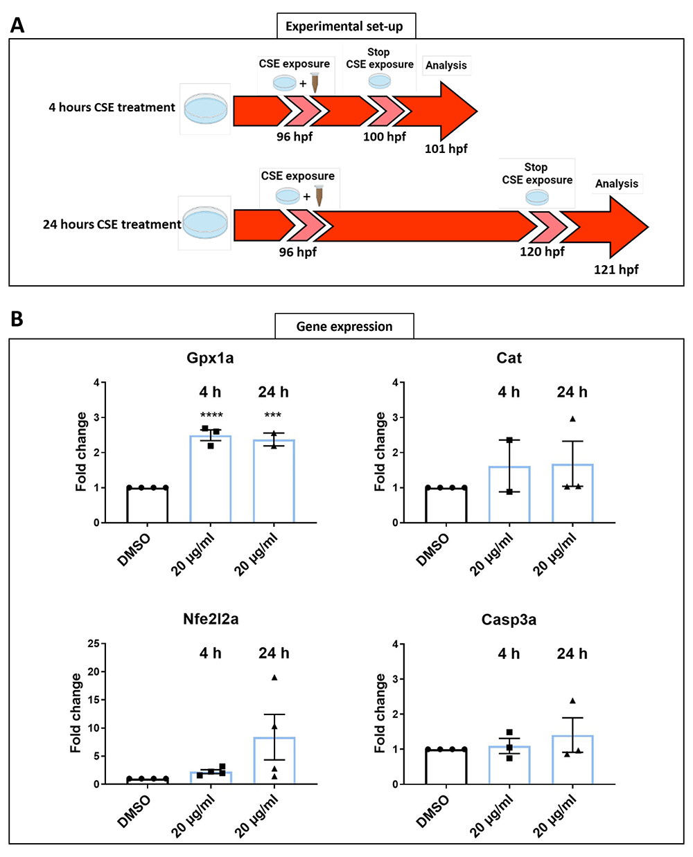
Figure 5. Shorter CSE treatment increases gpx1a expression.
A. Schematic representation of the timeline and experimental design of 4- and 24-hours cigarette smoke extract (CSE) treatment of zebrafish larvae. B. Gene expression profiles revealed a significant higher expression of gpx1a at 4- and 24- hours 20 µg/mL CSE treatment in zebrafish larvae eyes. Data was analysed by one-way ANOVA and a Dunnett’s multiple comparison test where ****=p<0.0001 and ***=p<0.001. 3 replicates of twelve larvae were carried out, n=72 larval eyes per group.
Hyaloid vessel diameter increases after 48-hours CSE exposure
To investigate the effect of CSE on developing intraocular vessels in zebrafish, Tg(fli1:EGFP) larvae were treated for 48 hours with 15 or 20 µg/mL CSE, prior to morphological analysis of the hyaloid vasculature (HV). The HV develops in zebrafish from 24 to 72 hpf and consists of four to five main branches radiating from the hyaloid artery which enters through the optic disc at the back of the lens. These primary vessels branch out multiple times and eventually drain to the inner optic circle at the anterior surface of the lens, which remains avascular. By 5 dpf, hyaloid vasculature constitutes an intricately branched network of intraocular vessels that nourish the developing lens and inner retina31,42,43. The number of main vessels radiating from the optic disc was not altered in larvae treated with 15 µg/mL CSE, whereas 20 µg/mL CSE modestly, but significantly decreased the number of primary hyaloid branches (p=0.002) compared to DMSO controls (Figure 6B). Direct observation under the microscope suggested the hyaloid vessels in both 15 and 20 µg/mL CSE-treated groups, appeared thicker than the DMSO vehicle controls (Figure 6A). Thus, hyaloid vessels were measured at the primary branch level, between the first bifurcation at the optic disc and the second bifurcation (Figure 6A). The diameter of the vessels significantly increased from 4.12 µm in 0.05% DMSO vehicle control group to 5.96 µm in 15 µg/mL CSE-treated larvae, and to 5.69 µm in the 20 µg/mL CSE-treated larvae (p≤0.0001 in both cases) (Figure 6C).
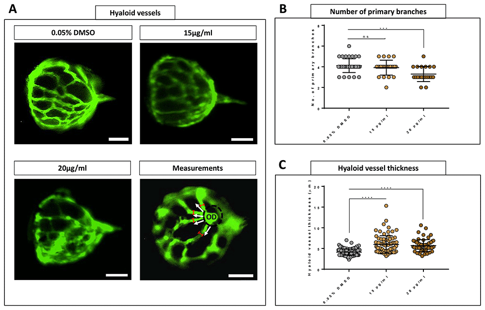
Figure 6. Hyaloid vessels are dilated with 48 hours Cigarette Smoke Extract Systemic Treatment.
A. Fluorescent images of hyaloid vasculature on 0.05% DMSO vehicle control group and 15 and 20 µg/mL CSE treated dissected lenses. Dilated and non-defined vessels are evident in 15 and 20 µg/mL CSE-treated lenses. White arrows indicate primary hyaloid branches. Delimiter red lines define where thickness of hyaloid vessels was measured. Scale bar= 50 µm. B. 20 µg/mL CSE-treated zebrafish larvae develop a smaller number of primary branches. C. Hyaloid vessel thickness plot graph revealed an abnormal increase of hyaloid vessel thickness in 15 and 20 µg/mL CSE-treated zebrafish larvae. Data was analysed by ordinary one-way ANOVA and a Tukey’s multiple comparisons where ****=p<0.0001, ***=p<0.001 and ns=no significant. Five biological replicates, minimum 63 measurements per group.
Ocular axial length and lens thickness increase with CSE exposure
Eye histology was imaged to see whether eye structures were affected in 121 hpf larvae treated with 0.05% DMSO or 20 µg/mL CSE for 48 hours (Figure 7). Retinal layers were well-defined and retinal cell morphology appear not be affected by 20 µg/mL CSE (Figure 7A). Interestingly, biometry analysis highlighted a statistically significant axial length elongation in 20 µg/mL CSE-treated larvae (216.9 µm, p<0.0001) compared to 0.05% DMSO vehicle control (205.1 µm) and a lens thickness increase, to 107.9 µm (p=0.0044) in CSE-treated larvae compared to 99.65 µm in the 0.05% DMSO vehicle control group (Figure 7B). No differences were found in the thickness of the ganglion cell, inner plexiform, inner nuclear, outer nuclear or retinal pigmented epithelium layers (Figure 7B).
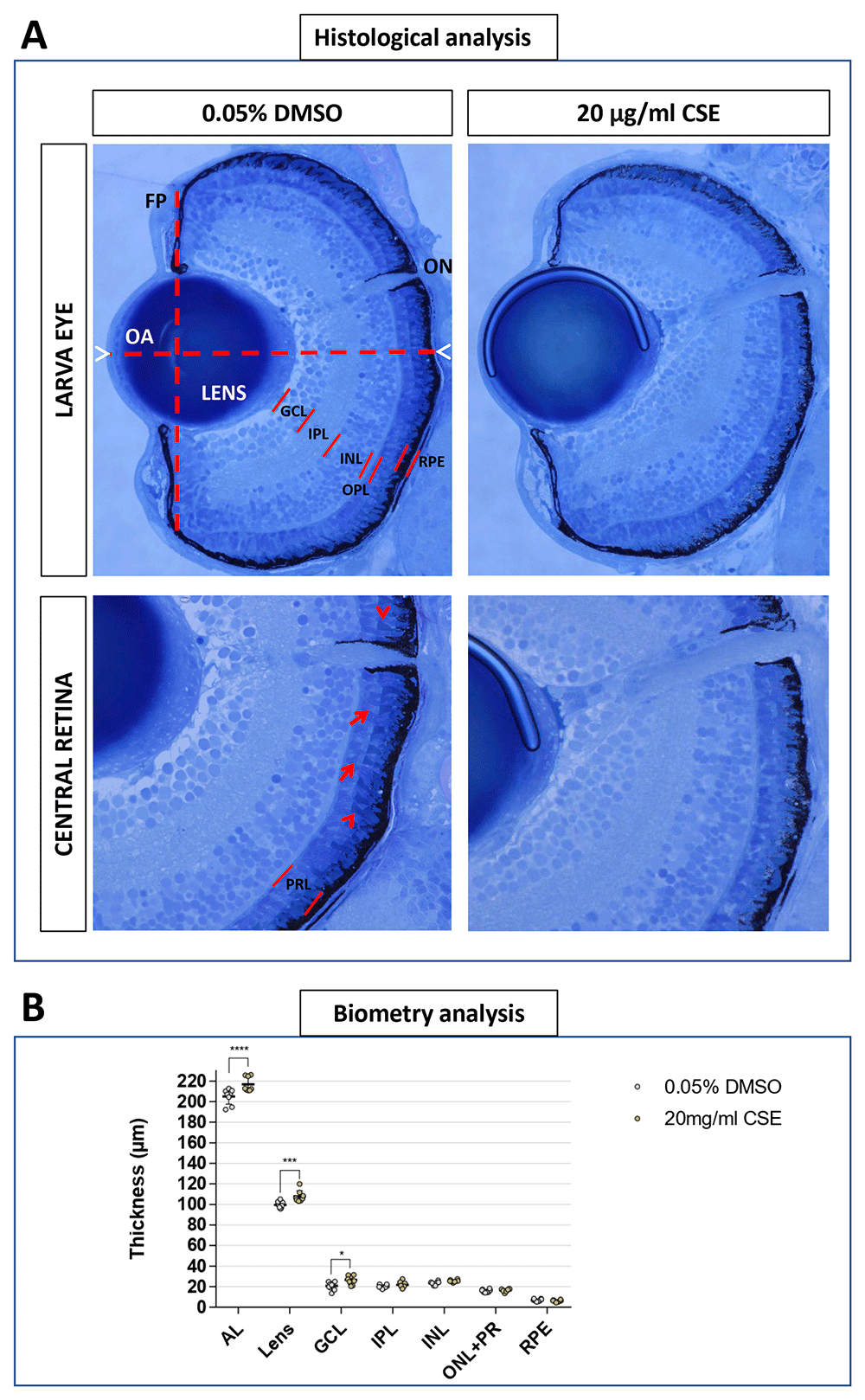
Figure 7. Axial Length, Lens and Ganglion Cell Layer appear increased with 48 hours Cigarette Smoke Extract Systemic Treatment.
A. 20 µg/mL CSE treated larvae do not display observable defects in eye morphology or retinal arrangement. Representative light microscopy images from 0.05% DMSO control and 20 µg/mL CSE-treated larvae. Front plane (FP) and Optic Axis (OA) traced for morphological analysis are defined by dashed lines. Axial length (AL) is delimited by white arrowheads. Retinal layers are defined by red lines. PRL is formed by both photoreceptors inner segments (red arrow) and outer segments (red arrowhead). B. Quantitative biometry of retinal layers using Olympus cellSens standard software, showed enlarged AL and lens in 20 µg/mL CSE-treated larvae, n=8 larvae analysed. Data was analysed by one-way ANOVA and a Bonferroni’s multiple comparison test where ****=p<0.0001 and **=p<0.01. GCL: ganglion cell layer; IPL: inner plexiform layer; INL: inner nuclear layer; OPL: outer plexiform layer; PRL: photoreceptors layer; RPE: retinal pigment epithelium; ON: optic nerve.
CSE increases the number of retinal pigment epithelium phagosomes
Previous studies in mammals showed CSE negatively affects the RPE26,27. As the RPE is one of the first retinal structures affected in AMD patients41 and given the relation between AMD and smoking3,13,14, we imaged the outer retina of 48 h CSE-exposed zebrafish by TEM. Larvae were fixed between 120-121 hpf, which coincides with the dawn peak of OSP phagocytosis in older zebrafish32,44 (Figure 8). Macroscopically, the outer retinae of 20 µg/mL CSE-treated larvae showed a slight disarray of the photoreceptor layer and RPE, and an elevated number of RPE phagosomes (Figure 8A). Interestingly, upon quantification, larvae exposed to CSE had significantly higher numbers of RPE phagosomes compared to vehicle controls (p=0.0002). 20 µg/mL CSE-treated larvae had an average of 0.1425 ± 0.009287 phagosomes/um RPE (n=4), a value ~50% higher than larvae treated as the vehicle control (0.093 ± 0.009 phagosomes/um RPE, n=3) (Figure 8B). This suggest that exposing zebrafish larvae to CSE causes dysregulation of OSP.
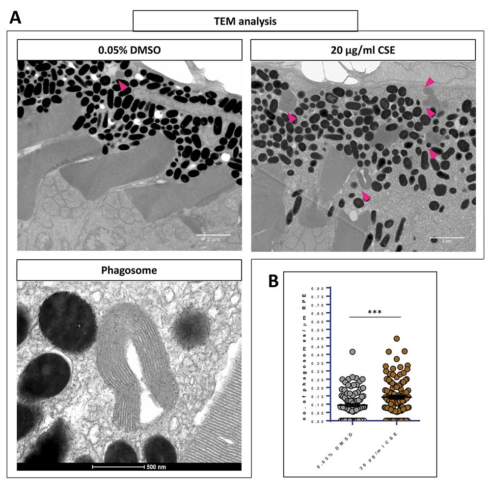
Figure 8. Exposure to cigarette smoke extract causes increased levels of RPE phagosomes.
A. Representative TEM images of wild-type retinae at 121 hpf showing the OS tip and RPE of larvae treated with 0.05% DMSO and 20 µg/mL CSE of CSE. Phagosomes are indicated (pink arrows). Scale bar; 2 µm. Magnified TEM image of a phagosome. Scale bar 500 nm. B. Scatter plots of phagosome/µm of RPE in 20 µg/mL CSE-treated and vehicle control (0.05% DMSO) retinae. Larvae exposed to CSE for 48 hours show a 50% increase in the number of RPE phagosomes containing engulfed OS material in comparison to the vehicle control controls. Statistical analysis carried out by student t-test between groups where ***=p<0.001. RPE of >3000 μm was measured per larva. N=3,4 larvae (6-8 retinae) per group.
Discussion
Cigarette smoke is well characterised as a serious health hazard1,2,45. Previous in vitro and in vivo studies (with human cell lines, mice and zebrafish) investigated the impact of cigarette smoke on the cardiorespiratory system, on vasculature24, and lungs46,47, as well as its relation with oxidative stress48,49. However, effects of cigarette smoke on the eye have only been researched in mice25–27,50. To our knowledge, this is the first study investigating adverse events of cigarette smoke on the developed zebrafish visual system. Zebrafish are an advantageous alternative vertebrate animal model to analyse cigarette smoke impact on the ocular system, with a high degree of structural and functional conservation between the human and zebrafish retina18,20.
CSE treatment of zebrafish larvae impairs vision
In our experimental design, we treated wild-type larvae at 72 hpf to rule out general developmental effects. At this time point, the zebrafish visual system is considered “mature”, with all retinal layers formed and the hyaloid vasculature network established18,31,51. The observed maximum tolerated dose of 20 µg/mL matches with response profiles reported in a previous teratogenic study induced by treating 6- to 72-hpf zebrafish larvae with CSE21. The teratogenic study by Ellis et al.21 showed a decreased behavioural response to light-dark changes, which the authors attributed to alterations in neuromuscular function or neurotoxicity.
To our knowledge, this is the first study reporting an abnormal visual behaviour in zebrafish treated with CSE. In our model, we observed uninflated swim bladders with 15 or 20 µg/mL CSE. Previous studies report that visually impaired zebrafish often fail to inflate their swim bladder34,52, conceivably due to difficulty to swim to surface to inhale air. To analyse if CSE affected larvae vision, we conducted variants of the optokinetic response assay (OKR), an efficient and well-established visual behaviour test which can be accurately measured in zebrafish from three days post-fertilization35. CSE significantly impaired zebrafish visual behaviour, visual acuity and contrast sensitivity. Responses to RGB-black coloured drums were also impaired by CSE treatment.
It was important to assess if CSE in our model was inducing broad systemic affects. Previous studies with nicotine or smoke show malformation of the zebrafish heart when treating with CSE at earlier embryonic stages22,24 and a 50% reduction of heart rate21,22. In our model, heart rate was not affected in CSE-treated larvae with similar values to the 0.05% DMSO vehicle controls and the untreated larvae. In summary, the reduced zebrafish optokinetic responses (e.g. VA, CS and RGB OKR drum assays) observed appear due to a selective defect in the visual system induced by CSE treatment at 3 dpf. The impaired optokinetic visual behaviour was corroborated by altered visualmotor responses, in which larvae display bursts of movement upon light changes16. Interestingly, previous locomotor activity assays on CSE-treated zebrafish23 found that 24 hpf embryos treated for 20 hours showed hyperactivity to 10 min light changes cycles23. In contrast, Ellis et al.21 reported that responses to short 5 min, lights ON and OFF cycles were not impacted by 20 µg/mL CSE (although higher concentrations did at) in 5 dpf larvae treated for 48 hours21. Here, when using longer light cycles (20 min intervals), providing zebrafish more time to adapt between acute light changes, we found overall locomotor activity was lower in zebrafish larvae exposed to 20 µg/mL CSE. For light to dark changes (peak OFF activity), the zebrafish showed a CSE concentration-dependent reduction in locomotor responses21.
CSE increases anti-oxidant gene expression in zebrafish eyes
Having identified CSE-induced visual behaviour defects that were not linked to widespread systemic effects, we sought to understand the molecular, cellular, and physiological changes in the eye linked to impaired vision. Smoking causes oxidative damage due to free radicals6,7,39 which imbalance the antioxidant system and generate reactive oxygen species (ROS) that underlies the pathogenesis of several human eye diseases53,54. Investigations into the pathophysiology of cigarette smoke-induced AMD revealed that cigarette smoke exposure can generate oxidative stress by producing free radicals, subsequently leading to a depletion of the antioxidant system39,55,56. A previous study treating zebrafish larvae from 2 hpf and analysing the activity of ROS biomarkers at 96 hpf found evidence of increased oxidative stress, however, gpx1a and cat activities were not significantly altered in CSE-treated larvae <96 hpf23. Here, we analysed antioxidant gene expression changes in zebrafish larval eyes treated with CSE from 96 to 101 and to 121 hpf. Cat gene expression was unchanged in agreement with the Massarsky et al. study23. However, in 101 and 121 hpf CSE-exposed zebrafish, a significantly higher expression of gpx1a was observed indicating CSE exposure induces oxidative stress in zebrafish larval eyes. Oxidative damage is a hallmark of AMD development27,53–55. Moreover, several studies39,48,49 report cigarette smoke-induced oxidative stress in animal models to show similar changes in the eye as AMD patients. Thus, CSE-exposed zebrafish provides an alternative model to study CSE gene expression changes in the eye and associated ocular pathologies.
CSE alters zebrafish retinal histology
Interestingly, axial length was significantly increased in zebrafish larvae exposed to the highest dose of CSE (20 µg/mL). Myopia is a refractive error where the eye has excessive axial length57. Myopia is a multifactorial condition so environmental factors are involved in its development57. The relationship of CSE exposure and smoking with myopia has been investigated, however, this association remains unclear57–59. Previous clinical studies found that children of smoking parents had a lower axial length compared with those of non-smokers57,58. However, in contrast, in chicks administered with nicotinic antagonist drugs59, researchers found that nicotinic receptors are involved in excessive ocular growth. This contrasts with clinical results in humans; nevertheless, nicotine only represents a small percentage in CSE composition60.
Analysis of the retinal layers revealed that the lens had the most significant increase in thickness upon CSE exposure. Smokers have an increased risk of developing nuclear cataracts40,61, an opacification in the middle of the lens, which is the leading cause of blindness worldwide61. Oxidative stress mechanisms and accumulation of cadmium, iron and other metals presents in the CSE play an important role in cataractogenesis61–63. For instance, the exposure of rats to cigarette smoke for two hours a day over 60 days, lead to an increase in the number, size and layering of cells in the lens epithelium62. Lens thickness is not usually a clinical practice to diagnose cataracts but it is known the lens becomes thicker in the presence of nuclear cataracts64. We speculate that the CSE zebrafish model may be experiencing an onset of similar biological effects in the lens, however, further histological and biomolecular analysis are needed.
Systemic CSE treatment induces enlarged hyaloid vessels
The zebrafish HV surrounds the larval lens and becomes attached to the inner surface of the retina in the adult (after 28 dpf)31. The HV support development of the lens and retina, and share many features with human retinal vasculature, making them a valuable model of human disease31,43,65. In 5-dpf larvae, exposure to CSE for 48 hrs, HV appear dilated and display a significantly enlarged thickness (Figure 6C). We also observed a reduction in the number of main HV branches (Figure 6B). As these were probably completely formed when CSE was administered at 72 hpf, this is likely a toxic effect rather than an antiangiogenic effect31. In mammals, systemic vascular tone dysfunction and vascular inflammation induced by CSE exposure (even short term) is described in the literature, and also associated with second-hand smoking66,67. Retinal vascular calibre is a well-established biomarker of cardiovascular disease (including diabetes)68,69, and in ophthalmology, age-related macular degeneration (AMD) is associated with arteriolar and venular dilation in the retina70. Many studies have researched the effects of cigarette smoking (including second-hand) on vessels in the retina and the choroid. Enlarged retinal calibre is extensively related to cigarette smoking in the literature71,72, although it has also been linked to narrower retinal arterioles and venules in certain cases72–74. The unanimous consensus in the literature is that cigarette smoke induces alterations in the microvascular calibre of the choroidal and retinal capillary plexus, which have been linked to oxidative stress, inflammation, and increased risk of vascular disease71–74.
Photoreceptor outer segment phagosomes increased by CSE
Outer segment phagocytosis (OSP) is a regulated process that maintains photoreceptor health and vision32. Pharmacological modulation can affect the OSP at any stage from recognition, binding or degradation75,76. Interestingly, exposing zebrafish larvae to CSE causes dysregulation of the OSP process, resulting in higher levels of RPE phagosomes. In general, increased phagosomes and phagolysosomes can indicate two possible mechanisms: either increased engulfment and phagocytosis and/or suppressed degradation. To our knowledge this is the first time CSE exposure has been linked to the OSP process in zebrafish. Cigarette smoke is associated with lysosomal dysfunction, with activity of the known OSP regulator cathepsin D being altered upon CSE exposure in both cell culture and murine models56,77. The lysosomal activity of cathepsin D is required for the degradation of the removed OS tip, the final stage of OS phagocytosis78. The molecular mechanisms involved in the increased level of RPE phagosomes observed in the CSE treated larvae remain to be elucidated.
Conclusions
We present here evidence that CSE disrupts visual behaviour, visual acuity and contrast sensitivity in larval zebrafish. Zebrafish eyes showed an increased oxidative stress as well as an abnormal HV. CSE induced axial length elongation, lens thickness increase and a disruption of photoreceptor outer segment phagocytosis. These findings provide zebrafish as a tool to investigate biological changes induced by cigarette smoke in the eye.
Data availability
Underlying data
Zenodo: Systemic Treatment with Cigarette Smoke Extract Affects Zebrafish Visual Behaviour, Intraocular Vasculature Morphology and Outer Segment Phagocytosis,
https://doi.org/10.5281/zenodo.772337379
This project contains the following underlying data:
Figure. 1C-D-F. Cigarette Smoke Extract Impairs Visual Behaviour..xlsx
Figure. 1D Figure 1. Cigarette Smoke Extract Impairs Visual Behaviour..pzfx
Figure. 1F Figure 1. Cigarette Smoke Extract Impairs Visual Behaviour..pzfx
Figure. 4B. Standard Optokinetic Response and Visual Motor Response are affected when CSE is applied for 24 hours..pzfx
Figure. 4C. Standard Optokinetic Response and Visual Motor Response are affected when CSE is applied for 24 hours..pzfx
Figure. 5. Shorter CSE treatment increases gpx1a expression.pzfx
Figure. 6. Hyaloid vessels are dilated with 48 hours Cigarette Smoke Extract Systemic Treatment..pzfx
Figure. 7. Axial Length, Lens and Ganglion Cell Layer appear increased with 48 hours Cigarette Smoke Extract Systemic Treatment..pzfx
Figure. 8. Exposure to cigarette smoke extract causes increased levels of RPE phagosomes.pzfx
Fig_1C_Cigarette Smoke Extract Impairs Visual Behaviour.csv
Fig_1D_Cigarette Smoke Extract Impairs Visual Behaviour.csv
Fig_1E_Cigarette Smoke Extract Impairs Visual Behaviour.csv
Fig_1F_Cigarette Smoke Extract Impairs Visual Behaviour.csv
Fig_2A_Visual Acuity and Contrast Sensitivity are affected by Cigarette Smoke Extract Treatment.csv
Fig_2B_Visual Acuity and Contrast Sensitivity are affected by Cigarette Smoke Extract Treatment.csv
Fig_3B_Cigarette Smoke Extract decreases the ability to discriminate colour patterns.csv
Fig_3C_Cigarette Smoke Extract decreases the ability to discriminate colour patterns.csv
Fig_4B_Standard Optokinetic Response and Visual Motor Response are affected when CSE is applied for 24 hours.csv
Fig_4C_MAX OFF peak activity_larva.csv
Fig_4C_MAX ON peak activity_larva.csv
Fig_4C_overall locomotor activity_larva.csv
Fig_4C_traces 1st OFF peak.csv
Fig_4C_traces 1st ON peak.csv
Fig_4C_traces 2nd OFF peak.csv
Fig_4C_traces 2nd ON peak.csv
Fig_4C_traces overall locomotor activity_larva.csv
Fig_5B_Caspase3.csv
Fig_5B_Catalase.csv
Fig_5B_gpx1.csv
Fig_5B_Nrf2.csv
Fig_6B_Hyaloid vessels are dilated with 48 hours Cigarette Smoke Extract Systemic Treatment.csv
Fig_6C_Hyaloid vessels are dilated with 48 hours Cigarette Smoke Extract Systemic Treatment.csv
Fig_7_Biometry.csv
Fig_8_Exposure to cigarette smoke extract causes increased levels of RPE phagosomes.csv
Figure 2. Visual Acuity and Contrast Sensitivity are affected by Cigarette Smoke Extract Treatment..pzf
Figure 3. Cigarette Smoke Extract decreases the ability to discriminate colour patterns..pzfx
Author contributions
Alicia Gomez Sanchez: Data Curation, Formal Analysis, Investigation, Validation, Visualization, Writing – Original Draft Preparation
Patrizia Colucci: Data Curation, Formal Analysis, Investigation, Validation, Visualization, Writing
Ailis Moran: Data Curation, Formal Analysis, Investigation, Validation, Visualization, Writing – Review & Editing
Alexandro Moya Lopez: Data Curation, Formal Analysis, Investigation, Validation, Visualization
Basilio Colligris: Supervision, Review & Editing.
Yolanda Álvarez: Data Curation, Formal Analysis, Validation, Visualization, Funding Acquisition, Project Administration, Resources, Supervision, Writing – Review & Editing
Brendan Kennedy: Conceptualization, Funding Acquisition, Methodology, Project Administration, Resources, Supervision, Writing – Review & Editing
Acknowledgments
The authors thanks to Niamh Stephens and Tiina O’Neill for imaging support and assistance. Authors have obtained permission from all those named in this section to include their names.
Faculty Opinions recommendedReferences
- 1.
Jha P:
Avoidable global cancer deaths and total deaths from smoking.
Nat Rev Cancer.
2009; 9(9): 655–64. PubMed Abstract
| Publisher Full Text
- 2.
World Health Organization: MPOWER: A Policy Package to Reverse the Tobacco Epidemic. Geneva;
2008. Reference Source
- 3.
National Center for Chronic Disease Prevention and Health Promotion (US) Office on Smoking and Health: The Health Consequences of Smoking—50 Years of Progress: A Report of the Surgeon General. 2014; 1–36. PubMed Abstract
- 4.
Composition, Exposure and Regulations:
Tobacco Smoke and Involuntary Smoking.
NCBI Bookshelf. [cited 2022 Apr 7]. Reference Source
- 5.
Paschke T, Scherer G, Heller WD:
Effects of Ingredients on Cigarette Smoke Composition and Biological Activity: A Literature Overview.
Beiträge zur Tab Int to Tob Res.
2002; 20(3): 107–247. Publisher Full Text
- 6.
Lois N, Abdelkader E, Reglitz K, et al.:
Environmental tobacco smoke exposure and eye disease.
Br J Ophthalmol.
2008; 92(10): 1304–10. PubMed Abstract
| Publisher Full Text
- 7.
Evaluating Exposure to Secondhand Smoke: Secondhand Smoke Exposure and Cardiovascular Effects.
NCBI Bookshelf. [cited 2022 Apr 7]. Reference Source
- 8.
IARC Monographs on the Identification of Carcinogenic Hazards to Humans.
INTERNATIONAL AGENCY FOR RESEARCH ON CANCER, [cited 2022 Apr 7]. Reference Source
- 9.
Reitsma MB, Kendrick PJ, Ababneh E, et al.:
Spatial, temporal, and demographic patterns in prevalence of smoking tobacco use and attributable disease burden in 204 countries and territories, 1990-2019: a systematic analysis from the Global Burden of Disease Study 2019.
Lancet.
2021; 397(10292): 2337–2360. PubMed Abstract
| Publisher Full Text
| Free Full Text
- 10.
Global Burden of Disease (GBD).
Institute for Health Metrics and Evaluation, 2019; [cited 2022 May 4]. Reference Source
- 11.
Centers for Disease Control and Prevention (US), National Center for Chronic Disease Prevention and Health Promotion (US), Office on Smoking and Health (US): How Tobacco Smoke Causes Disease: The Biology and Behavioral Basis for Smoking-Attributable Disease. A Report of the Surgeon General. 2010. PubMed Abstract
- 12.
Gidding SS, Xie X, Liu K, et al.:
Cardiac function in smokers and nonsmokers: the CARDIA study. The Coronary Artery Risk Development in Young Adults Study.
J Am Coll Cardiol.
1995; 26(1): 211–6. PubMed Abstract
| Publisher Full Text
- 13.
Klein BEK, Klein R:
Lifestyle exposures and eye diseases in adults.
Am J Ophthalmol.
2007; 144(6): 961–969.e1. PubMed Abstract
| Publisher Full Text
| Free Full Text
- 14.
Zhang X, Kahende J, Fan AZ, et al.:
Smoking and visual impairment among older adults with age-related eye diseases.
Prev Chronic Dis.
2011; 8(4): A84. PubMed Abstract
| Free Full Text
- 15.
Li JQ, Welchowski T, Schmid M, et al.:
Prevalence and incidence of age-related macular degeneration in Europe: a systematic review and meta-analysis.
Br J Ophthalmol.
2020; 104(8): 1077–1084. PubMed Abstract
| Publisher Full Text
- 16.
Deeti S, O’Farrell S, Kennedy BN:
Early safety assessment of human oculotoxic drugs using the zebrafish visualmotor response.
J Pharmacol Toxicol Methods.
2014; 69(1): 1–8. PubMed Abstract
| Publisher Full Text
- 17.
Teame T, Zhang Z, Ran C, et al.:
The use of zebrafish (Danio rerio) as biomedical models.
Anim Front.
2019; 9(3): 68–77. PubMed Abstract
| Publisher Full Text
| Free Full Text
- 18.
Richardson R, Tracey-White D, Webster A, et al.:
The zebrafish eye-a paradigm for investigating human ocular genetics.
Eye (Lond).
2017; 31(1): 68–86. PubMed Abstract
| Publisher Full Text
| Free Full Text
- 19.
Gómez Sánchez A, Álvarez Y, Colligris B, et al.:
Affordable and effective optokinetic response methods to assess visual acuity and contrast sensitivity in larval to juvenile zebrafish [version 2; peer review: 2 approved].
Open Res Eur.
2022; 1: 92. Publisher Full Text
- 20.
Hong Y, Luo Y:
Zebrafish Model in Ophthalmology to Study Disease Mechanism and Drug Discovery.
Pharmaceuticals (Basel).
2021; 14(8): 716. PubMed Abstract
| Publisher Full Text
| Free Full Text
- 21.
Ellis LD, Soo EC, Achenbach JC, et al.:
Use of the zebrafish larvae as a model to study cigarette smoke condensate toxicity.
PLoS One.
2014; 9(12): e115305. PubMed Abstract
| Publisher Full Text
| Free Full Text
- 22.
Massarsky A, Jayasundara N, Glazer L, et al.:
Outcomes of developmental exposure to total particulate matter from cigarette smoke in zebrafish (Danio rerio).
Neurotoxicology.
2018; 68: 101–14. PubMed Abstract
| Publisher Full Text
- 23.
Massarsky A, Jayasundara N, Bailey JM, et al.:
Teratogenic, bioenergetic, and behavioral effects of exposure to total particulate matter on early development of zebrafish (Danio rerio) are not mimicked by nicotine.
Neurotoxicol Teratol.
2015; 51: 77–88. PubMed Abstract
| Publisher Full Text
| Free Full Text
- 24.
Massarsky A, Prasad GL, Di Giulio RT:
Total particulate matter from cigarette smoke disrupts vascular development in zebrafish brain (Danio rerio).
Toxicol Appl Pharmacol.
2018; 339: 85–96. PubMed Abstract
| Publisher Full Text
- 25.
Li J, Zhang G, Nian S, et al.:
Dry eye induced by exposure to cigarette smoke pollution: An in vivo and in vitro study.
Free Radic Biol Med.
2020; 153: 187–201. PubMed Abstract
| Publisher Full Text
- 26.
Espinosa-Heidmann DG, Suner IJ, Catanuto P, et al.:
Cigarette smoke-related oxidants and the development of sub-RPE deposits in an experimental animal model of dry AMD.
Invest Ophthalmol Vis Sci.
2006; 47(2): 729–37. PubMed Abstract
| Publisher Full Text
- 27.
Fujihara M, Nagai N, Sussan TE, et al.:
Chronic cigarette smoke causes oxidative damage and apoptosis to retinal pigmented epithelial cells in mice.
PLoS One.
2008; 3(9): e3119. PubMed Abstract
| Publisher Full Text
| Free Full Text
- 28.
Zinflou C, Rochette PJ:
Absorption of blue light by cigarette smoke components is highly toxic for retinal pigmented epithelial cells.
Arch Toxicol.
2019; 93(2): 453–65. PubMed Abstract
| Publisher Full Text
- 29.
Grace Kennedy HS, UCD Biomedical Facility Staff BK: University College Dublin (UCD) Zebrafish Facility Environmental Parameters: 2019. Protocols.io. 2019; 1. Reference Source
- 30.
Institute NC: The FTC Cigarette Test Method for Determining Tar Nicotine, and Carbon Monoxide Yields of U.S. Cigarettes. Report of the NCI Expert Committee. Tob Control Monogr No 7 Bethesda, MD US Dep Heal Hum Serv Natl Institutes Heal Natl Cancer Institute NIH Pub. 1996; 96–4028. Reference Source
- 31.
Alvarez Y, Cederlund ML, Cottell DC, et al.:
Genetic determinants of hyaloid and retinal vasculature in zebrafish.
BMC Dev Biol.
2007; 7: 114. PubMed Abstract
| Publisher Full Text
| Free Full Text
- 32.
Moran AL, Carter SP, Kaylor JJ, et al.:
Dawn and dusk peaks of outer segment phagocytosis, and visual cycle function require Rab28.
FASEB J.
2022; 36(5): e22309. PubMed Abstract
| Publisher Full Text
| Free Full Text
- 33.
Follo C, Ozzano M, Mugoni V, et al.:
Knock-Down of Cathepsin D Affects the Retinal Pigment Epithelium, Impairs Swim-Bladder Ontogenesis and Causes Premature Death in Zebrafish.
PLoS One.
2011; 6(7): e21908. PubMed Abstract
| Publisher Full Text
| Free Full Text
- 34.
Daly C, Shine L, Heffernan T, et al.:
A Brain-Derived Neurotrophic Factor Mimetic Is Sufficient to Restore Cone Photoreceptor Visual Function in an Inherited Blindness Model.
Sci Rep.
2017; 7(1): 11320. PubMed Abstract
| Publisher Full Text
| Free Full Text
- 35.
Brockerhoff SE:
Measuring the optokinetic response of zebrafish larvae.
Nat Protoc.
2006; 1(5): 2448–51. PubMed Abstract
| Publisher Full Text
- 36.
Liu YC, Bailey I, Hale ME:
Alternative startle motor patterns and behaviors in the larval zebrafish (Danio rerio).
J Comp Physiol A Neuroethol Sens Neural Behav Physiol.
2012; 198(1): 11–24. PubMed Abstract
| Publisher Full Text
- 37.
Emran F, Rihel J, Adolph AR, et al.:
OFF ganglion cells cannot drive the optokinetic reflex in zebrafish.
Proc Natl Acad Sci U S A.
2007; 104(48): 19126–31. PubMed Abstract
| Publisher Full Text
| Free Full Text
- 38.
Emran F, Rihel J, Dowling JE, et al.:
A Behavioral Assay to Measure Responsiveness of Zebrafish to Changes in Light Intensities.
J Vis Exp.
2008; 3(20): 923. PubMed Abstract
| Publisher Full Text
| Free Full Text
- 39.
Bruno RS, Traber MG:
Vitamin E biokinetics, oxidative stress and cigarette smoking.
Pathophysiology.
2006; 13(3): 143–9. PubMed Abstract
| Publisher Full Text
- 40.
Solberg Y, Rosner M, Belkin M:
The association between cigarette smoking and ocular diseases.
Surv Ophthalmol.
1998; 42(6): 535–47. PubMed Abstract
| Publisher Full Text
- 41.
Somasundaran S, Constable IJ, Mellough CB, et al.:
Retinal pigment epithelium and age-related macular degeneration: A review of major disease mechanisms.
Clin Experiment Ophthalmol.
2020; 48(8): 1043–56. PubMed Abstract
| Publisher Full Text
| Free Full Text
- 42.
Kaufman R, Weiss O, Sebbagh M, et al.:
Development and origins of Zebrafish ocular vasculature.
BMC Dev Biol.
2015; 15(1): 18. PubMed Abstract
| Publisher Full Text
| Free Full Text
- 43.
Hartsock A, Lee C, Arnold V, et al.:
In vivo analysis of hyaloid vasculature morphogenesis in zebrafish: A role for the lens in maturation and maintenance of the hyaloid.
Dev Biol.
2014; 394(2): 327–39. PubMed Abstract
| Publisher Full Text
| Free Full Text
- 44.
Lewis TR, Kundinger SR, Link BA, et al.:
Kif17 phosphorylation regulates photoreceptor outer segment turnover.
BMC Cell Biol.
2018; 19(1): 25. PubMed Abstract
| Publisher Full Text
| Free Full Text
- 45.
Agents Classified by the IARC Monographs, Volumes 1 – 130. – IARC Monographs on the Identification of Carcinogenic Hazards to Humans. [cited 2022 Apr 7]. Reference Source
- 46.
Wright JL, Churg A:
Animal Models of Cigarette Smoke-Induced COPD*.
Chest.
2002; 122(6 Suppl): 301S–306S. PubMed Abstract
| Publisher Full Text
- 47.
D'hulst AI, Vermaelen KY, Brusselle GG, et al.:
Time course of cigarette smoke-induced pulmonary inflammation in mice.
Eur Respir J.
2005; 26(2): 204–213. PubMed Abstract
| Publisher Full Text
- 48.
Carnevali S, Petruzzelli S, Longoni B, et al.:
Cigarette smoke extract induces oxidative stress and apoptosis in human lung fibroblasts.
Am J Physiol Lung Cell Mol Physiol.
2003; 284(6): L955–63. PubMed Abstract
| Publisher Full Text
- 49.
Yao H, Edirisinghe I, Rajendrasozhan S, et al.:
Cigarette smoke-mediated inflammatory and oxidative responses are strain-dependent in mice.
Am J Physiol Cell Mol Physiol.
2008; 294(6): L1174–86. PubMed Abstract
| Publisher Full Text
- 50.
Nakayama M, Iejima D, Akahori M, et al.:
Overexpression of HtrA1 and Exposure to Mainstream Cigarette Smoke Leads to Choroidal Neovascularization and Subretinal Deposits in Aged Mice.
Invest Ophthalmol Vis Sci.
2014; 55(10): 6514–23. PubMed Abstract
| Publisher Full Text
- 51.
Chhetri J, Jacobson G, Gueven N:
Zebrafish—on the move towards ophthalmological research.
Eye (Lond).
2014; 28(4): 367–80. PubMed Abstract
| Publisher Full Text
| Free Full Text
- 52.
Ward R, Ali Z, Slater K, et al.:
Pharmacological restoration of visual function in a zebrafish model of von-Hippel Lindau disease.
Dev Biol.
2020; 457(2): 226–34. PubMed Abstract
| Publisher Full Text
- 53.
Nita M, Grzybowski A:
The Role of the Reactive Oxygen Species and Oxidative Stress in the Pathomechanism of the Age-Related Ocular Diseases and Other Pathologies of the Anterior and Posterior Eye Segments in Adults.
Oxid Med Cell Longev.
2016; 2016: 3164734. PubMed Abstract
| Publisher Full Text
| Free Full Text
- 54.
Beatty S, Koh H, Phil M, et al.:
The role of oxidative stress in the pathogenesis of age-related macular degeneration.
Surv Ophthalmol.
2000; 45(2): 115–34. PubMed Abstract
| Publisher Full Text
- 55.
Imamura Y, Noda S, Hashizume K, et al.:
Drusen, choroidal neovascularization, and retinal pigment epithelium dysfunction in SOD1-deficient mice: A model of age-related macular degeneration.
Proc Natl Acad Sci U S A.
2006; 103(30): 11282–7. PubMed Abstract
| Publisher Full Text
| Free Full Text
- 56.
Wang AL, Lukas TJ, Yuan M, et al.:
Changes in retinal pigment epithelium related to cigarette smoke: possible relevance to smoking as a risk factor for age-related macular degeneration.
PLoS One.
2009; 4(4): e5304. PubMed Abstract
| Publisher Full Text
| Free Full Text
- 57.
Iyer JV, Low WCJ, Dirani M, et al.:
Parental smoking and childhood refractive error: the STARS study.
Eye (Lond).
2012; 26(10): 1324–8. PubMed Abstract
| Publisher Full Text
| Free Full Text
- 58.
Stone RA, Wilson LB, Ying G, et al.:
Associations between Childhood Refraction and Parental Smoking.
Invest Ophthalmol Vis Sci.
2006; 47(10): 4277–87. PubMed Abstract
| Publisher Full Text
- 59.
Stone RA, Sugimoto R, Gill AS, et al.:
Effects of Nicotinic Antagonists on Ocular Growth and Experimental Myopia.
Invest Ophthalmol Vis Sci.
2001; 42(3): 557–65. PubMed Abstract
- 60.
Jaccard G, Djoko DT, Korneliou A, et al.:
Mainstream smoke constituents and in vitro toxicity comparative analysis of 3R4F and 1R6F reference cigarettes.
Toxicol Rep.
2019; 6: 222–31. PubMed Abstract
| Publisher Full Text
| Free Full Text
- 61.
Nizami AA, Gulani AC:
Cataract. In Treasure Island (FL); 2022. Reference Source
- 62.
Avunduk AM, Yardimci S, Avunduk MC, et al.:
Determinations of some Trace and Heavy Metals in Rat Lenses after Tobacco Smoke Exposure and their Relationships to Lens Injury.
Exp Eye Res.
1997; 65(3): 417–23. PubMed Abstract
| Publisher Full Text
- 63.
Avunduk AM, Yardimci S, Avunduk MC, et al.:
Cataractous Changes in Rat Lens Following Cigarette Smoke Exposure Is Prevented by Parenteral Deferoxamine Therapy.
Arch Ophthalmol.
1999; 117(10): 1368–72. PubMed Abstract
| Publisher Full Text
- 64.
Klein BE, Klein R, Moss SE:
Correlates of lens thickness: the Beaver Dam Eye Study.
Invest Ophthalmol Vis Sci.
1998; 39(8): 1507–10. PubMed Abstract
- 65.
Wiggenhauser LM, Qi H, Stoll SJ, et al.:
Activation of Retinal Angiogenesis in Hyperglycemic pdx1−/− Zebrafish Mutants.
Diabetes.
2020; 69(5): 1020–31. PubMed Abstract
| Publisher Full Text
- 66.
Adams T, Wan E, Wei Y, et al.:
Secondhand Smoking Is Associated With Vascular Inflammation.
Chest.
2015; 148(1): 112–9. PubMed Abstract
| Publisher Full Text
| Free Full Text
- 67.
Sevilla-Montero J, Munar-Rubert O, Pino-Fadón J, et al.:
Cigarette smoke induces pulmonary arterial dysfunction through an imbalance in the redox status of the soluble guanylyl cyclase.
Free Radic Biol Med.
2022; 193(Pt 1): 9–22. PubMed Abstract
| Publisher Full Text
- 68.
Wong TY, Kamineni A, Klein R, et al.:
Quantitative retinal venular caliber and risk of cardiovascular disease in older persons: the cardiovascular health study.
Arch Intern Med.
2006; 166(21): 2388–94. PubMed Abstract
| Publisher Full Text
- 69.
Guo S, Yin S, Tse G, et al.:
Association Between Caliber of Retinal Vessels and Cardiovascular Disease: a Systematic Review and Meta-Analysis.
Curr Atheroscler Rep.
2020; 22(4): 16. PubMed Abstract
| Publisher Full Text
- 70.
Jabs DA, Van Natta ML, Trang G, et al.:
Association of Systemic Inflammation With Retinal Vascular Caliber in Patients With AIDS.
Invest Ophthalmol Vis Sci.
2019; 60(6): 2218–25. PubMed Abstract
| Publisher Full Text
| Free Full Text
- 71.
Daien V, Carriere I, Kawasaki R, et al.:
Retinal Vascular Caliber Is Associated with Cardiovascular Biomarkers of Oxidative Stress and Inflammation: The POLA Study.
PLoS One.
2013; 8(7): e71089. PubMed Abstract
| Publisher Full Text
| Free Full Text
- 72.
Kifley A, Liew G, Wang JJ, et al.:
Long-term Effects of Smoking on Retinal Microvascular Caliber.
Am J Epidemiol.
2007; 166(11): 1288–97. PubMed Abstract
| Publisher Full Text
- 73.
Zhu X, Yang K, Xiao Y, et al.:
Association of cigarette smoking with retinal capillary plexus: an optical coherence tomography angiography study.
Acta Ophthalmol.
2022; 100(7): e1479–88. PubMed Abstract
| Publisher Full Text
- 74.
Gokmen O, Ozgur G:
The Effects of chronic smoking on retinal vascular densities and choroidal thicknesses measured by optical coherence tomography angiography.
Eur J Ophthalmol.
2023; 33(1): 455–462. PubMed Abstract
| Publisher Full Text
- 75.
Hall MO, Abrams TA, Mittag TW:
The phagocytosis of rod outer segments is inhibited by drugs linked to cyclic adenosine monophosphate production.
Invest Ophthalmol Vis Sci.
1993; 34(8): 2392–401. PubMed Abstract
- 76.
Mannerström M, Mäenpää H, Toimela T, et al.:
The Phagocytosis of Rod Outer Segments is Inhibited by Selected Drugs in Retinal Pigment Epithelial Cell Cultures.
Pharmacol Toxicol.
2001; 88(1): 27–33. PubMed Abstract
| Publisher Full Text
- 77.
Dalvi S, Galloway CA, Winschel L, et al.:
Environmental stress impairs photoreceptor outer segment (POS) phagocytosis and degradation and induces autofluorescent material accumulation in hiPSC-RPE cells.
Cell death Discov.
2019; 5: 96. PubMed Abstract
| Publisher Full Text
| Free Full Text
- 78.
Bosch E, Horwitz J, Bok D:
Phagocytosis of outer segments by retinal pigment epithelium: phagosome-lysosome interaction.
J Histochem Cytochem.
1993; 41(2): 253–63. PubMed Abstract
| Publisher Full Text
- 79.
Sánchez G, Alicia C, Patrizia M, et al.:
Systemic Treatment with Cigarette Smoke Extract Affects Zebrafish Visual Behaviour, Intraocular Vasculature Morphology and Outer Segment Phagocytosis [Data set]. Zenodo. 2023. http://www.doi.org/10.5281/zenodo.7723373
- 80.
Sánchez G, Alicia C, Patrizia M, et al.:
ARRIVE guidelines - Systemic Treatment with Cigarette Smoke Extract Affects Zebrafish Visual Behaviour, Intraocular Vasculature Morphology and Outer Segment Phagocytosis. Zenodo. 2023. http://www.doi.org/10.5281/zenodo.7653425