Introduction
Material defects on rails and wheels are the main cause of maintenance costs in rail traffic1. With increasing train frequency in recent years, increased wear and degradation of rails and wheels are expected, which emphasizes the importance of understanding rail and wheel defects to ensure safety and reliability of rail transportation. The terminology of rail and wheel defects is diverse and has been the subject of numerous studies in recent decades1–4. However, it is evident that high mechanical and thermal loads affect the near-surface microstructure in a significant way, leading to a microstructure evolution over time in service, depending on the loading history experienced5–7. A well-known microstructure found on rail and wheel surfaces is the so-called “white etching layer” (WEL), named after its white appearance under optical microscopy when etching with an ethanol nitric acid. Its formation is described either mechanically by severe plastic deformation and/or thermally by increased temperature and rapid cooling8–11. WELs are related to the stud defects on rails and presumed to affect the initiation of rolling contact fatigue clusters in wheels2,12–16. In rolling-sliding contacts, WELs are commonly described as martensitic microstructure with thicknesses from several micrometers up to several hundreds of micrometers, high hardness values (700 – 1000 HV) and low fracture toughness9,17–21. In recent studies a related near-surface microstructure evolved in wheel-rail contact is observed, the so-called “brown etching layer” (BEL)22–25, coming up with a brownish appearance under an optical microscope. The concurrent observation of WEL and BEL pictures a stratification of the near-surface microstructure, hence, the name “stratified surface layer” (SSL) is introduced25. However, not many studies are done on BELs, especially on wheel samples. Due to the gradual wear of rails and wheels and the unknown local loading history of rail and wheel samples from the field, as well as the further influence of the degree of etching on the staining of the microstructure, a clear definition of WEL or BEL is rarely possible.
Within this work, we detected variations of WELs, BELs, and SSLs on an ER7 wheel from service with a milage of ~200,000 km. The evolved near-surface microstructures are characterized to improve the understanding of specific evolved near-surface microstructures on rail wheels, with focus on the less studied BEL. The aim of this work is to gain better knowledge about mechanically and thermally affected microstructures to reduce maintenance costs in rail transportation.
Methods
A rail wheel (0.95 m in diameter) after ~200,000 km in service is provided by the Austrian Federal Railways. The material is an ER7 grade wheel steel with a composition of Fe-0.52C-0.8Mn-0.4Si-0.3Cr-0.3Cu-0.3Ni (in weight%), which is widely used on European railway networks26. The worn tread surface of this wheel is investigated by cutting out a slice (thickness of ~3 mm) of the wheel using a band saw (FMB Pegasus G; FMB s.r.l., Italy) (Figure 1a). Then, a laboratory cutting device (Struers Secotom-50; Struers ApS, Denmark) is used to cut the surface region of the slice in several cubic samples of ~1x1x1 mm. To create cross-sectional cuts in rolling direction, the cubic samples are embedded in conductive compounds (CitoPress-30, Resing: PolyFast; Struers ApS, Denmark). The embedded samples are then grinded and polished (Tegramin-30; Struers ApS, Denmark) in various steps (SiC Paper #220, MD-Largo 9µm, MD-Dac 3µm and MD-Nap 1µm; Struers ApS, Denmark). Finally, the cross-sectional cuts are etched with diluted nitric acid (3% HNO3, 97% ethanol).
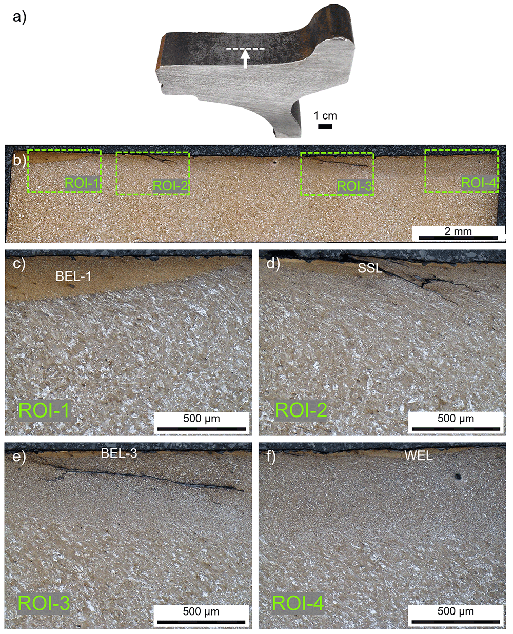
Figure 1.
(a) The investigated ER7 wheel sample from the field and the related cross-sectional cut (b) in rolling direction. Four different regions of interest (ROI) with white etching layer (WEL)/brown etching layer (BEL) variations are investigated in more detail, optical microscope images are given for ROI-1 (c), ROI-2 (d), ROI-3 (e), and ROI-4 (f).
Microstructural characterizations are primarily performed by optical microscopy (OM) (Axio Imager M2m, Carl Zeiss AG, Germany) in bright-field mode, images are captured and thicknesses of layers are measured by using IMS Client (Imagic Bildverarbeitung AG, Switzerland). Further, a scanning electron microscopy (SEM) (Jeol JIB 4700F, Jeol Ltd., Japan) is performed, where the secondary electron (SE) detector is used to investigate the characteristics of the observed microstructures, 15 kV acceleration voltage is applied. Low-load Vickers hardness measurements are executed using a Future-Tech FM-700 hardness tester (Future-Tech FM-700, Future Tech Corp., Japan) using a load of 0.01 kp (0.098 N). The diagonals of the indents are measured with an OM.
Results
Along the tread surface of the investigated wheel four specific regions of interest (ROIs) with mechanically and thermally affected microstructures are investigated, see Figure 1b. Discrepancies concerning the degree of etching can be excluded since all the regions are detected on the same cross-sectional sample in the middle of the tread. It can be seen how different the evolved microstructures are within several millimeters. Hence, the local contact situation and the environmental influences vary widely. Within ROI-1 and ROI-3 a brownish-appearing microstructure can be seen at the current magnification (Figures 1c and e), therefore these layers are indicated as BELs. In ROI-2 a stratification containing WEL and BEL can be seen, stated as an SSL where severe cracking is evident (Figure 1d). Finally, the layer observed in ROI-4 appears brighter, hence it is named WEL. Howsoever, a more detailed analysis is presented in the following paragraphs for each ROI to evaluate the microstructure and its potential naming.
ROI-1 shows a massive brownish-appearing layer (named BEL-1) with an underlying deformed ferritic-pearlitic microstructure (Figure 1c). The thickness of the BEL-1 is up to 250 µm, with decreasing extent towards a transition to the deformed wheel material. On top of the BEL-1 a bright-appearing thin layer (WEL-1) can be observed (Figure 2a). Within the WEL-1 small breakouts can be seen, as well as crack initiation (Figure 2b), while the average layer thickness is ~20 µm. The morphology of the BEL-1 just underneath the WEL-1 is presented by the SE image in Figure 2c, showing a severely plastically deformed (SPD) microstructure with an alignment under a certain angle to the surface. Further, globular particles can be identified as randomly distributed. Due to the high degree of deformation, the cementite lamellae of the pearlite of the ER7 microstructure break and thermal activation have led to the spherodization of the cementite lamellae fragments. The low-load hardness measurements executed within the area of the BEL-1 region shown in Figure 2c come up with hardness values of 537±71 HV0.01.
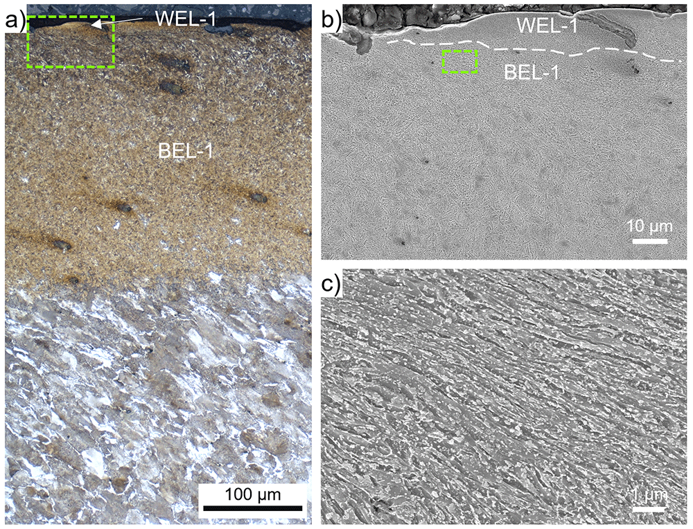
Figure 2.
(a) shows the evolved microstructure within ROI-1 (see Figure 1b, c) consisting of a thin white etching layer (WEL) at the top, a ~250 µm thick brown etching layer (BEL) underneath, segueing into a deformed ER7 microstructure. (b) presents a scanning electron (SE) image of the WEL and BEL area framed in (a). A higher magnification SE image of the BEL-1 microstructure is given in (c).
Within ROI-2, an SSL can be seen in Figure 1d, accompanied by a crack initiation and propagation along an angle similar to the shear-deformed near-surface microstructure into depth. The SSL is shown by higher magnification OM in Figure 3a. The SSL consists of an almost featureless topmost layer with a thickness of ~30 µm (indicated as WEL-2) and an underlying brownish-appearing layer with a thickness of ~50µm, designated as BEL-2. Micro-spalling can be seen in the WEL-2, a SE image of the SSL is given in Figure 3b. To analyze the different microstructural characteristics, high magnification SE images are presented: Figure 3c shows the microstructure of WEL-2, coming up with a fine mesh-like structure without any preferred orientation detectable. The image indicates some nanometer-sized globular particles fine dispersed. Hardness values of 1184±28 HV0.01 are obtained within this region. In the region BEL-2 (Figure 3d), a coarser deformed microstructure aligned under an angle of ~30° to the surface can be identified, containing some globular particles with significantly larger proportions with respect to the WEL-2. The low load hardness measurements reveal results of 500±42 HV0.01. Comparable microstructure can be seen in the microstructure underneath the BEL-2 (Figure 3e), but with a slight change in the alignment angle of the deformed microstructure. Comparable hardness values are determined with 521±47 HV0.01. The globular particles are suggested to result from broken cementite lamellae (originating from the pearlite of the ER7 wheel microstructure), spherodized due to thermal influences.
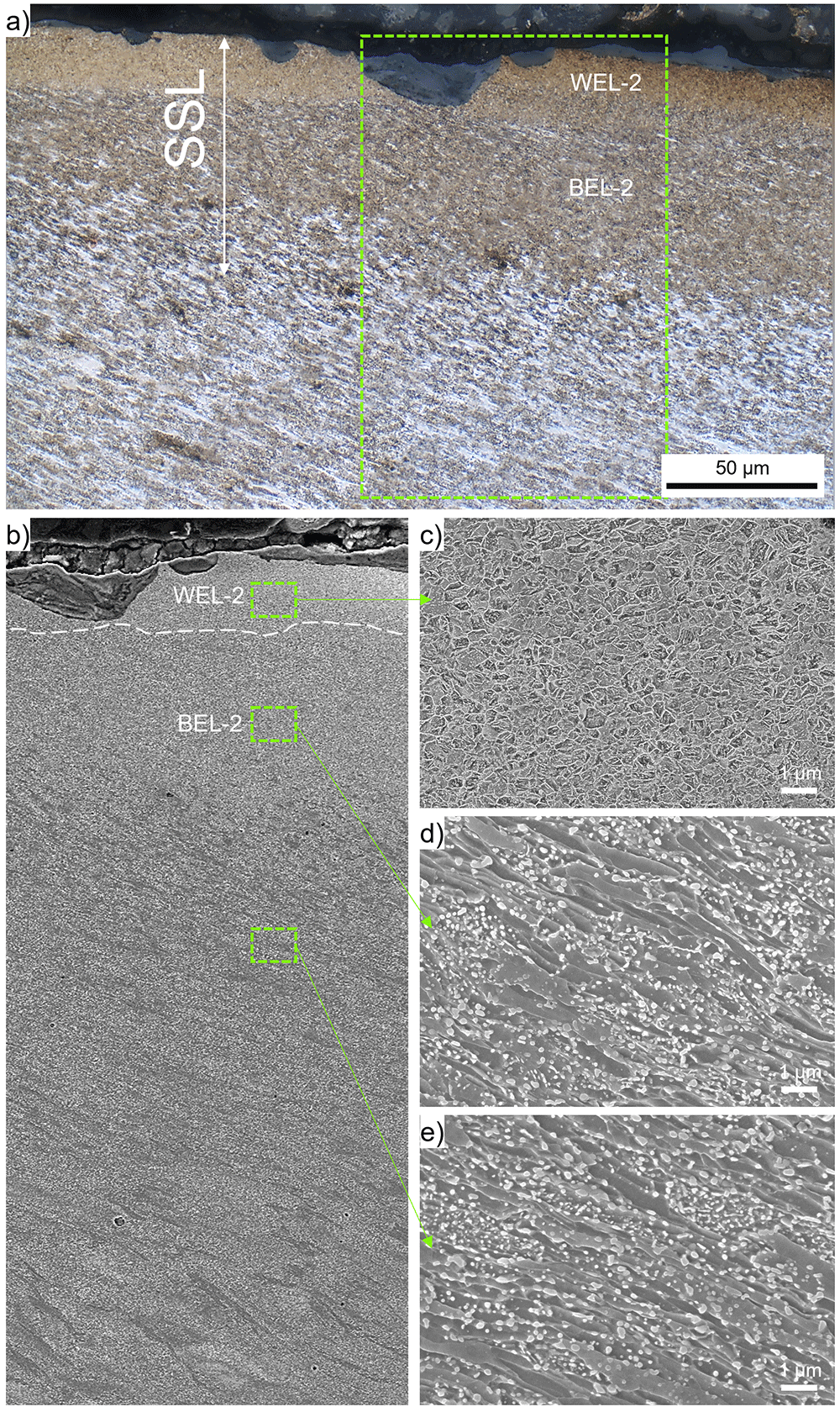
Figure 3.
(a) shows the stratified surface layer (SSL) (white etching layer-2 (WEL-2) and brown etching layer-2(BEL-2) within ROI-2 (see Figure 1b, d). (b) presents a scanning electron (SE) image of the region framed in (a). For more details, higher magnification SE images of certain locations are given: WEL-2 (c), BEL-2 (d), and the underlying deformed ER7 microstructure (e).
The brownish-appearing layer within ROI-3, where a severe crack network is visible (Figure 1e), is analyzed in more detail by higher magnification OM and SEM (Figure 4). The near-surface microstructure shows some very bright appearing flakes at the surface, named WEL-3 (Figure 4a). Crack initiation can be seen along the surface, propagating into a brownish appearing layer with a thickness of ~50 µm (BEL-3). On the right side of the SE image (Figure 4b) a spalled BEL region can be detected. The cracks initiating at the surface seem to stop at the interface of the BEL-3 to the underlying SPD microstructure. However, severe horizontal cracks are observed at a depth of ~100 – 200 µm from the surface. The SPD microstructure gradually changes to a deformed and aligned ER7 microstructure with increasing grain sizes. The focus of this study, the microstructure of the BEL, is further investigated by high magnification SE images presented in Figure 4c. A randomly orientated mesh-like microstructure can be seen, with a certain degree of spheroidization. The low load Vickers hardness measurements in this area show hardness values of 509±24 HV0.01.
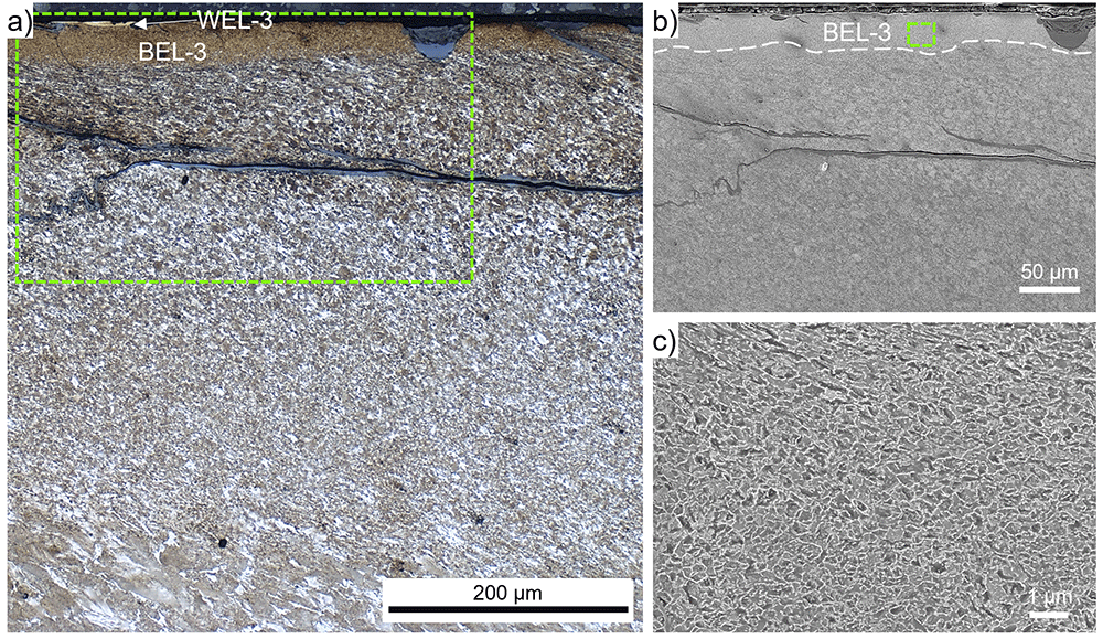
Figure 4.
(a) shows the evolved microstructure within ROI-3 (see Figure 1b, e), where a very thin white etching layer (WEL) is on top of a brown etching layer (BEL) with a thickness of ~50 µm (BEL-3). This layer is situated on a severely plastically deformed (SPD) microstructure accompanied by severe cracking. (b) presents a scanning electron (SE) image of the WEL and BEL area framed in (a). A higher magnification SE image of the BEL-3 microstructure is given in (c).
With respect to the equal etching conditions of the investigated regions, the layer observed at the wheel surface in ROI-4 is designated as WEL (Figure 1f), since it appears bright and is situated on an SPD ferritic-pearlitic microstructure. Micro-spalling, crack initiation, and crack propagation through the WEL under almost 90° to the surface can be observed (Figure 5a), the layer is named WEL-4. The alignment of the underlying ER7 microstructure is visible (Figures 5a and b). The microstructure within WEL-4 is shown by SE imaging in Figure 5c, indicating a fine-grained mesh-like structure. Further, a certain degree of spherodization can be identified. The results of the hardness measurements come up with pronounced scattering, howsoever, high hardness values are indicated with 979±141 HV0.01.
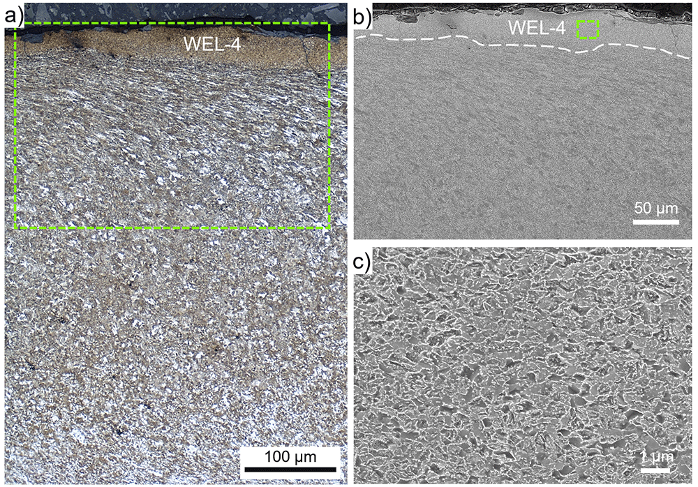
Figure 5.
(a) shows the microstructure within ROI-4 (see Figure 1b, f), where a white etching layer (WEL) with a thickness of ~50 µm (WEL-4), is situated on an severely plastically deformed microstructure. (b) presents a scanning electron (SE) image of the WEL area framed in (a). A higher magnification SE image of the WEL-4 microstructure is given in (c).
Discussion
The characterization of different evolved near-surface microstructures on the ER7 rail wheel from service outpoints the wide range of microstructural variations with respect to the location along the tread surface. The naming WEL and BEL is based on the appearance under the OM after etching with Nital, hence, based on the degree of etching and the image settings. Therefore, a common naming based on OM images is hardly possible. The information of the characterization by SEM and hardness measurements may enable a more consistent naming. The results within this work show the WEL as a fine-grained mesh-like microstructure without globular particles and high hardness (~1000 HV0.01), see WEL-2. In comparison, WEL-4 shows almost 1000 HV0.0.1, but a more dissolved microstructure with spheroidization. The naming is in this case arguable since the brownish appearance under the OM may suggest this region as BEL. Further, the BEL-3 shows comparable microstructure in the SEM, but the hardness is much lower (~500 HV0.0.1). Evolved microstructures with hardness in the range of 400 – 600 HV0.01, brownish appearance in the LOM, and pronounced spherodized cementite lamellae leading to globular particles can be indicated as BEL. However, even within a BEL, the microstructure can locally differ, as shown in BEL-1. OM images and hardness results are in accordance to investigations on WELs on rails22,25. Regarding the SEM images, no comparable literature is present since the presented work gives the first detailed microstructural investigation on WEL, BEL and SSL on wheel materials. In most cases, when observing a BEL, a WEL can also be identified, representing an SSL as presented by Messaadi et al.25. The thicknesses of the WEL and BEL is varying, hence, the SSL often is detectable only with higher magnification, see ROI-1 and ROI-3. Samples from field are worn and the analysis is always a snapshot, which additionally complicates a common identification and classification. For instance, the so-called WEL-4 in this work may be a BEL, with the WEL already broken off the wheel tread surface.
Conclusions
The broad variety of evolved near-surface microstructures on rail wheels makes a common terminology hardly possible, but with increasing numbers of published studies certain comparison is possible. A rough classification can be made for the ER7 wheel material:
■ WEL appears white in OM after etching with Nital, shows a fine mesh-like microstructure without globular particles in SEM, and hardness in the range of 900 – 1200 HV0.01.
■ BEL looks brownish in OM after etching with Nital, shows globular particles (spherodized cementite lamellae) within SPD microstructure in SEM, and hardness in the range of 400 – 600 HV0.01
In most cases, a BEL is always accompanied by a WEL, forming an SSL. Crack initiation and crack networks are observed in the presence of SSLs on the ER7 wheel from service, indicating a relation to fatigue crack initiation and possible failure of the wheel.
Data and software availability
Zenodo, Microstructural characterization of near-surface microstructures on rail wheels in service – an insight into "stratified surface layers": Supplementary Data. https://doi.org/10.5281/zenodo.783667027.
This project contains the following underlying data:
- fig_ORE_Freisinger (1).JPG
- fig_ORE_Freisinger (1).tif
- fig_ORE_Freisinger (10).jpg
- fig_ORE_Freisinger (10).tif
- fig_ORE_Freisinger (2).jpg
- fig_ORE_Freisinger (2).tif
- fig_ORE_Freisinger (3).jpg
- fig_ORE_Freisinger (3).tif
- fig_ORE_Freisinger (4).jpg
- fig_ORE_Freisinger (4).tif
- fig_ORE_Freisinger (5).jpg
- fig_ORE_Freisinger (5).tif
- fig_ORE_Freisinger (6).jpg
- fig_ORE_Freisinger (6).tif
- fig_ORE_Freisinger (7).jpg
- fig_ORE_Freisinger (7).tif
- fig_ORE_Freisinger (8).jpg
- fig_ORE_Freisinger (8).tif
- fig_ORE_Freisinger (9).jpg
- fig_ORE_Freisinger (9).tif
- hardness_ORE_Freisinger.xlsx
Data are available under the terms of the Creative Commons Attribution 4.0 International license (CC-BY 4.0).
Acknowledgments
We thank DI Roman Schmid and Andreas Schachner from the Austrian Federal Railways Holding Stock Company for providing ex-service material and their help in this work. We are also thankful to Iñigo Sarries (CAF, Construcciones y Auxiliar de Ferrocarriles) for useful discussion and the supply of rail wheel materials.
Faculty Opinions recommendedReferences
- 1.
Ekberg A, Åkesson B, Kabo E:
Wheel/rail rolling contact fatigue - Probe, predict, prevent.
Wear.
2014; 314(1–2): 2–12. Publisher Full Text
- 2.
Grassie SL:
Studs and squats: The evolving story.
Wear.
2016; 366–367: 194–199. Publisher Full Text
- 3.
Ahlstrom J, Karlsson B:
Microstructural evaluation and interpretation of the mechanically and thermally affected zone under railway wheel flats.
Wear.
1999; 232(1): 1–14. Publisher Full Text
- 4.
Steenbergen M:
On the genesis of squat-type defects on rails - Toward a unified explanation.
Wear.
2021; 478–479: 203906. Publisher Full Text
- 5.
Eden HC, Garnham JE, Davis CL:
Influential microstructural changes on rolling contact fatigue crack initiation in pearlitic rail steels.
Materials Science and Technology.
2005; 21(6): 623–629. Publisher Full Text
- 6.
Cvetkovski K, Ahlström J:
Characterisation of plastic deformation and thermal softening of the surface layer of railway passenger wheel treads.
Wear.
2013; 300(1–2): 200–204. Publisher Full Text
- 7.
Molyneux-Berry P, Davis C, Bevan A:
The Influence of Wheel/Rail Contact Conditions on the Microstructure and Hardness of Railway Wheels.
ScientificWorldJournal.
2014; 2014: 209752. PubMed Abstract
| Publisher Full Text
| Free Full Text
- 8.
Nguyen BH, Al-Juboori A, Zhu H, et al.:
Formation Mechanism and Evolution of White Etching Layers on Different Rail Grades.
Int J Fatigue.
2022; 163: 107100. Publisher Full Text
- 9.
Al-Juboori A, Zhu H, Wexler D, et al.:
Characterisation of White Etching Layers formed on rails subjected to different traffic conditions.
Wear.
2019; 436–437: 202998. Publisher Full Text
- 10.
Freisinger M, Rojacz H, Pichelbauer K, et al.:
Comparative study on the influence of initial deformation and temperature of thermally induced white etching layers on rail wheels.
Tribol Int.
2023; 177: 107990. Publisher Full Text
- 11.
Pan R, Chen Y, Lan H, et al.:
Investigation into the evolution of tribological white etching layers.
Materials Characterization.
2022; 190: 112076. Publisher Full Text
- 12.
Steenbergen M:
Squat formation and rolling contact fatigue in curved rail track.
Eng Fract Mech.
2015; 143: 80–96. Publisher Full Text
- 13.
Ekberg A, Kabo E:
Fatigue of railway wheels and rails under rolling contact and thermal loading—an overview.
Wear.
2005; 258(7–8): 1288–1300. Publisher Full Text
- 14.
Liu C, Zhang G, Chen C, et al.:
Formation mechanism for the white etching microstructure in the subsurface of the failure pearlite wheel steel.
Wear.
2022; 494–495: 204243. Publisher Full Text
- 15.
Zhang YM, Xiao ZM, Fan M:
Fatigue Investigation on Railway Wheel Steel with White Etching Layer.
Int J Steel Struct.
2020; 20(1): 80–88. Publisher Full Text
- 16.
Kato T, Kato H, Makino T:
Effect of elevated temperature on shelling property of railway wheel steel.
Wear.
2016; 366–367: 359–367. Publisher Full Text
- 17.
Carroll RI, Beynon JH:
Rolling contact fatigue of white etching layer: Part 1: Crack morphology.
Wear.
2007; 262(9–10): 1253–1266. Publisher Full Text
- 18.
Bernsteiner C, Meierhofer A, Trummer G, et al.:
Simulation and experiment based investigations of squat formation mechanisms.
Wear.
2019; 440–441: 203093. Publisher Full Text
- 19.
Pan R, Ren R, Chen C, et al.:
The microstructure analysis of white etching layer on treads of rails.
Eng Fail Anal.
2017; 82: 39–46. Publisher Full Text
- 20.
Freisinger M, Zauner L, Hahn R, et al.:
In-situ micro-cantilever bending studies of a white etching layer thermally induced on rail wheels.
Mater Sci Eng A.
2023; 869: 144805. Publisher Full Text
- 21.
Kumar A, Saxena AK, Kirchlechner C, et al.:
In situ study on fracture behaviour of white etching layers formed on rails.
Acta Materialia.
2019; 180: 60–72. Publisher Full Text
- 22.
Li S, Wu J, Petrov RH, et al.:
“Brown etching layer”: A possible new insight into the crack initiation of rolling contact fatigue in rail steels?
Eng Fail Anal.
2016; 66: 8–18. Publisher Full Text
- 23.
Kumar A, Agarwal G, Petrov R, et al.:
Microstructural evolution of white and brown etching layers in pearlitic rail steels.
Acta Materialia.
2019; 171: 48–64. Publisher Full Text
- 24.
Tung PY, Zhou X, Morsdorf L, et al.:
Formation mechanism of brown etching layers in pearlitic rail steel.
Materialia.
2022; 26: 101625. Publisher Full Text
- 25.
Messaadi M, Steenbergen M:
Stratified surface layers on rails.
Wear.
2018; 414–415: 151–162. Publisher Full Text
- 26.
Lewis R, Olofsson U:
Wheel-rail interface handbook. Woodhead Publishing Limited, 2009. Publisher Full Text
- 27.
Freisinger M:
Microstructural characterization of near-surface microstructures on rail wheels in service - an insight into "stratified surface layers": Supplementary Data. [Dataset]
Zenodo.2023. http://www.doi.org/10.5281/zenodo.7836670