Plain language summary
There are life-threatening situations of patients due to insufficient oxygen supply. Here we propose and test a method to increase oxygen uptake by the skin, similar as reptiles do. The trick is to wet the skin, but instead of water using a special substance, best described as 'liquid Teflon', that can take up 20 times more oxygen than water can do.
This idea was tested on nude mice that were ventilated with a low amount of oxygen (comparable to the oxygen concentration at the highest mountains of the Alps) and at the same time brushed with the 'liquid Teflon'. Then the oxygen saturation in the blood was measured with a special mouse oximeter. We could not find differences with or without the 'liquid Teflon' treatment.
There might be small improvements but if they are below 2% of oxygen saturation, we could not detect it with the method we applied, because there is a limited accuracy of the oximeter and also a considerable variation from mouse to mouse. However, a relevant improvement for the patients would require an increase in the range of 5%.
Introduction
The major cause of death in coronavirus disease 2019 (COVID-19) patients is severe atypical pneumonia and respiratory distress (Hui et al., 2020). Severe acute respiratory distress syndrome in these patients is treated mainly with mechanical ventilation, which supports O2 delivery through the lungs, and with anti-inflammatory and anti-viral medication or even targeting the receptor used by the virus to invade the lungs and the epithelium, angiotensin-converting enzyme 2 (Zhang et al., 2020). Unfortunately, the increasing number of patients and the finite number of healthy and operationally trained medical care personnel and machines limits the choice of therapeutic strategies that are available for all that require assistance. The efficacy of the oxygen supply of blood provided by the ventilation of the lungs varies depending on the amount of fluid in the lungs, as the Krogh diffusion coefficient of oxygen in water is 2×10⁵-fold lower than that in the gas phase (Dejours, 1989). Therefore, approaches used for the treatment of high-altitude pulmonary edema were recently suggested for patients with COVID-19 infection associated with severe pneumonia (Solaimanzadeh, 2020). Other strategies use complementary O2 delivery options that bypass the lungs, such as extracorporeal membrane oxygenation (ECMO) (Matthay et al., 2020). However, ECMO is associated with thromboembolic and hemolytic complications and, when performed, requires constant attention of skilled medical care experts (Ramanathan et al., 2020).
Here, we propose and test a simple method to increase the systemic oxygen supply by bypassing the lungs and boosting transdermal oxygen delivery. Evolutionary vertebrates that were equally efficient in aquatic and terrestrial habitats used lungs, skin and gills for oxygen uptake (Dejours, 1989; Jorgensen, 2000). In frogs, transcutaneous respiration was shown to be responsible for approximately 10–20% of oxygen uptake, the amount sufficient to support animal survival during dives (Vitalis, 1990).
In humans, transcutaneous respiration mediates oxygen uptake of 0.53 ml O2/(m2 min), contributing 0.4% to the total oxygen supply under normoxic conditions (Stucker et al., 2002). A decrease in oxygen uptake through the lungs (e.g., under hypoxic conditions) results in a compensatory increase in transcutaneous oxygen flow that may be boosted up to 25-fold, reaching 10% of total oxygen uptake under certain conditions. This oxygen uptake pathway could support patients with severe respiratory distress on or off mechanical ventilation, allowing them to avoid barotrauma to the lungs (Ioannidis et al., 2015).
Studies on healthy humans revealed that transdermal oxygen travels 400 µm, reaching well into the dermis and thus to blood capillaries (Stucker et al., 2002). Apart from the oxygen gradient between the skin surface and dermisa that supports diffusion, physical parameters in favor of transdermal respiration are temperature (Jaszczak, 1988) and skin humidity (Vaupel, 1976).
However, the crucial intervention will be the increase in available oxygen by replacing a putative aquatic phase with a liquid that could provide much more oxygen. Such compounds are fluorocarbons, which can dissolve 20-fold the amount of oxygen compared to aqueous solutions (Riess, 2005).
If humidity favors oxygen uptake by the skin, i.e. wet skin has a better oxygen uptake than skin in a gas phase (air), a liquid dissolving 20-fold the oxygen of water could result in an increased oxygen uptake by the skin. However, the entire process is multifactorial. In addition to the diffusion step at the skin surface, there is the diffusion deeper into the tissue, the putative uptake of the oxygen by the red blood cells, the role of vasodilation, possible differences at different parts of the body, just to name a few obstacles. Therefore, it seems impossible to estimate an expectable effect size.
Fluorocarbons are not new in circulation and respiration research as well as in dermatological and ophthalmological applications. Three major applications were suggested and have partly been tested decades ago. (i) Perfluorocarbons have already been proposed and tested as blood substituents in the 1980s (Waxman, 1986). This approach never made it into clinical practice mainly because of emulsion issues. In the future, the situation may improve by combining oxygen carrier compounds with a metal-organic framework (Gao et al., 2018). (ii) Fluorocarbons were tested in liquid ventilation either as total liquid ventilation or as partial liquid ventilation (Kolla et al., 1997). Although initial tests on humans showed beneficial effects in patients with acute respiratory distress syndrome (Hirschl et al., 1996) and acute hypoxemic respiratory failure (Kolla et al., 1997), this treatment was never approved for routine clinical treatment. (iii) Suggested for human use are hydrogels containing fluorinated methacrylamide. Chitosan has been proposed to support wound healing (Patil et al., 2016). Furthermore, perfluorodecalin is used to support wound healing (Jalani et al., 2017) (and as an ingredient in cosmetics) and is applied in ophthalmic surgery (Dk-line®, Bausch+Lomb). Perfluoro-n-octane (e.g., Auro octane®, Aurolab; Okta-line®, Bausch+Lomb) is approved for application in the treatment of traumatic ocular trauma and retinal detachment or in ophthalmic surgery. However, topical whole-body skin application has to our best knowledge not been tested. Therefore, the goal of this work was to test if oxygen supply through the skin enforced with applications of oxygen-concentrating fluorocarbons may result in better oxygenation of blood under conditions of poor absorption of oxygen through the lungs. This aim was tackled as a proof-of-principle studies in mice.
Methods
Animal experiments
Experiments with mice were carried out in strict accordance with the recommendations in the Guide for the Care and Use of Laboratory Animals of the National Institutes of Health. The animal experiment protocol was approved by the State Office for Health and Consumer Protection (permit number: 08/2020). All efforts were made to ameliorate harm to the animals. This was achieved by performing the entire experimental procedure with the mice in anesthesia and by providing an additional pain therapy (details are provided below).
For this study, we used sex mixed population of CD1nu/nu mice (Charles River, Wilmington, MA, USA) at an age of 50.3±8.5 weeks with a bodyweight of 29.9±3.7 g, which were housed under a 12h/12h day-night rhythm in the animal husbandry of the Institute for Clinical & Experimental Surgery (Homburg, Germany). The animals were kept in individually ventilated cages (IVCs) in groups of 5 animals per cage. They had free access to drinking water and standard pellet food (Altromin, Lage, Germany). No additional inclusion criteria were set. Animals were randomly distributed in two groups (control group and treated group). Randomization was done by choosing lots and experiments were performed by alternating measurements of control and treated mice with a cage of animals per (experimental) day. The number of animals per group could not be precalculated because we established this kind of measurements and had no reliable information about the variances of the measured parameters. Therefore, we performed a statistical analysis on a daily base and as such decided after we reached a group size of five animals to stop the experiments of protocol 1 and after reaching four mice for protocol 2.
During the experiment, the mice were anesthetized with isoflurane (5%: initiation, 1%: maintenance of anesthesia). Immediately after anesthesia initiation, the mice received 5 mg/kg carprofen (Rimadyl; Zoetis, Berlin, Germany) i.p. for intraoperative pain therapy. Afterwards, they were fixed on a heating pad in supine position and tracheotomized for the insertion of a catheter for ventilation. Breathing rate and stroke volume could be set on the mouse ventilator (Minivent; Harvard Apparatus, March-Hugstetten, Germany) and was adapted according to the body weight of the mice in the range of 200–270 per minute and 120–140 µL, respectively. The body temperature was kept constant in the range between 37–38°C, which was monitored with an anal probe (GTH1170, Greisinger Messtechnik, Regenstauf, Germany). The heart rate was documented with the help of a pulse oximeter (MouseSTAT Jr. with Paw sensor, Kent Scientific, Torrington, CT, USA). The control group contained 5 animals at an age of 47.4±2.4 weeks with a body weight of 28.8±3.7 g and the test group contained the same number of animals (n=5, age: 49.3±3.3 weeks, body weight: 29.4±4.9 g; no significant difference to control group).
Experimental protocol
All mice in all groups and all protocols were given an initial acclimatization phase as outlined in Figure 1B (-10 to 0 min). Mice of control and treatment groups experienced the same hypoxic protocol. Control mice received no additional treatment whereas mice of the treatment group were brushed with prewarmed (30°C) perfluorodecalin (medical grade, Pharmpur, Königsbrunn, Germany). This included a full-body application with the exception of the area of the head and the paw to which the sensor of the pulse oximeter was attached. The procedure was continuously renewed in the course of the experiment to compensate the perfluorodecalin evaporation. The oxygenation of the blood was monitored and documented (in addition to the heart rate) with the pulse oximeter. After the oxygen saturation had reached a stationary level, the oxygen content of the ventilation was reduced according to the test protocol (see below). For this purpose, a gas mixer (KM10–2 FLEX, Witt-Gastechnik, Witten, Germany) was used, which mixed oxygen and nitrogen in an adjustable ratio. In addition, the setting was monitored with an oxygen meter (GOX100, Greisinger Messtechnik, Regenstauf, Germany). The mouse group used for protocol 2 consisted of 4 mice at an age of 55±15 weeks with a body weight of 31.9±1.1 g.
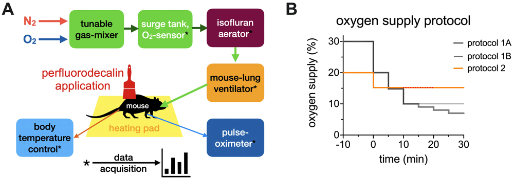
Figure 1. Experimental design.
(A) Experimental setup: A tunable gas mixer was connected to oxygen and nitrogen gas bottles. The percentage of oxygen could be adjusted. This gas mixture was collected in a surge tank, where the oxygen content was continuously measured. The required amount of isoflurane was added in an appropriate aerator and finally connected to the mouse-lung ventilator. The mouse was monitored for body temperature, heart rate and oxygenation level using a pulse-oximeter. (B) Time line of the oxygen supply in the different protocols applied. Protocols 1A and 1B are performed in the presence and absence of topical application of 100% pure perfluorodecalin. The red dots in protocol 2 indicate the period of perfluorodecalin application.
The initial idea (represented as protocol 1A in Figure 1B) was to reduce the available oxygen in steps from 20% - 15% - 10% - 9% - 8% -7%, each step lasting for 5 min. While oxygen supply, heart rate and oxygen saturation were recorded, the rationale was to detect differences in the survival time between perfluorodecalin treated mice and control mice. However, it turned out that it was impossible to properly determine the time point of death of the mice. The pulse-oximeter was thought to provide the information of the termination of the heart beat but it turned out that the pulse-oximeter stopped working at a weak pulse before the death of the animal (alternating periods of no signals and measured signals). Since the animals were ventilated stop of breath was neither a criterion. A mouse electrocardiogram was not available. Therefore, after the measurement of 6 animals, the protocol was modified to protocol 1B in Figure 1B, i.e. the available oxygen was reduced in 2 steps of 5 min from 20% to 10% and then kept constant at 10%, still with the intention to determine the survival time expecting a better reliability of the pulse oximeter at higher percentage of oxygen supply. Because this was not the case, we stopped to apply protocol 1B after further 4 animals and changed the concept of measurements. Instead of recording the survival time at decreasing oxygen availability, we measured the blood oxygen saturation (as well as the heart rate) under hypoxic conditions (15% oxygen supply) before, during and after termination of the perfluorodecalin application, each period lasting for 10 min.
Statistical analysis
All data were initially tested for normal distribution using the Shapiro-Wilk test. Figure 1 does not contain any statistical data. When the normality test was passed, an unpaired Student’s t-test (Figure 2) or a paired one way-ANOVA (Figure 3) was performed to determine the p-value. P-values below 0.05 we regard as significant. When data are presented as box-plots, whiskers depict the minimal and maximal value (Figure 2). Single points show medians with error bars representing the standard error of mean (Figure 3). Age and weight of mouse groups are given as mean value ± standard deviation. All statistical tests were performed in Prism 9 (GraphPad Software, San Diego, CA, USA).
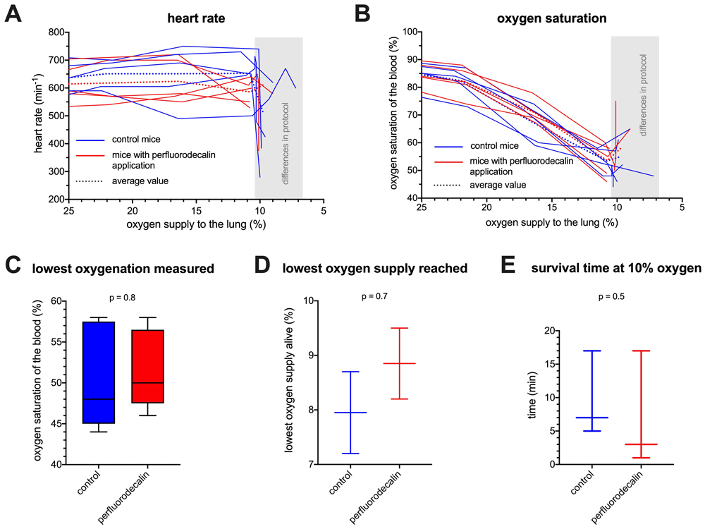
Figure 2. Results of a comparison of perfluorodecalin-treated mice with untreated controls.
(A) Heart-rate plotted against the percentage of oxygen supply for 5 control mice and 5 perfluorodecalin-treated mice. When an oxygen supply of 10% is reached, mice follow two different protocols (compare Figure 1B). (B) Oxygen saturation of the blood plotted against the percentage of oxygen supply for 5 control mice and 5 perfluorodecalin-treated mice. When an oxygen supply of 10% is reached, mice follow two different protocols (compare Figure 1B). (C) Lowest blood oxygenation measured during the experiment for each mouse. (D) Lowest oxygen supply reached before the mice died (protocol 1A). (E) Survival time of the mice at 10% oxygen supply (protocol 1B).
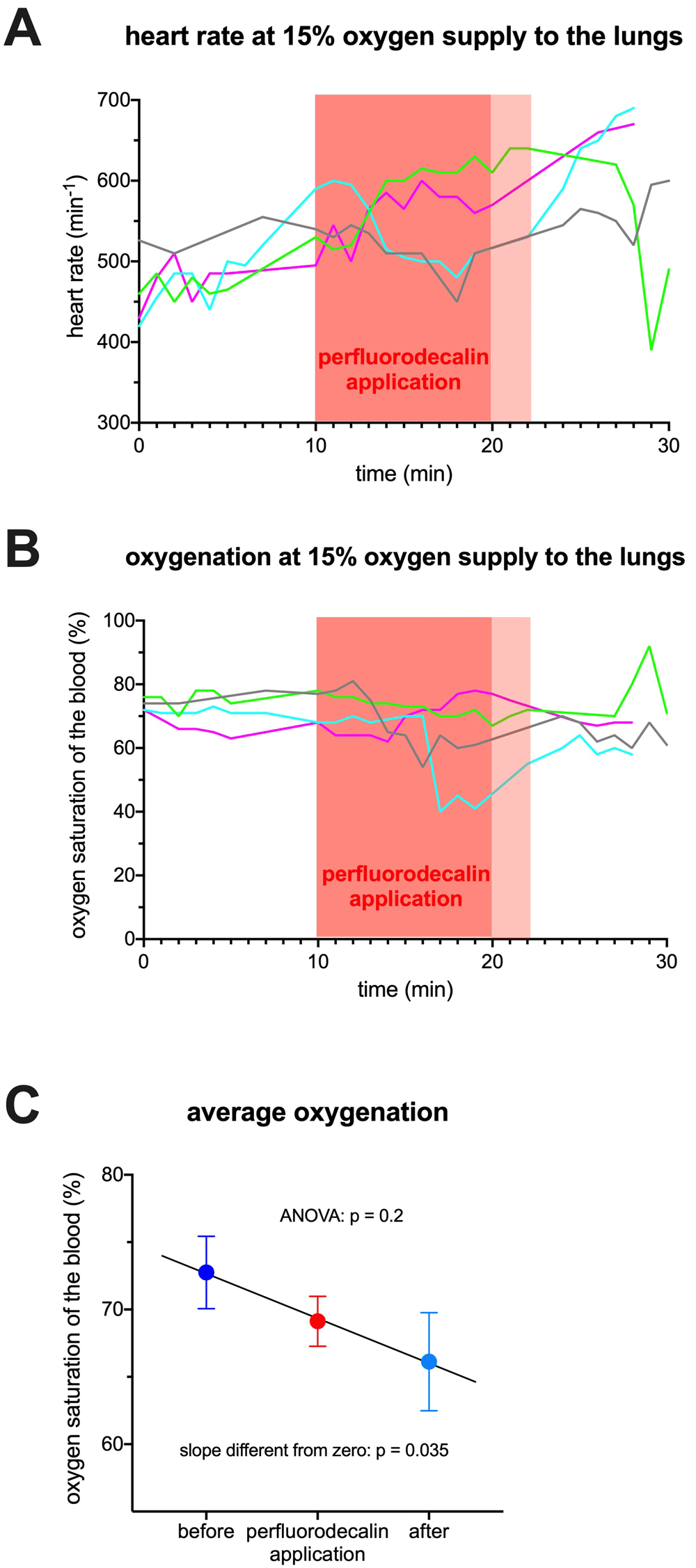
Figure 3. Results of the comparison of hypoxic mice before, during and after treatment with perfluorodecalin.
(A) Heart-rate plotted against the time at 15% oxygen supply for 4 mice. The region highlighted with a red background indicates the perfluorodecalin application. The light red region indicates the interval, when active perfluorodecalin application was stopped but still not the entire substance evaporated and remains were present on the skin. (B) Blood oxygenation plotted against the time at 15% oxygen supply for 4 mice. The region highlighted with a red background indicates the perfluorodecalin application. The light red region indicates the interval, when active perfluorodecalin application was stopped but still not the entire substance evaporated and remains were present on the skin. (C) Statistical analysis of the blood oxygenation before, during and after perfluorodecalin application.
Results
Comparison of perfluorodecalin-treated mice with untreated controls
The results of the measured heart rate and the oxygen saturation as a function of oxygen supply using the pulse oximeter in the presence and absence of topically applied perfluorodecalin are depicted in Figure 2A and 2B, respectively, following the timing protocols 1a and 1b (Figure 1B) (Kaestner, 2021). It turned out that the heart rate and oxygenation readouts were unstable at the lowest oxygen concentrations in the gas phase used for ventilation. Therefore, protocol 1A was adapted and instead of determining the lowest tolerable gaseous oxygen concentration, we measured and compared the survival time at 10% oxygen supply (protocol 1B in Figure 1B).
Figure 2A and 2B reveal a high variability between the mice (heart rate between 490 and 750 per minute and an oxygen saturation between 58% and 75% at a value of 15% oxygen supply). No obvious differences was observed between perfluorodecalin-treated and untreated mice. Figure 2C shows the lowest oxygenation measured independent of the particular amount of oxygen supply. Despite a huge variability between the mice, there is a lack of a significant difference between the 2 groups. Such a difference was not expected and therefore this parameter can be seen as an internal quality control. Panels 2D and 2E show the envisaged final read-out parameters of protocol 1A and 1B, respectively. However, their value is limited due to the low number of animals (n = 4 and 6, respectively) and the huge variation between the mice. Based on these results (Figure 2), experiments following protocol 1 were stopped, because it was judged that the variability of the results did not justify continuing measurements.
Comparison of hypoxic mice before, during and after treatment with perfluorodecalin
A new protocol was designed, where the oxygen supply was set to 15% and then the oxygenation (and heart rate) was measured before, during and after a 10 min period of perfluorodecalin treatment, as outlined in Figure 1B (protocol 2). This approach has the advantage that presence and absence of perfluorodecalin can be analyzed on a paired basis within the same animal. Figure 3A and 3B depict the heart rate and the oxygenation, respectively, at 15% oxygen supply before, during and after perfluorodecalin application. These measurements reveal a high variability even within the defined experimental intervals of a particular mouse, i.e., the measurement itself shows a high 'noise' level. To approach this effect the median of each experimental period was determined for every mouse and then the mean of the four measured mice for the three experimental intervals were plotted in Figure 3C. A paired one-way ANOVA test showed no significant differences. However, a linear fit of the oxygenation over time revealed a goodness of fit R2 of 0.997 with a significant non-zero slope (p = 0.035). This shows that (i) indeed a considerable portion of the variation is likely to be measurement noise and (ii) in our experimental approach, we could not improve the blood oxygenation level. Instead, a decline of the oxygen saturation along the experimental periods was detected, which is most likely a temporal effect of the low oxygen supply duration.
Discussion and conclusions
Conceptual considerations
An experimental setting had to be developed to assess a possible transdermal oxygen delivery. Although measurements on human volunteers would be attractive, a pilot animal study was regarded as more appropriate. For the animal experiments, we considered young house-pigs and nude mice, because in both cases a fur-less skin is available. While porcine skin is anatomically and physiologically very similar to human skin (Avci et al., 2013), it is thicker and less vascularized. Murine skin, with an average body epidermal thickness of approximately 8.5 µm and a cutaneous blood flow of 17.8 mL/min/100 g (Monteiro-Riviere et al., 1990), is an ideal candidate. Accordingly, we used nude mice (CD1nu/nu, Charles River, Wilmington, MA, USA) in the present proof-of-principle study.
Next, a fluorocarbon had to be selected. The physico-chemical property to dissolve about 20 times more oxygen than water (Riess, 2005) applies to more or less all fluorocarbons. However, it has previously been shown (ECETOC, 2008) that adverse effects of fluorocarbons when topically applied are mainly caused by impurities of the preparations. Therefore, it was compulsory to use a fluorocarbon with medical purity. Pharmpur (Königsbrunn, Germany), a manufacturer of liquid medical products, has the fluorocarbon perfluorodecalin (also known as perflunafen) in its portfolio and kindly provided it for this study. It has a boiling point of approximately 142°C, which is less than other long-chain fluorocarbons depict and leads to a considerable evaporation on the 37°C skin surface of mice. However, this problem can be handled by repetitive/continuous brushing of the mouse skin with perfluorodecalin.
Our approach was that transdermal oxygen is only relevant, if a difference can be detected on the systemic level. As a major read-out device, a dedicated rodent-optimized pulse oximeter (MouseSTAT Jr. with Paw sensor, Kent Scientific, Torrington, CT, USA) was chosen. Preliminary experiments revealed that the maintenance of the body temperature at 37°C or slightly above is vital to obtain reliable read-outs of the pulse oximeter. Therefore, body temperature was continuously monitored and subsequently adjusted with help of a heating pad (Figure 1A).
To mimic a hypoxic condition that can also be found in the COVID-19 disease situation, an artificial reduction of the oxygen supplied to the lungs was envisaged, while the skin of the body was exposed to the normal atmospheric oxygen concentration of above 19%. This was realized by the combination of a tunable gas-mixer and a dedicated mouse lung-ventilator as detailed in Figure 1A. The initial concept was to subsequently reduce the oxygen supply and compare the survival time of mice at a reduced oxygen supply in dependence of dermal whole-body perfluorodecalin application, as outlined in Figure 1B (protocol 1A).
Transdermal oxygen delivery to increase systemic oxygen
Using CD1nu/nu mice and medical grade perfluorodecalin we were not able to measure a significant increase in the oxygenation of the blood. We have to admit that the resolution of our blood oxygenation measurements was not very high. The display resolution is 1% but we have the impression that the hypoxic conditions, the mice are exposed to, are not in favor of pulse oximeter reliability (jumping values, i.e. noise). Measurement variations even on the same mouse over time require a certain averaging. Therefore, we conclude that increases in blood oxygenation below 2% cannot be detected with the method applied and the number of animals investigated. However, since our goal was a proof of principle, which may have helped to improve the situation of COVID-19 patients, a dermal oxygen supply of 5% of the total would be a minimal requirement for a practical application. Accordingly, we falsified the hypothesis that perfluorodecalin improves the transdermal oxygen supply to be relevant for a clinical application. On the other hand, we like to stress that this does not generally disprove an increase in transdermal oxygen delivery by topical application of fluorocarbons.
Methodological inferences
The manipulative part of the setup introduced in the upper part of Figure 1A seems to be a reliable approach for a (hypoxic) stress model for anesthetized animals with full manual access to the animal, i.e. without the use of hypoxia cages or boxes.
For the detection part of the system we experienced shortcomings and for future studies we propose to complement pulse-oximetric measurements with a simple ECG for a more reliable detection of the vital function. Furthermore, a more sophisticated measurement of tissue oxygenation, e.g., by applying fibre-optic micro-sensor technology, could be a valuable extension of the measured parameters.
Data availability
Underlying data
Dryad: Test of perfluorodecalin to increase transdermal oxygen delivery in mice. https://doi.org/10.5061/dryad.931zcrjjp (Kaestner, 2021).
This project contains the following underlying data:
Heart_rate_protocol_1.csv (data provide the heart rate of the mice (per minute) in dependence of the oxygen supplied (%) according to protocol 1, described in the methods)
Oxygenation_protocol_1.csv (data provide the oxygenation of the mice (%) in dependence of the oxygen supplied (%) according to protocol 1, described in the methods)
Heart_rate_protocol_2.csv (data provide the heart rate of the mice (per minute) in dependence of the experimental time (min) according to protocol 2, described in the methods)
Oxygenation_protocol_2.csv (data provide the oxygenation of the mice (%) in dependence of the experimental time (min) according to protocol 2, described in the methods)
Data are available under the terms of the Creative Commons Zero "No rights reserved" data waiver (CC0 1.0 Public domain dedication).