Introduction
Quality sperm separation is essential in accomplishing the practices associated with artificial reproductive techniques (ART). In order to prevail over the health issues related to human infertility, ART practices include intracytoplasmic sperm injection (ICSI), intrauterine insemination (IUI), and in vitro fertilization (IVF) treatments.
Since the last decade, the market for ART has been growing unceasingly (Aderaldo et al., 2023). Increasing infertility rates, technological advancements, government policies, and the involvement of private actors appeared as chief market drivers (Patrizio et al., 2022). On the contrary, the higher cost and failure rates emerged as potential marketing constraints. According to the “European Society of Human Reproduction and Embryology (ESHRE)”, the average possibility of pregnancy and delivery per embryo transfer is 37% and 21%, respectively (De Geyter et al., 2018). Substandard in vitro conditions, quality of male/female gametes, and damages related to embryos are the potential factors leading to the failures of ART. The separation of high-quality spermatozoa from semen samples is a significant step, and the efficacy of ART is majorly correlated with it (Oseguera-López et al., 2019; Pérez-Cerezales et al., 2018; Sakkas et al., 2015).
The “World Health Organization (WHO)” protocol for sperm preparation involves standard centrifugation-based practices, including sperm wash, density gradient centrifugation (DGC)”, and sperm swim-up (World Health Organization, 2021), which causes DNA fragmentation in spermatozoa (Alvarez et al., 1993). Fernandez-Gonzalez et al., validated the side effects of utilizing damaged male gametes in a mouse model where the substandard cells were proficient in fertilizing oocyte; however, it leads to an alteration in gene expression and promotes a defective fetal/placental development (Fernández-Gonzalez et al., 2008).
Regardless of these technological drawbacks, the DGC and swim-up have remained the most practiced protocols for quality sperm separation over the past 40 years (Rappa et al., 2016). Consequently, there is an enormous possibility of technological upgradation, which can assist in advancing ART outcomes. Sakkas et al. recommended connecting the missing elements in quality sperm cell selection and encouraged to replicate the natural selection process (Sakkas et al., 2015). Three main mechanisms, explicitly rheotaxis, chemotaxis, and thermotaxis, are known to direct sperm cells toward oocytes. Rheotaxis justifies the swimming and rolling of sperm cells against the flow direction. Thermotaxis —migration of sperm cells induced by temperature gradient— is theorized for directing the swimming of sperm cells through the follicular tube. Chemotaxis evokes the redirection of sperm cells towards oocytes and triggers sperm cell accumulation (Giojalas & Guidobaldi, 2020; Pérez-Cerezales et al., 2018; Suarez, 2016; Suarez & Pacey, 2006). Emphatically, the female reproductive tract facilitates the microenvironment, enabling the selection of high-quality spermatozoa for in vivo conception.
The in vivo mechanisms of spermatozoa swimming are complex phenomena; henceforth, translating these mechanisms to in vitro settings is not straightforward. Nonetheless, lab-on-a-chip (LoC) researchers have successfully leveraged the advantages of microfluidic technology and established the proof-of-concept (PoC) in relation to the selection of high-quality sperm cells.
Microfluidic engineering involves the manipulation of small volumes ranging from nanoliters (nL to milliliters (mL). The small-volume scale and sub-millimeter channel dimension comprise a unique feature: the fluid motion in parallel streams, known as laminar flow, where the ratio of inertial and viscous forces is meager. This dimensionless ratio is known as Reynolds (Re) number, and it computes the predisposition of the fluid motion to develop turbulence (Bruus, 2008). The laminar flow through the microchannel enables a high degree of control, and this characteristic brings numerous advantages compared to conventional laboratory practices. The microfluidic system utilizes low sample and reagent volumes, which reduces the operational cost and improves the sensitivity and rapidness of the associated biological protocol. Microfluidic offers parallel processing, which results in high yields; moreover, technology can be integrated with external manipulation, including acoustics (Clark et al., 2019), optics (Schiffer et al., 2020), magnetic (Xu et al., 2020), electric (Chen et al., 2013a; De Wagenaar et al., 2016a) for cell analysis.
In application to sperm cell separation, microfluidic-based approaches are capable of producing biomimetic environments, primarily associated with temperature and chemical changes, to examine the navigation of sperm cells under conditions that resemble the in-vivo landscape. Additionally, the technology can replicate the anatomical confinement of the female reproductive tract, allowing to unravel the impact of fluidic shear and geometrical constraints on swimming of sperm cells. However, the commercial translation of LoC-based devices —particularly for the mentioned field— is inferior. Furthermore, post-commercialization of a few microfluidic devices, the clinical outcomes of the ART practices remain unchanged.
This review endeavors to project an extensive overview of microfluidic devices, unravelling the natural swimming mechanisms of sperm cells and propose potential advancements for their incorporation in fertility treatments, specifically for quality sperm selection. We have created a reference database through Pubmed® and reviewed the last ten years of research articles attributed to keywords including rheotaxis, chemotaxis, thermotaxis, and sperm separation concerning microfluidic technology. This study sheds light on the primary factors influencing the low commercial translation of microfluidic-based technologies for sperm separation despite their demonstrated capabilities in exploring natural swimming mechanisms. Subsequently, we propose a pathway for commercializing microfluidic technologies and translating them from the laboratory into the clinical environment.
Microfluidics: a potential toolbox to unravel sperm cell dynamics
Rheotaxis
Sperm cells entering the vagina have to endure an acidic microenvironment, as the pH level of vaginal discharge typically falls within the range of 4 to 4.5 (Prins, 2010). Subsequently, cervical mucus promotes rheotaxis, resulting in the separation of non-spermatozoa cells, pathogens, and non-motile sperm cells. The swimming of sperm cells through the cervical canal is not entirely understood. However, the animal model suggests that the microgrooves expedite the upstream swimming of sperm cells toward the uterus (Suarez, 2016).
The in vivo locomotion of the sperm cells occurs at a low Reynolds (Re) number, where they create fluid flow patterns by repelling the adjacent fluid at the back and front while propelling themselves forward through their beating flagellum. The migration of sperm cells through a microchannel is a close replication of in vivo conditions. A microfluidic device featuring a rectangular or circular microchannel connected to a fluid inlet and an outlet can exhibit rheotaxis. The flow control along the microchannel is established through the “active” or “passive” methods. The “active” approach enables controlling of fluid flow via an external fluid-injector unit like a syringe, peristaltic, or pressure pump (Bukatin et al., 2015; Kantsler et al., 2014; Miki & Clapham, 2013; Nishina et al., 2019; Romero-Aguirregomezcorta et al., 2018; Schiffer et al., 2020; Tung et al., 2014; You et al., 2019a; Zaferani et al., 2019; Zhang et al., 2016). Conversely, the “passive” method involves the flow induction through the hydrostatic pressure difference between the inlet-outlet of the connected microchannels (Abdel-Ghani et al., 2020; El-sherry et al., 2017; El-Sherry et al., 2014; EL-sherry et al., 2020; Hyakutake et al., 2021). The active flow system facilitates a high degree of flow precision but augments the expense and complexity of the experimental setup. On the contrary, the passive flow unit offers a relatively inexpensive and standalone system; however, the flow precision can be compromised.
The laminar fluid streams across the microchannel exhibit a parabolic profile, and the transportation of spermatozoa in the upstream direction abides by a helical trajectory. The spiral pattern is the most apparent swimming pattern for sperm cells, enabling them to cover a larger oviductal region. The shear flow through the microchannel complements the apparent and curvilinear velocities, which augment the progressiveness and chirality of swimming sperm (Kantsler et al., 2014). Reviewed studies have demonstrated that sperm cells exhibit rheotaxis in human and bovine models, with observed velocity ranges of 20 µm/s to 150 µm/s (Kantsler et al., 2014; Nishina et al., 2019). On the other hand, increased viscosity (1 mPa s to 20 mPa s) deaccelerates progressiveness and chirality (Kantsler et al., 2014; Zaferani et al., 2021a).
Additionally, sperm cells showed a high propensity to move along the wall or micro-pockets (El-Sherry et al., 2014; Heydari et al., 2023; Hyakutake et al., 2021; Nosrati et al., 2016; Sarbandi et al., 2021; Yaghoobi et al., 2022; Zaferani et al., 2018). The conical envelope of the flagella beating promotes the wall and spermatozoa interaction. Adjacent to the wall, the magnitude of the flow streams is minimal due to no-slip boundary conditions, where the sperm head is hardly affected by the fluid streams, and the flagellum encounters a greater force. The studies claim that the confinement provokes rheotaxis and helical turns in the human and bovine models (El-Sherry et al., 2014; Nishina et al., 2019). Hence, the interaction between the reproductive tract and spermatozoa appeared as the principal machinery in upstream navigation. Zaferani et al. implemented the double-cone structure and unraveled butterfly swimming trajectories as a result of potential conservation by spermatozoa while they swim upstream through the narrow regions, where they are exposed to high-magnitude laminar streams (Zaferani et al., 2019). Tung et al. studied the coupling of rheotaxis and surface topography, explicitly focusing on the use of microgrooves. The findings indicate that surface anomalies support the upstream movement of sperm cells under in vivo conditions. Quantitatively, a higher subpopulation was observed on the micro-grooved surface compared to the flat surface (Tung et al., 2014; Tung et al., 2015). Additionally, micro-structures combined with rheotaxis can tether the single sperm cell and facilitate the beating analysis (You et al., 2019b). Raveshi et al. replicated the complex topography of epithelial tissue and encapsulated the sperm cells into different-sized droplets to observe the role of surface curvature in promoting capacitation (Raveshi et al., 2021). Figure 1 encompasses a 2-dimensional illustration of reviewed studies. This depiction elucidates the responses exhibited by swimming spermatozoa in the context of rheotaxis concomitant with microstructure geometries.
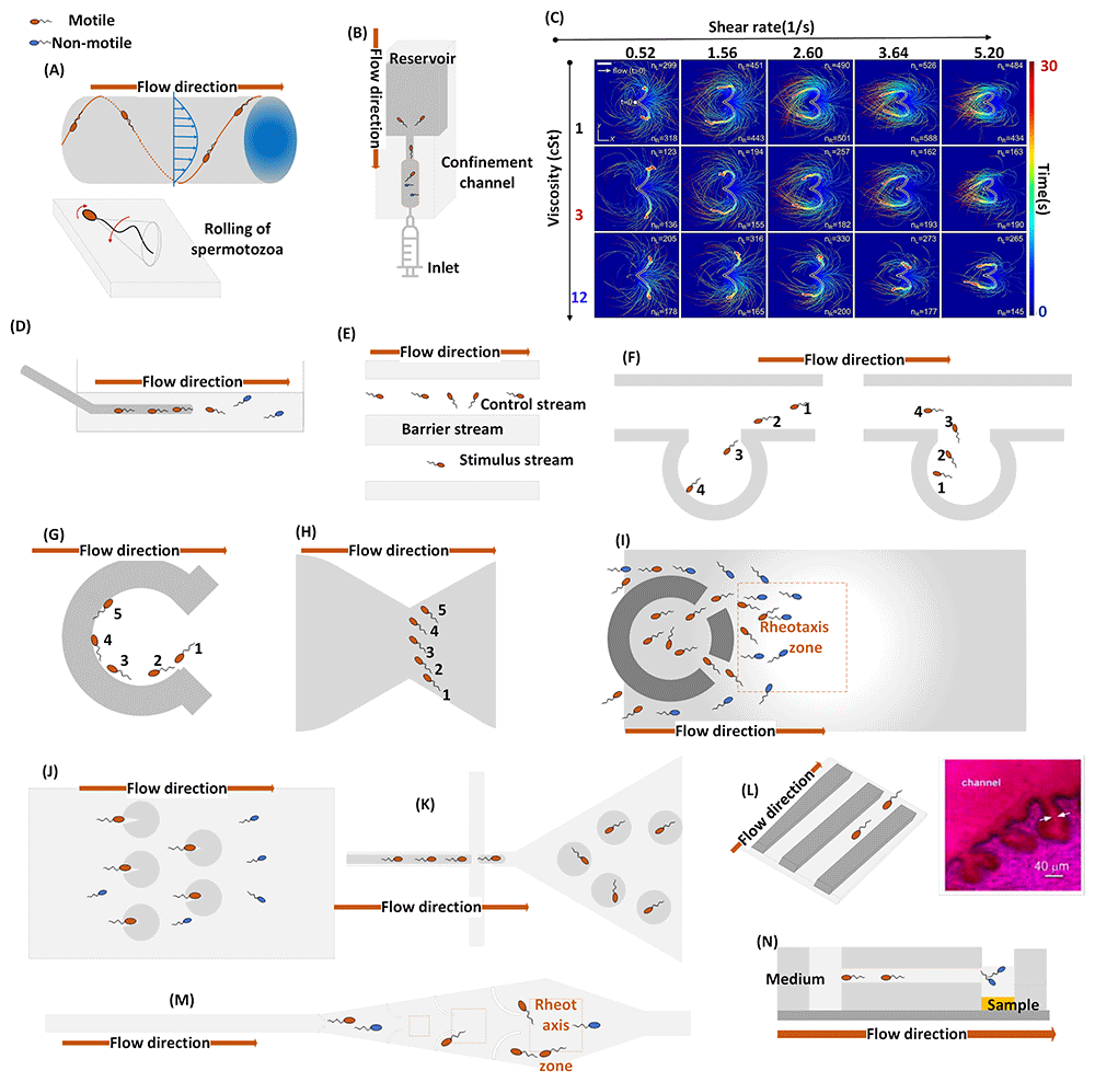
Figure 1. Microfluidic models for the demonstration of sperm rheotaxis.
Figure (A) illustrates the investigation conducted by (Kantsler et al., 2014) on rheotaxis (at 20-60 µm/s) induced chirality in a circular microchannel (0.34mm × 3mm) for the bovine and human model. Figure (B) depicts the experimental setup of (Nishina et al., 2019) for studying the antigravity and confined swimming of human sperm using a circular microchannel (50, 70, 100 µm). Figure (C) is copied from (Bukatin et al., 2015), demonstrates the reorientation of bovine sperm cells in response to shear rates (0.52 - 5.2 s-1) in a simple microchannel (W×H: 0.29mm ×3mm). Figure (D) replicates the rheotaxis setup (50µm/s and 90 µm/s) for mouse and human cells using a glass dish and an 80µm and 125 µm capillaries, respectively (Miki & Clapham, 2013; Zhang et al., 2016). Figure (E) represents an optical trapping setup integrated with microfluidic capillaries (H×W: 0.4µm×0.33µm) for CatSper induced reorientation study in human sperm (Schiffer et al., 2020). Figure (F) illustrates the rheotactic behaviors (33–101 µm/s) of bovine sperm cells through microchannels (W×H: 200µm × 20µm and 50µm × 20µm) with side pouches (El-Sherry et al., 2014). Figure (G) explains rheotaxis spermatozoa through curved wall coral design (D: 500µm, H: 25µm) in viscous flow (1.ml/h) (Zaferani et al., 2018; Zaferani et al., 2021a). A similar approach was utilized for cell entrapment in (Yaghoobi et al., 2022). Figure (H) demonstrates the butterfly-like upstream swimming (0-2ml/h) of human/bovine sperm in narrowed microchannels (W×H:40×30µm) (Zaferani et al., 2019). Figure (I) reproduced from (Zaferani et al., 2018) device comprises the corral design, which facilitates the rheotaxis zone (40–90 µm/s) for bovine/human and the entrapment of the motile cell. Figure (J) demonstrates the rheotaxis-based human sperm cells trapping micro-system (0.5ml/min) with 32µm channel depth and 2µm depth for a circular array (You et al., 2019b). Figure (K) mimics the complex topography of epithelial tissue by trapping the bovine sperm cells into droplets sized 90–120µm (Raveshi et al., 2021). Figure (L) represents microgrooves (8mm × 20µm × 20µm) integrated into microchannels (4cm × 300µm × 120µm) to replicate the bovine sperm rheotaxis (1-3µl/min) with surface inhomogeneities (Tung et al., 2014; Tung et al., 2015). Figure (M) illustrates the capturing of motile human sperm cells using a microchannel (1.5mm × 450µm ×100µm) containing micro-pockets with curvature of 150–300 µm (Sarbandi et al., 2021). Figure (N) shows a passive-flow rheotaxis (60µm/s) using a microchannel (5mm × 200 µm × 20 µm) to unravel bovine sperm-wall interaction (Hyakutake et al., 2021).
The upstream turning, rolling, and spirality of the spermatozoa is arguable, as proposed by Miki and Clapham, suggesting that the turning and rolling of spermatozoa is an outcome of “Cation channel of Sperm (CatSper)” induced [Ca++] signaling cascade (Miki & Clapham, 2013). However, Schiffer et al. and Zhang et al. disproved the role of CatSper activation in rheotaxis (Schiffer et al., 2020; Zhang et al., 2016). Bukatin et al. also confirmed the rolling and reorientation of the spermatozoa, which occurs due to the asymmetry in the midpiece. The resultant shear force along the head and flagellum attributes to the reorientation (Bukatin et al., 2015). These findings substantiate that rheotaxis is a hydrodynamic physical phenomenon, while CatSper-induced motion of the sperm cells may involve additional swimming mechanisms.
Chemotaxis and thermotaxis
Only capacitated sperm cells exhibit enhanced and more frequent asymmetrical flagellum beating, frequently known as hyperactivated (Zaferani et al., 2021b). Capacitation is a highly complex phenomenon and is essential for sperm cells to leave the viscous oviductal reservoir, subsequently enabling them to penetrate through cumulus cells and extracellular matrix contiguous to the oocyte. Exclusion of the cholesterol from the sperm membrane, activation of protein kinases, regulation of intracellular pH and membrane potential, increase in intracellular ions level, and production of reactive oxygen species (ROS) are fundamental biomolecular adaptations required for sperm cell capacitation (Jin & Yang, 2017; Molina et al., 2018). The altered ovarian follicular environment during ovulation has been recommended as the primary driver for sperm capacitation (Ravaux et al., 2016).
Chemotaxis has been postulated and established as a possible mechanism to explain hyperactivation (Suarez, 2008). The oocyte secretes specific biochemicals, and the diffusion of released chemicals through a fallopian tube promotes a concentration gradient. The secreted biochemical is called follicular fluid (FF), which contains complex components including heparin and hormones: progesterone, estrogens, atrial natriuretic peptide (ANP), adrenaline, prolactin, vasopressin, oxytocin, calcitonin, and acetylcholine. The binding of chemoattractant molecules stimulates the receptors on flagella and initiates the proliferation of cyclic adenosine monophosphate (cAMP). An increment in cAMP causes the activation of the protein kinase A (PKA). PKA phosphorylates various proteins essential for hyperactivation and fecundation. The cAMP-PKA pathway induces the efflux of K+ ions through the Slo3 potassium channel and promotes hyperpolarization of the plasma membrane. Subsequentially, the hyperpolarization promotes the opening of calcium channels, which re-settles the hyperpolarization of the membrane—this back-and-forth signaling cascade results in an augmentation of [Ca++] through the flagellar length and altering the beating pattern from symmetrical to asymmetrical (Molina et al., 2018). Consequently, the chemotaxis is an outcome of the intracellular signaling pathway; the irregularities in associated receptors and in sperm acrosome might potentially alter the fertilization success.
The primary challenges encountered in conducting the chemotaxis study lie in the manipulation of low concentrations of chemoattractant and in establishing a stable gradient with it. Nonetheless, high-degree flow control through microfluidic devices attributes the molecular diffusion, which results in linear/no-linear, precise, and controlled chemical gradients (Jaberi et al., 2020).
The attainment of linear molecular diffusion can be accomplished with a microfluidic device featuring a central channel and two side branches. Hussain et al. correlated individual fertilization with chemotaxis in sea urchins and validated the impact of precise chemoattractant secretion in female fertilization (Hussain et al., 2016; Hussain et al., 2017). These studies established chemotactic behavior as a marker to assess the fertilizing capability of male and female individuals. However, the presence of fabrication defects and the occurrence of flow-induced pressure imbalances at the contact zone result in uneven mixing (Deekshith & Jadhav, 2018). Additionally, the excessive width of the principal channel affects the linearity, repeatability, and stability of the gradient.
Eisenbach suggested a qualitative characterization of chemotaxis, which involves the wide-ranging sperm cell trajectories in the chemical gradient field (Eisenbach, 1999). Indeed, hydrogel-based devices can be potentially exploited for linear gradient generation. The crosslinked network of hydrophilic polymers enables controlled molecular diffusion in such devices. Berendsen et al. and Chang et al. have studied the trajectories discrepancy within the mouse and sea urchin model utilizing these approaches. The studies signify the relevance of the model selection for sperm chemotaxis. Nonetheless, the repeatability and stability of hydrogel-based devices are questionable, as the molecular absorption of material leads to irregular diffusion in different laboratory environments (Berendsen et al., 2020; Chang et al., 2013).
Bhagwat et al., reconstituted a ladder-based design with active flow control where one extra outlet was introduced at the contact zone. The additional outlet compensated for the irregular mixing and pressure imbalance at the contact zone. The device was exploited to assess the acetylcholine (Ach)-based chemotactic behaviors in the mouse model and further utilized in discovering novel chemoattractant like N-formyl-L-aspartate (Bhagwat et al., 2018; Bhagwat et al., 2021; Panchal et al., 2022). However, the ladder-based design involves multiple orthogonally positioned channels that can trigger the bubble formation and micro-vertices at the junction corner. Alternatively, the non-linear gradient can be utilized, like Zhang et al. established a non-linear concentration ramp for the progesterone characterization (Zhang et al., 2015). Figures 2A to 2F represent the microfluidic-based approaches that have been exploited for the generation of chemical gradients.
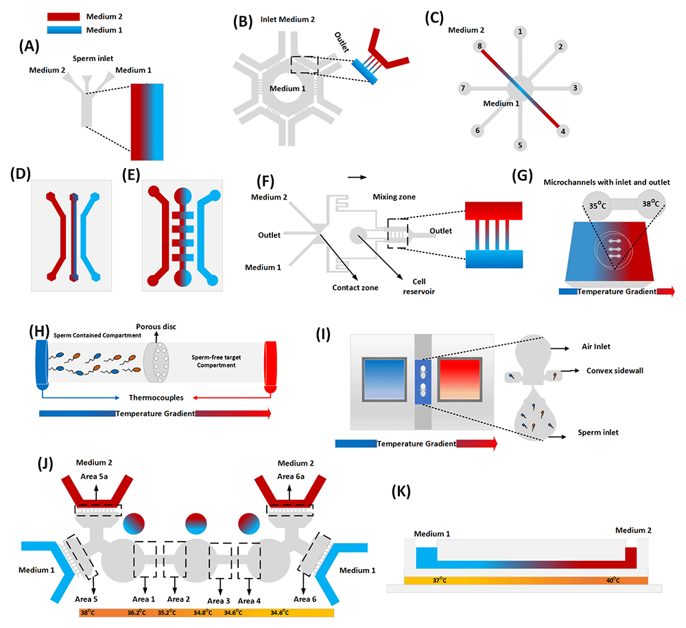
Figure 2. Microfluidic designs for sperm chemotaxis and thermotaxis.
Figure (A) reproduces the structure of (Hussain et al., 2016; Hussain et al., 2017), where a chemical gradient is generated across the centered microchannel (4cm × 1020µm × 99µm) for the sea urchin model. Figure (B) is the adapted design of (Zhang et al., 2015), where the progesterone gradient occurs in transfer channels (W×H: 5 × 2µm2) that connect the hexagonal pool surrounded and U-shape microchannels (W×H: 700 × 50µm2) connected with the inlet-outlet media. Figure (C) shows the combination of 8 microchannels (10mm×500µm×100µm) that intersect each other at the cell reservoir, with an inlet/outlet positioned at an octagonal configuration, enabling the generation of linear Ach gradient for mouse sperm chemotaxis (Ko et al., 2012). Figures (D) & (E) illustrate the hydrogel-based chemotaxis devices. The molecular diffusion through the hydrogel facilitates the linear progesterone gradient. Berendsen et al. used parallel channels (depth 350 µm) to study boar model (D), while Chang et al. used different channel dimensions (W×H: 400 ×167 µm2) to study mouse and sea urchin respectively (Berendsen et al., 2020; Chang et al., 2013). Figure (F) is the adaptation of (Bhagwat et al., 2018; Bhagwat et al., 2021), which utilizes the ladder-based design high-fluidic resistance microchannel (1000 × 50 × 50 µm3) to investigate the chemotaxis in the mouse model. Figure (G) reproduces the idea of (Pérez-Cerezales et al., 2015), where the terminals of a capillary tube are kept at different temperatures (35–38°C) to demonstrate thermotaxis in mouse and human models. Figure (H) recreates the setup of (Bahat et al., 2012), where a lucite microtube attached with two thermistors at the terminal causes the temperature gradient for the human model (0.5–2 OC). Figure (I) summarizes thermotaxis assays of (Li et al., 2014) for the human model, where a microfluidic chip like a Dumble-shape is sandwiched between tanks of aluminum alloy (source and sink) filled with glycerol. Figure (J) shows a combined chemotaxis and thermotaxis study (Yan et al., 2020) in the human model. Three circular chambers facilitate linear and no-linear concentration gradients. Figure (K): presents the temperature and Ach gradient on mouse models (Ko et al., 2018). The study used simple microchannels (13mm × 500µm × 100µm) integrated with ITO tapes to execute the temperature gradient (~2°C).
The molecular mechanism underlying hyperactivation remains elusive. For instance, chemotaxis predominantly occurs in relatively small regions near the oocyte, whereas hyperactivation commences far from the oocyte location. Thermotaxis is postulated to be the long-distance directing mechanism in the follicular tube (Bahat et al., 2003). Sperm cells comprise G-proteins receptors, including opsins and transient receptor potential (TRP) channels, which are fundamentally involved in the regulation of phototransduction. TRP channels and opsin are involved in increasing intracellular calcium levels and guiding the hyperactivated swimming of sperm cells through the oviductal funnel (Pérez-Cerezales et al., 2015; Xiao et al., 2022). Figure 2G to 2K demonstrates the microfluidic designs employed in thermotaxis studies.
It is not so straightforward to reproduce the in vivo conditions related to thermotaxis, as the magnitude of the gradient is very small (~2°C), and the range of the gradient is not comprehended adequately (Bahat et al., 2003; Bahat & Eisenbach, 2006). Bahat et al. developed a biological assay to characterize thermotactic sperm response. The study reported the kinematics of progressive sperm cells along temperature gradients of ~2°C, noting that the instantaneous velocity and motility of sperm cells deteriorate above 40°C (Bahat et al., 2012). Li et al., developed an assay to isolate thermotactic sperm cells using a microfluidic device placed between two heat sources that exploit glycerol for higher gradient stability (Li et al., 2014).
Pérez-Cerezales et al., established the clinical relevance of thermotaxis. The developed assay comprises a capillary set at ~3°C temperature gradient. The temperature-based migration was recorded, and trajectory analysis of sperm cells unveiled that thermotactic cells exhibit higher progressivity in human model, whereas progressivity diminishes in mice. Additionally, the immunocytochemical assay revealed the heterogeneity in rhodopsin location in non-selected sperm cells, affecting thermo-sensing capabilities. The additional biological validation included in vitro development of the rat embryos utilizing thermotaxis and conventional swim-up assays. The morphological analysis of the morula, blastocyst, and cell division stage confirmed the extended hatching of zygotes and validated the significance of thermotaxis (Pérez-Cerezales et al., 2015).
Reviewed studies have substantiated the efficacy of microfluidics-based devices in elucidating the complexities related to with in-vivo interactions between sperm cells and the female reproductive tract. The outcomes can be potentially translated into developing efficient sperm separation devices. However, the capability to characterize the physiological parameters like pH, temperature, and concentration of chemoattractant(s) for the development of reproductive medicine has not been sufficiently exploited for the human model. Few studies investigated the optimum physiological environment for the bovine model and analyzed the impact of Kisspeptin-10 on the Ram model (Abdel-Ghani et al., 2020; El-sherry et al., 2017; EL-sherry et al., 2020). Nonetheless, sperm physiology differs even within mammal specimens (Molina et al., 2018; Romero-Aguirregomezcorta et al., 2018), and the lack of studies on the human model is evidential.
Indeed, microfluidic-based devices offer promising potential for concurrent biomimicking of rheotaxis, thermotaxis, and chemotaxis. A few studies have demonstrated the possibilities and analyzed the swimming migration and tail beating in bi-gradient space (temperature and chemical) (Ko et al., 2018; de Wagenaar et al., 2016b; Yan et al., 2021). Nonetheless, translating these studies for quality sperm selection requires an improved representation that involves biological and clinical validations.
Chemotaxis and thermotaxis have been correlated with the fertilization capability of spermatozoa. However, there has been no significant advancement in studying human sperm chemotaxis. For example, the high degree of control and the geometric versatility of microfluidics should be better exploited to improve the characterization of the biochemical composition of FF and its interaction with spermatozoa. Indeed, progesterone is the only widely tested chemoattractant in the human model, but its role as a chemoattractant is debatable, as the absence of progesterone from FF did not diminish the chemotactic behavior but altered the hyperactivation of spermatozoa (Jaiswal et al., 1999). Few studies identify crucial signaling proteins, such as epidermal growth factor (EGF), brain-derived neurotrophic factor (BDNF), vascular endothelial growth factor (VEGF), insulin-like growth factor (IGF1), and leptin, which plays a vital role in oocyte maturation and might contribute to the spermatozoa signal transduction pathway (Lakshminarasimha et al., 2022; Li et al., 2020; Prochazka et al., 2017). However, there is a lack of studies establishing mentioned proteins as chemoattractant(s). Nonetheless, for sperm cells, the Ach triggers EGF receptor phosphorylation, leading to increased calcium ions during the acrosome reaction in the mouse model (Jaldety et al., 2012; Ko et al., 2012). As of now, there is a dearth of empirical evidence supporting the existence of such mechanisms in the human model.
Microfluidics-based sperm selection
The motility of the spermatozoa is likely the most exploited marker for microfluidics-based sperm cell separation. A microfluidic device containing a straight microchannel that bifurcates into sub-microchannels near the inlets and outlets was employed, characterized, and optimized for the quality subpopulation (Matsuura et al., 2012; Shirota et al., 2016). Chen et al., delivered a microfluidic device inspired by the conventional swim-up method; however, the study did not report analysis for quality parameters against the conventional centrifugation-based methods (Chen et al., 2013b). Riordon et al., delivered the DNA-intact subpopulation by separating the plane swimmer. Nonetheless, the recovery of the subpopulation was very low (Riordon et al., 2019). Eamer et al., and Nosrati et al. exploited the biophysics of sperm-wall interaction and delivered rapid qualitative selection (Eamer et al., 2016; Nosrati et al., 2014). Subsequently, Vasilescu et al., modified the device and introduced microgrooves and a magnetic beads chamber to increase the concentration and quality of the subpopulation collection (Vasilescu et al., 2023).
Moon et al., invented a simple microchannel for sperm separation (Moon et al., 2009), later adopted by Tasogulu et al., who reported the significance of assay duration (Tasoglu et al., 2013a). The device, known as Fertile®, has been commercialized by Zymot Fertility© and Koek biotechnology©. Several studies claim that Fertile® can effectively separate high DNA intact cells (Anbari et al., 2021; Gode et al., 2019; Gonzalez-Castro & Carnevale, 2019; Quinn et al., 2018), although clinical studies have shown no improvement in fertilization success rates (Yalcinkaya Kalyan et al., 2019). Chinnasamay et al., modified the device by introducing cylindrical obstacles for enhanced quality recovery (Chinnasamy et al., 2018).
Motility alone does not exclusively determine natural fertilization. Despite the unresolved physiological mechanisms of sperm cells in human models, previously discussed microfluidic-based approaches can be potentially exploited to deliver quality sperm separation. The rheotaxis-based devices combined with biomimicking of reproductive tract anatomy have emerged as a prominent approach (Ataei et al., 2021; Rappa et al., 2018; Romero-Aguirregomezcorta et al., 2021; Sharma et al., 2022; Wu et al., 2017; Zeaei et al., 2023). A detailed description, findings, advantages, and disadvantages can be found in Table 1. Additionally, Figure 3 elucidates the design and protocol conceptions outlined in the cited scientific journals that were employed for quality sperm selection. It is evident from the table that reviewed technology does not fully address the unmet needs of the reproductive clinicians, primarily the lack of separated concentrations and the laborious protocol. Additionally, the lack of extensive research on chemotaxis and thermotaxis has significantly affected the employability of microfluidics approaches for separating capacitated cells. To date, (Doostabadi et al., 2022) is the only study that has demonstrated improved quality metrics by utilizing chemotaxis and thermotaxis to separate human sperm cells. However, it is worth noting that despite the improved quality, the separated cells still exhibit significant DNA fragmentation (Figure 3).
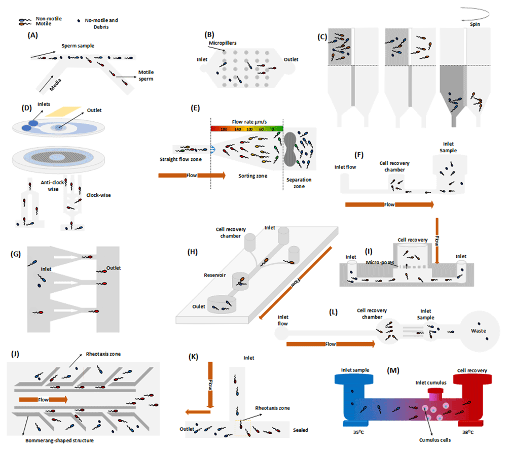
Figure 3. Quality subpopulation extraction utilizing microfluidic models.
The device descriptions and the corresponding findings are in supplementary table 1. Figure (A) illustrates the device concept utilized in the studies conducted by (Matsuura et al., 2012; Shirota et al., 2016). Figure (B) depicts the device (without cylindrical obstacles), initially introduced by (Moon et al., 2009) and further tested on a human model by (Tasoglu et al., 2013b; Zhang et al., 2011). The device, commercially available as Fertile®, was subsequently modified by (Chinnasamy et al., 2018) by incorporating micro-obstacles to improve its quality metrics. Figure (C) demonstrates the design and protocol developed by (Chen et al., 2013b), which is inspired by the conventional swim-up method for quality sperm cell separation. Figure (D) shows the radial fluidic design invented by (Nosrati et al., 2014) for rapid, high-DNA intact cell separation. (Eamer et al., 2016) modified the device to separate clockwise and anti-clockwise swimmers, while (Vasilescu et al., 2023) introduced microgrooves and magnetic beads chamber to enhance the separation efficacy of the device. Figure (E) is adapted from (Wu et al., 2017) and showcases a dumbbell-like structure that effectively obstructs debris and non-motile sperm cells, thereby promoting rheotaxis-based cell separation. Figure (F) represents the cell separation concept of (Rappa et al., 2018). The active flow driven system promotes rheotaxis, while a chamber positioned between flow inlet and outlet (sample inlet) facilitates the recovery of cells. Figure (G) demonstrates the device of (Riordon et al., 2019) for planer-swimmer sorting. Figure (H) shows the rheotaxis based cell sorting by (Romero-Aguirregomezcorta et al., 2021). Figure (I) replicates passive-flow driven device based on rheotaxis and a membrane for quality cell separation (Ataei et al., 2021). Figure (J) illustrates the design proposed by (Zeaei et al., 2023), where boomerang-shaped structure is employed to promotes rheotaxis for the separation of quality sperm cells. Figure (K) showcases the T-shaped design, where sample insertion generates flow and promotes rheotaxis for the quality sperm collection (Mane et al., 2022). Figure (L) represents an active flow-driven device for the separation of the cells (Sharma et al., 2022). Figure (M) represents a device for the separation of sperm cells based on chemotaxis and thermotaxis in the human model (Doostabadi et al., 2022).
Table 1. Summary of reviewed microfluidic devices for quality sperm selection employed in the human model.
Journal
publications | Method | Material | Device description | Advantages/
outcomes | Disadvantages |
|---|
| Mechanism | Flow
Control |
|---|
| (Matsuura et al., 2012) | Motility | No-flow | COP1 | The symmetric microfluidic device
contains a straight microchannel
that bifurcates into two sub-
microchannels at both terminals
(Cho et al., 2003). The cells with
higher motility showed proficiency
in swimming across the main
channel. On the contrary, debris
and non-motile cells flow away
with the streams. Channels with
different configurations (H × W:
0.3mm × 0.5mm: 0.1mm × 0.6mm)
were tested to improve the efficacy
of the device. | -Recovered cells
with ~90%. motility
-Validated DNA
integrity in a
previous study
(Schulte et al., 2007) | -Low recovery
subpopulation
(0.2-0.3%) |
| (Tasoglu et al., 2013a) (Zhang et al., 2011) | Motility | No-flow | PMMA2 | The “Space Constrained Microfluidic
Sorting (SCMS)” was invented by
(Moon et al., 2009) and was adapted
to optimize assay length. The
microchip comprising 50 µm deep
microchannels with various lengths
was implemented to characterize
sperm kinematics. More extended
channels yield less subpopulation
of sperm cells with increased
dynamics. Chip was also tested for
the human model. | -Significance of the
length of the assay
-Recovered yield of
~1.6×10⁶ (Zhang et al., 2011).
-Clinical validation
-Commercialized as
Fertile® Chip | -Low % recovery
against
conventional
-Not
recommended
for cryopreserved
sample
-Unhandy
protocol |
| (Chen et al., 2013b) | Motility | No-flow | PDMS3 | The study is inspired by the
conventional swim-up method. The
chip encompasses two miniaturized
compartments with small pellets.
The left-side compartment holds
the semen sample, and the right
contains the sperm preparation
medium. The Brownian motion
of motile cells encourages the
swimming of the subpopulation
(~50% of motile sperm cells)
towards the compartment
filled with buffer. Subsequently,
density centrifugation assists
in the sedimentation of sperm
concentrations into pellets. | - Semen analysis
validated against
microscopic Makler
chamber method
(<5%). | -Centrifugation-
based separation
-Lack of DNA
fragmentation
test |
| (Nosrati et al., 2014) | Motility | No-flow | PDMS | The radial shape design includes
two layers: the bottom-part hosts
500 microchannels (100µm × 75µm)
and a top-layer structure containing
the inlet and outlet connected with
the round channel. The coupling of
both fluidic layers accomplished the
fluidic circuits. The channel lengths
were characterized to optimize the
device’s efficacy. | - Validation against
conventional
methods
-Subpopulation
vitality (~98%) and
concentration
reported (~0.6%)
-Chromatin test-
based DNA integrity
validation (~98%). | -Low
subpopulation
recovery in
comparison to
conventional
methods |
| (Shirota et al., 2016) | Motility | No-flow | COP | Device design and functioning are
explained in (Matsuura et al., 2012). | -Clinical
validation against
conventional
methods
-Obtained
subpopulation
motility >95%
with 0.8% DNA
fragmentation. | -Low
subpopulation
recovery in
comparison to
the centrifugal
method (~3.6 ×
106)
-Not
recommended
for IUI4 |
| (Eamer et al., 2016) | Motility | No-flow | PDMS | As explained by (Nosrati et al., 2014),
the radial-chip concept was re-
implemented for sperm selection.
The chip has been modified,
allowing the subpopulation
recovery of clockwise, anti-
clockwise, and straight micro-
swimmers. The radial-shape
design contains 52 channels
(7.0cm×100µm×60µm). Additionally,
three chips comprising the
microchannels with side branches
at different angles (45°,90°, and
135°) were demonstrated to
optimize the angle of attack for
boundary swimmers. | -High-yield
(~4million/ml)
->80% vitality
-DNA fragmentation
validation (~98%
DNA intact cells)
-Junction angles
optimization (45°) | -Lack of validation
against currently
practiced
methods |
| (Wu et al., 2017) | Rheotaxis | Passive | PDMS | The study considered the
physiology of the female
reproductive tract while sperm cells
swim from the vagina toward the
uterus, perceiving a downstream
flow of the vaginal discharge. A
diffusive structure microchannel
(L×H: 1cm ×100µm) and hydrostatic
pressure-driven flow were
combined to mimic the rheotaxis. | -Clinical Validation
-Proved efficacy
against swim-up
and DGC methods
-Reported highly
motile sperm
cells separation in
infertile patients
(~22%)
-Subpopulation
recovery ~2×10⁶
cells/ml with ~90%
motility. | -Lack of DNA
integrity validation
- Low recovery
of the separated
cells |
| (Chinnasamy et al., 2018) | Motility | No-flow | PDMS | A simple periodic micro-cylindrical
(D: 10µm, H:50µm) obstacle was
employed in simple microchips as
explained n (Tasoglu et al., 2013a).
The recovered subpopulation
was validated against the plane
channels and swim-up method. | -Quality validation
against swim-up
-Morphology
validation (>55%)
-The chromatin
test validates a
higher percentage
of mature sperm
(>43.5%)
-~5% DNA
fragmentation | ~Low recovery
efficiency (<2%) |
| (Rappa et al., 2018) | Rheotaxis | Active | PMMA | A simple microchannel of
dimension (22.4mm × 4mm
× 76µm) connected to the
inlet, reservoir, and a midway
collection chamber. Rheotaxis
experimentation was conducted at
2,4,5,6,8 and 10µl/min. Hyaluronic
and morphological assays did not
indicate a significant difference
with executed cell separation
assays, contrary to (Romero-Aguirregomezcorta et al., 2021). | -Quantitative
analysis of
recovered
subpopulation
(<20% recovered
cells with ~80%
motility). | -Lack of validation
against practiced
methods
-Missing DNA
fragmentation
test
-Comparatively
less motility
compared to the
Fertile® |
| (Riordon et al., 2019) | Motility | No-flow | Glass | Two parallel zigzag microchannels
(30cm long) were designed for
cell separation. The channels were
connected through 3250 channels
(L×H: 400µm × 2µm). The channel
height is in order of cell head
thickness, resulting in only the
plane swimming of spermatozoa.
The study found that the sperm
cells with planner swimming carry
better DNA integrity and validated
against the density gradient and
swim-up methods. | -Clinical validation
against swim-up
and DGC5.
-Recovered cell DNA
integrity is ~90% | -Low
subpopulation
recovery ~0.1%
-Discarded helical
swimming |
| (Romero-Aguirregomezcorta et al., 2021) | Rheotaxis | Active | PMMA | A microchannel of dimension
(10mm × 300 µm × 100 µm) was
exposed to the inlet-outlet of the
channel. The flow was induced
through a syringe pump (30µm/s),
and 40µl of subpopulation
was accumulated midway through
the channel. The motility, DNA
integrity, and morphology of the
separated specimen were validated
against conventional methods. The
rheotaxis-based selection improves
the ICSI6 outcomes. | -Recovered 100%
motility and >80%
viability
-DNA fragmentation
<5%
>50% normal
morphology
More than 20%
higher fetal
development | - Processing
of low semen
volume
-Unhandy
protocol
-Not convenient
for other ART
methods like IUI
and IVF7. |
| (Ataei et al., 2021) | Rheotaxis | Passive | PMMA | A passive flow device comprises
a microchannel with two inlets
at the terminal and an outlet
in the middle. The outlet of the
channel facilitates the hydrostatic
pressure difference for flow and
holds a membrane with holes for
separation. Morphology and DNA
fragmentation was validated, and
a flow velocity of 25µl/min was
reported for the optimized cell
separation. | -Isolation efficacy
~30%
-Morphological,
DNA, and CASA8
validation
->45% normal
morphology
-~5% DNA
fragmentation | - Lack of validation
against practiced
methods
-45 minutes long
assays |
| (Doostabadi et al., 2022) | Chemotaxis/
Thermotaxis | No-flow | 3D-
printing
resin | A simple microtube with a 2mm
diameter connects two cylindrical
chambers (H×D: 9mm × 5mm). The
length of the microtube is 2cm,
including a hole near one chamber
for the insertion of cumulus cells.
Both chambers are kept at different
temperatures, respectively 35°C
and 38°C, to provoke thermotaxis
and chemotaxis-based sperm
migration. | - Validation against
direct swim-up
- High concentration
separation (>30%)
-Better DNA
integrity against
swim-up
-toxicity validation | -Separated cells
comprise >10%
fragmented cells
-1hrs long
protocol
-Instability in
temperature
gradient |
| (Sharma et al., 2022) | Rheotaxis | Active | PDMS | A device with a 96 µm height
comprises a 20 mm long
microchannel (W: 300 µm) that
connects the collection chamber
and the flow inlet unit. The
collection chamber is connected
to the sample inlet via multiple
sub-microchannels (W×L:
300 µm × 5 mm) integrated into a
2.4 mm channel width. The same
channel is extended without sub-
microchannels to connect to the
waste chamber. | ->95% motility of
the separated cell
-~1.5% separation
efficacy
-DNA fragmentation
validation (<2.5%) | - Lack of
validation against
conventional
practice
-Low-
concentration
recovery
-Missing protocol
timeline |
| (Mane et al., 2022) | Rheotaxis | Passive | PDMS | A pre-filled T-shaped microfluidic
device (W×H: 200 µm ×50 µm),
sealed at one side of the horizontal
channel, is used to utilize a 40 µl
semen sample and generate
gravity-assisted diffuse flow.
Rheotaxis occurs adjacent to the
junction due to the inlet-outlet
pressure imbalance, causing motile
cells to migrate toward the sealed
channel. | -Inexpensive
protocol
- Nitro-blue
tetrazolium (NBT)
validation
-~100% motile cells
recovery | -2hrs long
protocol
-The separated
concentration was
not mentioned
-Lack of
validation against
conventional
practices |
| (Zeaei et al., 2023) | Rheotaxis | Active | PDMS | The device comprises a central
channel connected to two
parallel channels (500 µm) with
boomerang-shaped structures.
The optimal shear rates (3.5-6 s-1)
were achieved in the contact region
(rheotaxis zone). Different depths of
the designs were tested, including
500 µm, 200 µm, and 100 µm. | ~160 000 motile
cells in 20 min
>95% motility of
separated cells
-ICSI favorable | -Meager sample
collection for
normal mother
sample
-Not
recommended for
IUI and IVF
-More than
>3% of cells are
fragmented |
| (Vasilescu et al., 2023) | Motility | No-flow | 3D-
printing
resin | The device, inspired by (Nosrati et al., 2014), underwent
modifications to improve its efficacy.
Multiple microgrooves were
introduced to the connection wall
in the z-direction. Additionally, a
magnetic microbeads chamber was
created to trap phosphatidylserine-
positive sperm. | ->5% of separation
recovery
-~100% vitality
-~100% motility
-very low DNA
fragmentation
(<1.4%)
-Short-duration
protocol | -Not appropriate
for IUI |
Recommended commercialization pathway
As established in this review, microfluidic-based sperm selection approaches have shown the potential to facilitate high-quality sperm cells. However, the viability of microfluidics in life science applications is debatable, as the technology has not lived up to the initial hype during its early developmental phase (1990–2009) (Mukhopadhyay, 2009). Materials, modeling, and machinery play a significant role in upsurging the technology's readiness level (TRL). In the aspects of microfluidic application for sperm selection, the rate of technology translation is still meager. The authors can allude to three possible reasons: 1) the developed technologies are still in the early stage of development; 2) there is a lack of communication between the end-users and the researchers; and 3) there is a potential lack of interest by the industry to change current methods unless costs, efficacy, and usability are maintained or improved.
An innate communication disparity among academia, industry, and the end-user is the most critical impediment towards commercialization readiness of the developed technologies. For example, most microfluidic engineers practice rapid prototyping using Polydimethylsiloxane (PDMS). Our review analysis indicated that approximately 70% of researchers opt for PDMS and hydrogel-based prototyping. However, the materials are not very well-suited for biological applications. Fluidic chips made of these materials comprise several shortcomings, including their tendency to absorb small hydrophobic molecules, release tiny polymeric fragments, and are relatively expensive for large-scale production (Mukhopadhyay, 2007). Furthermore, the conventional UV-lithography for the SU-8 mold fabrication is associated with repeatability issues, high maintenance requirements, a sophisticated environment, and a specialized workforce.
Hence, it is evident that researchers should consider adopting a mitigation plan or show the possibility of using alternative materials to enhance the readiness level of prototyping. High-resolution biocompatible resin-based 3D printing can serve as a viable alternative for prototyping fluidic devices, offering improved repeatability and scalability for device fabrication. Nevertheless, 3D-printing-based prototyping faces limitations regarding the printing of microchannels dimensions printing and tolerances. Therefore, these prototypes should incorporate feasible printing dimensions and further promote user-friendly protocol. Subsequently, thermoplastics-based devices are recommended for industrialization, especially cyclic olefin copolymer (COC/COP) and polymethyl methacrylate (PMMA). Both polymers have FDA approval for medical applications; moreover, lower biomolecular absorption, ease of fabrication, and cost-effectiveness with industry-ready methods and sustainability of these polymers encourage their utilization in large-scale fabrication of microfluidic chips.
In the context of clinical implementation of microfluidic-based approaches, low throughput appears as a principal hurdle. The associated scientific community has undoubtedly discovered exceptionally engineered microfluidic approaches for quality sperm selection. However, clinical screening requires a statistical analysis over a wide range of human semen samples. The limited recovery of sperm cell concentration poses a constraint on reproductive laboratories, hindering their ability to execute reproductive procedures like IUI and IVF. As a result, reviewed microfluidic-based methods lack clinical validation, where the biological validation associated with quality sperm selection has not been correlated with the pregnancy rate and the quality of embryos. Additionally, centrifugation-based protocols offer rapid processing of multiple samples, while microfluidics-based methods require multiplexing for single-sample processing, potentially resulting in a less user-friendly protocol. Despite the better-quality outcomes offered by microfluidics-based approaches, they have not gained much attention due to their long and tedious protocols. The emphasis on developing proof-of-concept without analyzing the unmet needs of end-users has influenced the translation of technology.
The commercial execution of medical devices —especially for the class 1-3 category— necessitates regulatory approvals. The ascending numeric assignment (1 to 3) correlates with a proportional escalation in the associated risk level with the implication of the device (Marešová et al., 2020a). In the context of the assisted reproduction, where the disposable devices correspond to the class 2a to 3, which incur substantial costs and labor efforts (Heinemann, 2021; Marešová et al., 2020b). Additionally, the cost of production in ISO environments and logistics accumulate the expenses associated with the developed medical disposable. Such significant investments cannot be solely supported by academia. On the contrary, end-users have low willingness-to-pay (WTP), as the current conventional methods, including DGC and swim-up, offer low processing costs and high throughput. Hence, the commercial translation of such technology (high investment demand with low WTP) is only feasible if the involved industry or academia is supported by investors/business angels/venture capitalists.
Ethics and consent
Ethics approval and consent were not required.









