Introduction
Hepatitis B virus (HBV) and Plasmodium falciparum infections are endemic in low- and middle-income countries like Ghana. In 2019, it has been estimated that 296 million individuals were chronically infected with Hepatitis B Virus (HBV). This report also revealed that 1.5 million people get infected annually, and 820,000 deaths are attributed to CHB1. Although a universal HBV vaccine and the use of anti-viral agents for disease management exists, the WHO reports that 6.1% of the adult population in sub-Saharan Africa are infected with HBV2. Ghana being one of the endemic countries has an estimated seroprevalence of 8.36% and 14.3% among adults and adolescents respectively3.
The persistent exposure of T lymphocytes to antigens of the infecting organism in chronic infections leads to the reduction in the quality of T cell function: termed ‘exhaustion’4–7. Effector functions of these exhausted T lymphocytes are lost. Some of these include the production of cytokines. This impairment consequently reduces the capacity of the immune system to fight the infection. This promotes the development and establishment of the pathogen causing an infection8. However, chronic cytokine expression by T lymphocytes promotes progression of the infection to a chronic state9,10. Thus, whereas IFNα and β signals enhance viral control during early infection by promoting effector CD8+ T cell differentiation11,12, chronic exposure to IFN-α and β can be detrimental and enhance T cell dysfunction10.
Additionally, evidence suggests that the Th1 response to chronic infection is down-regulated by virus-specific regulatory T cells (Tregs). This activity of Tregs, in an attempt to down-regulate inflammation and prevent liver pathology, may also promote long term persistence of the virus13,14. Transforming Growth Factor (TGF)-β and IL-10 signalling facilitates viral persistence by promoting immune suppression15. In contrast, other inflammatory pathways including those induced by IL-6 may be required for controlling chronic viral infection and avoiding CD8+ T cell exhaustion9,16. Pro-inflammatory cytokines are a precipitating factor for the onset of liver fibrosis, cirrhosis, and carcinoma in CHB infection15,17,18. Most of what have been described above have been observed in animal models and or hepatoma cell lines that were infected with different viruses11,12,16,19 and persons with advanced chronic HBV infection on treatment20–23. Most studies that have assessed cytokine response in CHB participants (not requiring antiviral therapy), have been cross-sectional studies8,17,24,25, with one prospective study conducted among children26.
Malaria and HBV infections are amongst the most common infections in many developing countries. The WHO African Region, with an estimated 233 million cases in 2022, accounted for about 94% of cases globally27. Exhaustion of T cells in these CHB individuals may consequently affect the immune response to, and clearance of other pathogens such as Plasmodium. Additionally, malaria vaccine candidate trials usually exclude individuals infected with immune modulating pathogens such as HBV. Most of what have been described above have been observed in persons with advanced chronic HBV infection, usually requiring treatment. However, it is not known whether vaccine induced responses could be compromised in chronic HBV-infected individuals who have recently entered chronicity (and do not require antiviral therapy yet), if vaccines are later approved for routine immunization in HBV-endemic populations of sub-Saharan Africa, as currently the WHO recommends the broad use of malaria vaccines; RTS,S/AS0128 and R21/Matrix-M27 for the prevention of P. falciparum malaria in children living in malaria endemic areas, prioritising places with moderate to high transmission.
This study, therefore, investigated how early this T cell exhaustion state establishes, since most of the available data relates to CHB cases that have persisted for much longer time, using ex-vivo cytokine production to selected Plasmodium falciparum antigens.
Methods
Study design and population
This is part of the HEPMAL study, a prospective case-control study with monthly follow-up of two groups of participants for up to one year, based at the Noguchi Memorial Institute for Medical Research (NMIMR), University of Ghana. Individuals between the ages of 18 to 60 years, diagnosed with chronic HBV infection, reside within 20 kilometres (km) of the Korle-Bu Teaching Hospital (KBTH) and the Legon Hospital Public Health Unit (LHPHU), available during the one-year study period and willing to give informed consent were recruited as study participants. However, subjects with chronic HBV infections with viral load more than 2000 IU/ml or 10,000 copies/ml) that require treatment were excluded. In addition, subjects with anaemia, (Hb < 11 g/dl) and other chronic inflammatory conditions HIV/AIDS, cancer, liver diseases, and pregnant women were excluded. Therefore, the experimental or exposed group (cases) was a cohort of CHB-infected individuals who have not yet progressed to the point where treatment was necessary, and the unexposed group included healthy individuals with no evidence of liver or chronic infections. For each participant recruited in the experimental group, an HBV uninfected individual living in the same locality (within a 2 km radius of the exposed person’s place of residence) was recruited as a matching control. This is to ensure that the exposure to infectious mosquito bites, and hence the propensity to be infected with Plasmodium, is similar between the exposed and unexposed groups.
The GPower 3.1.9.7 online software (available at https://www.psychologie.hhu.de/arbeitsgruppen/allgemeine-psychologie-und-arbeitspsychologie/gpower.html) was used to estimate the sample size of 118 (59 cases and controls) for this study. The parameters: test (means: Wilcoxon-Mann-Whitney test-two groups), two-tailed, effect size of 0.54, and power (1-β) of 0.81 were used. Samples used in this study were therefore obtained from 59 cases and 59 location-matched controls, who were recruited at the Gastroenterology Clinic of the KBTH and the LHPHU, both in Accra, Ghana.
Heparinized blood samples were obtained from each participant at four time points. Samples were taken between October 2020 to May 2022 at three-month intervals -baseline (V0), month 4 (V3), month 7 (V6) and month 10 (V9) over a period of one year from participants who gave written informed consent. Finger prick blood was also used to prepare DBS every month for the measurement of eluted plasma anti-CSP antibody levels.
Whole blood ex-vivo stimulation and Multiplexed measurement of cytokine levels
Heparinised fresh whole blood from our participants were simulated using the whole blood assay technique as previously described29. An equal volume of fresh heparinised whole blood was added to 250 µl each of and 20 µg/ml of Plasmodium antigens, Circumsporozoite Protein (CSP), Apical Membrane Antigen 1 (AMA-1), and Thrombospondin-related Adhesive Protein (TRAP). The stimulants were prepared using plain Roswell Park Memorial Institute (RPMI) 1640-based culture medium. For every sample in addition to a positive control (2.5 µg/ml of Phytohemagglutinin A (PHA), two negative controls were included, that is, 500 µl of undiluted blood and the second one, 250 µl each of whole blood and plain RPMI-1640. Samples were mixed well and plates were incubated at 37 °C, 5% CO2 humidified incubator. Culture supernatants were harvested 24 hours after initiation of culture and stored immediately at -80 °C until required for analysis in multiplexed assays.
A commercial multiplexed kit from Invitrogen was used to quantify 13 cytokines: Interferons (IFN) - α, β, and γ, Tumour Necrosis Factor (TNF)-α, and Interleukins (IL) -1β, 2, 4, 6, 10, 12p70, 13, 17A, and 21 in plasma and supernatants from whole blood assays. Procedures described in the Human ProcartaPlex Mix & Match 13-Plex kit was followed. Briefly, 50 μl of diluted microparticle cocktail were added to each well of a 92-well plate. Fifty (50) μl of sample was then added and the plate was incubated for 30 minutes at room temperature on a plate shaker at a speed of 600 ± 50 rpm. The plates were then transferred onto a flat horizontal surface at 4 °C overnight. The next day, the plates were further incubated for 30 minutes at room temperature on a plate shaker at a speed of 600 ± 50 rpm. The plates were washed two times by the addition of 150 μl/ of the wash buffer to each well. Fifty microliters (50 μl) of diluted biotin-antibody cocktail were added to each well and the plates were incubated for 1 hour on a plate shaker at 600 ± 50 rpm, followed by washing. Fifty microliters (50 μl) of diluted streptavidin-PE were added to each well, and the plate was incubated for 30 minutes on a shaker at 600 ± 50 rpm. Hundred and twenty microliters (120 μl) of reading buffer were added to each well and incubated for 5 minutes on the shaker at 600 ± 50 rpm. Data was acquired using the Luminex MAGPIX analyzer (Luminex Corporation). The CSV files containing the MFIs were then uploaded onto an online analysis app (ProcartaPlex app, ThermoFisher scientific) which then converts the MFIs into concentrations in pg/ml.
Indirect ELISA for the measurement of plasma anti-CSP antibody levels as evidence of recent infectious mosquito bites
Antibodies were eluted from Dried Blood Spots (DBS). Briefly, 2 punches (3 mm in diameter) of each DBS sample were transferred into 2.0 ml 96-wells deep well plates (Greiner Bio-One GmbH, Germany), 150 μl of 1% BSA in phosphate buffered saline (PBS), 0.05% sodium azide solution was added to each well and incubated overnight on a plate shaker (Thermo Scientific, USA). The eluate, containing antibodies with an equivalent final serum dilution of 1:100 was used for ELISA. Antibody titres were measured from eluted samples from dried blood spots by indirect ELISA using the P. falciparum 3D7 strain CSP recombinant protein, and two 24-mer peptides (PEP1 and PEP2) from the middle region of CSP. NUNC Maxisorp 96-well ELISA plates were coated with 0.5 μg/ml, 5 μg/ml and 5 μg/ml concentrations of CSP, PEP1 and PEP2 respectively. The plates were covered with a microplate sealer and incubated overnight at 4 °C. The next day, the coated plates were washed 3 times with Phosphate-Buffered Saline with Tween (PBST) and patted dry. The plates were blocked with 200 μl of blocking buffer (5% w/v skimmed milk in 1X PBS) and incubated for one hour at room temperature after which the plates were washed and patted dry.
In duplicates, standards were pipetted into the plates followed by a 3-fold serial dilution. The standard was made up of a pool of semi-immune sera used in a study by Kusi, et al.30. Samples (eluted serum) were also added in duplicates in a 1:10 dilution. The plates were sealed and incubated for an hour. After washing and drying, 100 μl of polyclonal rabbit anti-human IgG-horse-radish peroxidase diluted in the ratio of 1: 3000 in 1X PBS was added to each well and incubated for 1 hour at room temperature. After three rounds of washing, 100 µl of tetramethylbenzidine substrate solution (TMB PLUS 2) was applied to each well and followed by a 15-minute incubation period. To stop the reactions, 100 μl of 2M H2SO4 was added per well and a MULTISKAN FC plate reader was used to read the optical density of each plate at 450 nm.
Data handling/statistical analyses
GraphPad Prism 9.0.0 was used for all statistical analyses. A trial version which can perform an equivalent function is available here https://www.graphpad.com/demos/ The participants' ages were presented as median (Interquartile Range: IQR) and compared using the Mann–Whitney U test. Sex was presented as proportions and compared using the Chi-square test between HBV-positives and HBV-negatives. Luminex data contained in CSV files were analysed using the GraphPad Prism 9 .0.0 software. Plasma and culture supernatant cytokine levels were compared across the four time points within/between groups by Kruskal-Wallis tests.
Ethics statement
Ethical clearance was obtained from the Institutional Review Board (IRB) of the Noguchi Memorial Institute for Medical Research and the Scientific and Technical Committee (STC) of Noguchi Memorial Institute for Medical Research (NMIMR) on the 8th of January, 2020 and 13th of December, 2019 respectively The ethics and protocol review committee of the Korle-Bu Teaching Hospital (KBTH) also gave approval on the 9th of April, 2020. The NMIMR-IRB has a United States Government Federal-wide Assurance (FWAA00001824) from the Office for Human Research Protections and has approved thousands of biomedical research protocols. The approval numbers, FWAA00001824(NMIMR-IRB/046/19-20) and KBTH-IRB/00024/2020) were given by the above institutions respectively. Before recruitment, trained study personnel thoroughly explained the purpose of the study to potential participants and informed them about their rights as participants in the study. Participants completed and signed informed consent forms after the consenting process. The principles outlined in the Declaration of Helsinki were adhered to in every activity carried out in this study.
Results
Socio-demographic characteristics of participants
One hundred and eighteen (118) participants; 59 each of CHB individuals (cases) and controls participated in this study. The median age of the cases was 32 years and that of the controls was 34 years. Majority (54.2%) of the CHB individuals were females and 52.5% of the HBV-negative individuals were males, but these proportions were similar between the cases and controls (Table 1).
Table 1. Socio-demographic characteristics of participants.
| Variable | Level | CHB
(n=59) | HBV-negatives
(n=59) | ꭕ2 (p-value) |
|---|
| Age | Median
(25th – 75th Percentile) |
32.0
(27.0 – 38.0) |
34.0
(28.0 – 46.0) | 0.086 |
| Sex | Males (%) | 27 (45.8) | 31 (52.5) | 0.543 (0.461) |
Chronically infected HBV individuals showed similar increasing trends in levels of some plasma cytokines as controls across the year. Plasma cytokine levels at four time points (three months apart) over a period of one year were compared between cases and controls. The levels of IL-10, TNF-α, IL-1β, and IL-6 increased across the four time points in both groups (Figure 1B–E), but these cytokines were not different statistically, between cases and controls. Interleukin 6 was the cytokine produced at the highest levels by the two groups (Figure 1D).
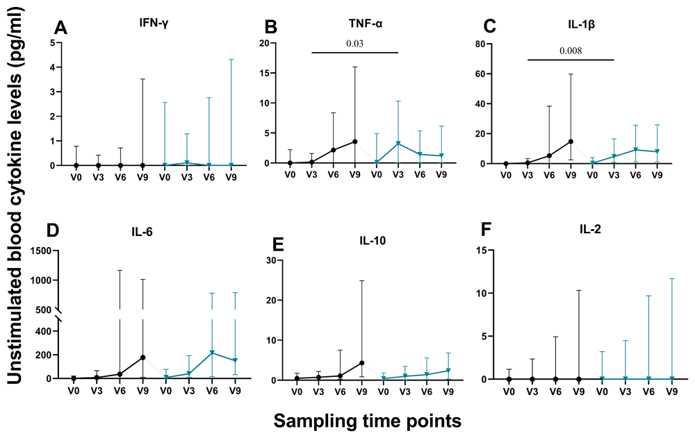
Figure 1. Plasma cytokine levels.
Levels of plasma cytokines were estimated over four time points using a Human ProcartaPlex Mix & Match 13-Plex kit. The graphs display levels of (A) IFN-γ, (B) TNF-α, (C) IL-1β, (D) IL-6, (E) IL-10, and (F) IL-2 levels in (pg/ml). V0: baseline, V3: Visit 3, V6: Visit 6, V9: Visit 9.
Chronically infected HBV individuals exhibited similar ex-vivo cytokine expression patterns as controls in response to the Plasmodium antigens. To determine whether chronic HBV infection elicits a strong regulatory T cell environment that dampens induction of the needed pro-inflammatory response against Plasmodium, we stimulated the participants’ blood cells with Circumsporozoite Protein (CSP), Apical Membrane Antigen 1 (AMA-1), and Thrombospondin-related Adhesive Protein (TRAP) in whole blood assays at four time points (three months apart) over a period of one year. We indirectly determine T cell exhaustion status using the expression of cytokines ex-vivo. Findings from this showed that chronically infected participants exhibited similar ability to express TNF-α, IFN-α, IFN-γ, IL-17A, IL-1β, IL-6, IL-2, and IL-10 cytokines (Figure 2–Figure 4). Also, T regulatory function (IL-10 levels) was similar among the two groups across the four time points (Figure 2H, Figure 3F, Figure 4H).
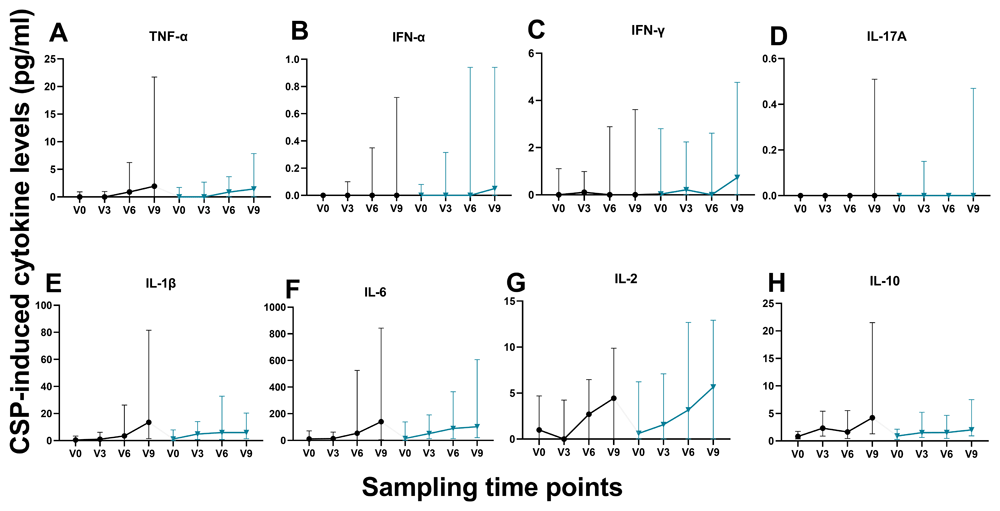
Figure 2. CSP-induced cytokine levels.
Levels of Plasmodium falciparum CSP induced-cytokines were estimated using a Human ProcartaPlex Mix & Match 13-Plex kit. The graphs display levels of (A) TNF-α, (B) IFN-α, (C) IFN-γ, (D) IL-17A, (E) IL-1β, (F) IL-6 (G) IL-2, and (H) IL-10 levels in (pg/ml).
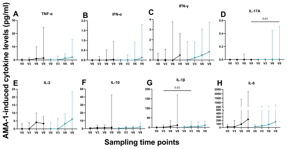
Figure 3. AMA-1-induced cytokine levels.
Levels of Plasmodium falciparum AMA-1 induced-cytokines were estimated using a Human ProcartaPlex Mix & Match 13-Plex kit. The graphs display levels of (A) TNF-α, (B) IFN-α, (C) IFN-γ, (D) IL-17A, (E) IL-2, (F) IL-10 (G) IL-1β, and (H) IL-6 levels in (pg/ml).
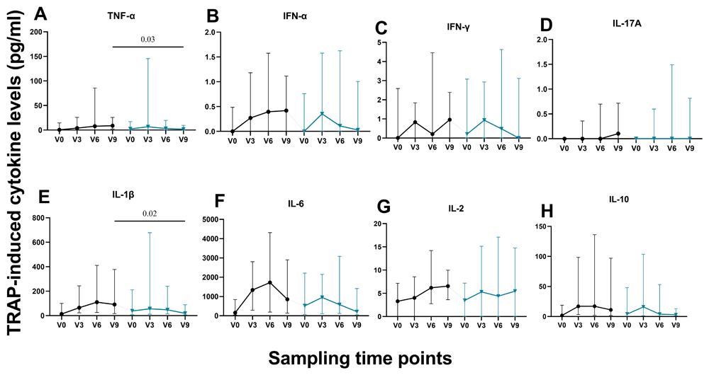
Figure 4. TRAP-induced cytokine levels.
Levels of Plasmodium falciparum TRAP induced-cytokines were estimated using a Human ProcartaPlex Mix & Match 13-Plex kit. The graphs display levels of (A) TNF-α, (B) IFN-α, (C) IFN-γ, (D) IL-17A, (E) IL-1β, (F) IL-6 (G) IL-2, and (H) IL-10 levels in (pg/ml).
Increasing levels of TNF-α, IL-1β, IL-6, IL-2 and IL-10 were observed in both groups, in response to CSP. IL-17A was produced in very minute quantities (Figure 2D). Similar increasing trends were observed in the levels of TNF-α, IFN-α, IL-1β, IL-6, IL-2, and IL-10 in response to AMA-1 (Figures 3A, B, E, F, G & H).
However, this trend was not observed with TRAP stimulation of cells. Cytokine levels decreased after visit 4, except for IL-1β, and IL-6 where higher levels were observed at visit 7 (Figures 4 E & F).
TRAP elicited a stronger cytokine response in both study groups. To determine which of the three antigens (AMA-1, CSP, and TRAP) induces the production of cytokines in highest amounts, we compared various cytokines across the four time points. Our results indicated that TRAP induced the highest levels of cytokines compared to CSP and AMA-1 (Figure 2, Figure 3 & Figure 4). Within each group, TRAP-induced significantly higher levels of IL-1β, IL-6, IL-10, and TNF-α compared to the values observed at baseline, and those induced by CSP and AMA-1 (Figure 5A–G, Figure 6A–G, Figure 7A, B, C, E, F, & G and Figure 8B, C, E, F & G) but there was no significant difference between the two groups. At visit 9, only TRAP-induced IL-1β and IL-6 showed a significantly higher levels compared to baseline, CSP and AMA-1 values respectively among HBV-positives (Figure 5D & Figure 6D).
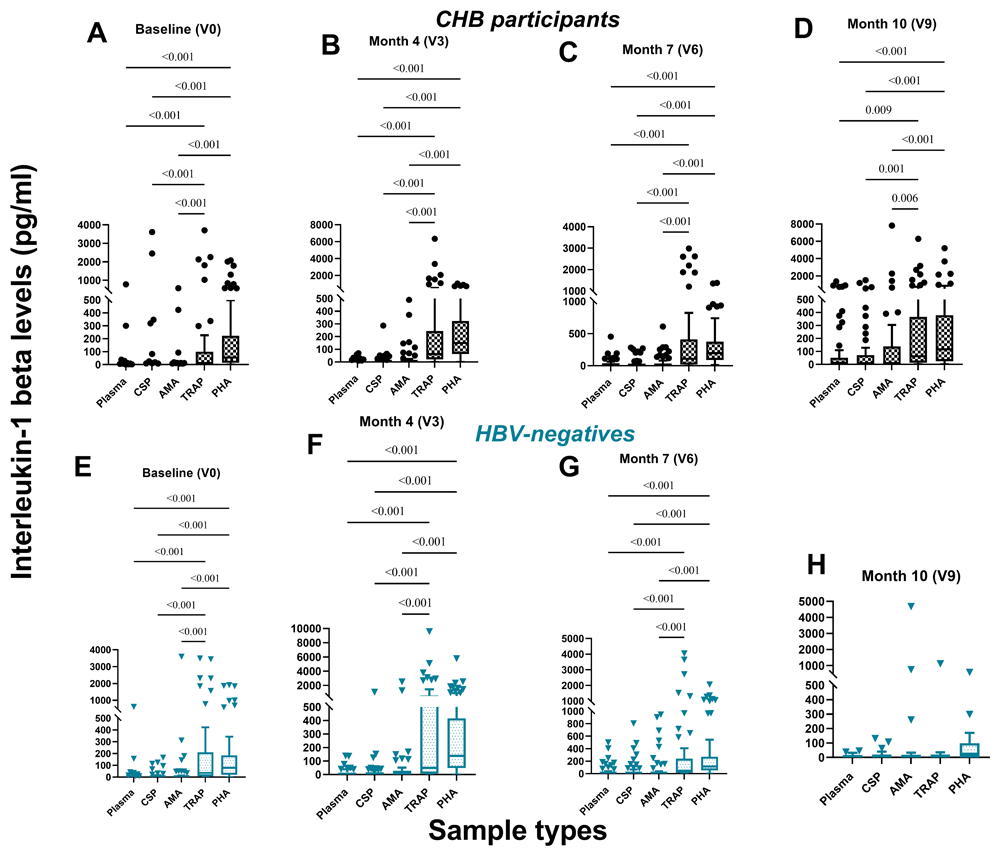
Figure 5. Ex-vivo production of IL-1β by chronic HBV (black) and HBV-negative (sea blue) individuals.
Levels of IL-1β in unstimulated and stimulated plasma were estimated over four time points.
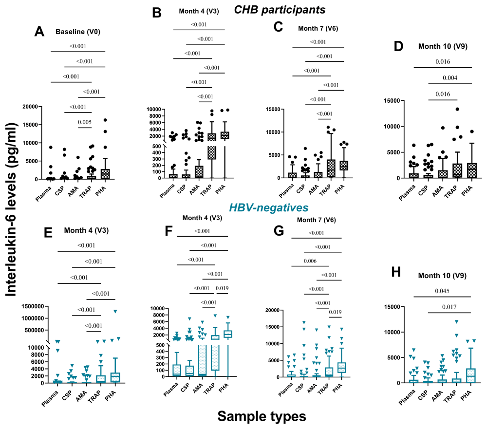
Figure 6. Ex-vivo production of IL-6 by chronic HBV (black) and HBV-negative (sea blue) individuals.
Levels of IL-6 in unstimulated and stimulated plasma were estimated over four time points.
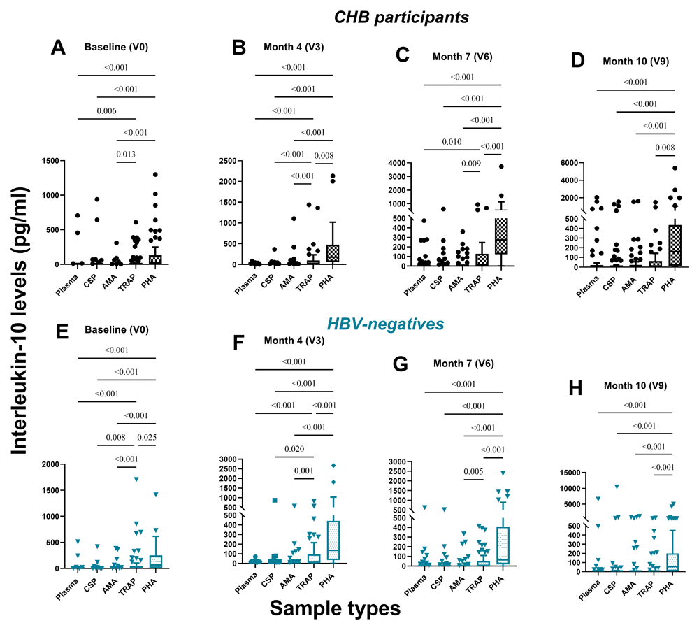
Figure 7. Ex-vivo production of IL-10 by chronic HBV (black) and HBV-negative (sea blue) individuals.
Levels of IL-10 in unstimulated and stimulated plasma were estimated over four time points.
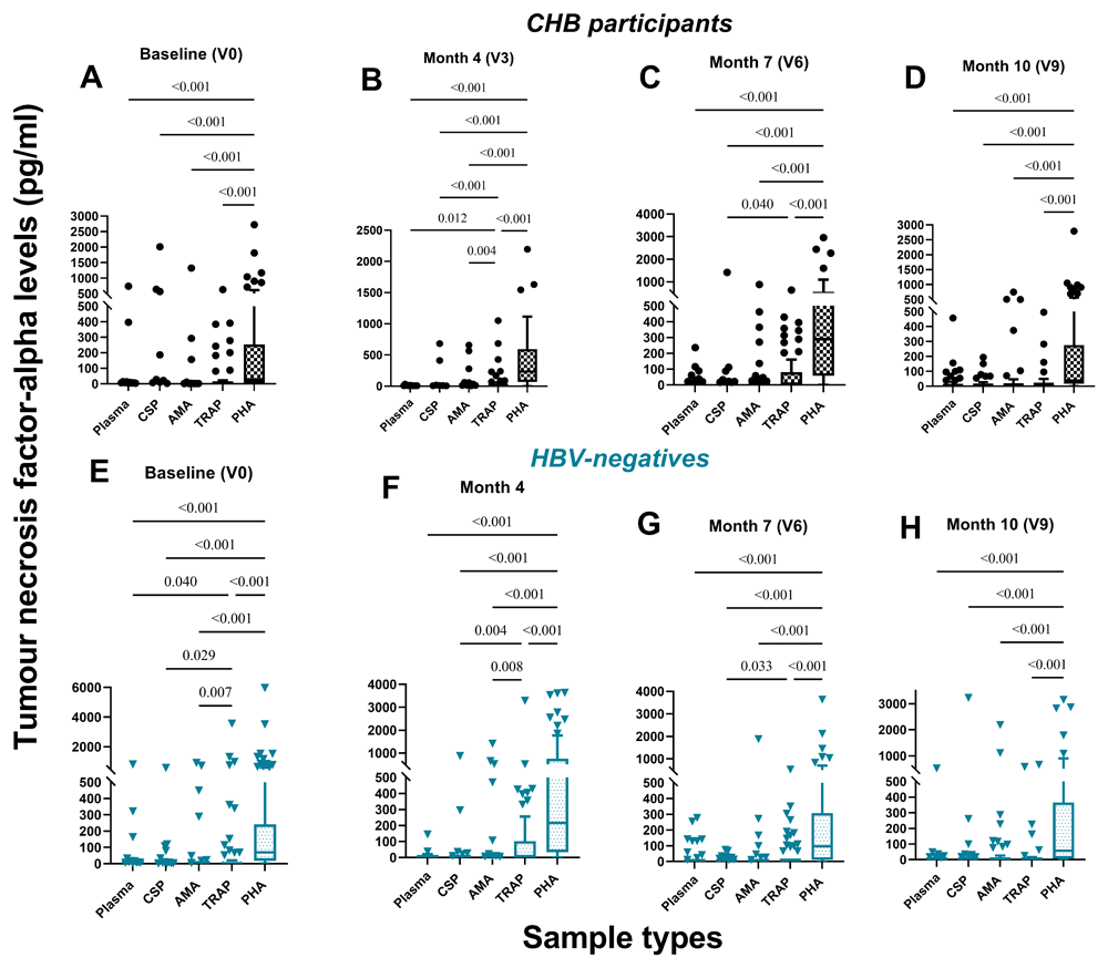
Figure 8. Ex-vivo production of TNF-α by chronic HBV (black) and HBV-negative (sea blue) individuals.
Levels of TNF-α in unstimulated and stimulated plasma were estimated over four time points.
Pro-inflammatory and T-regulatory function is similar in both study groups. To determine the level of immune system regulation (a balance between pro-inflammatory and T-reg cytokines), we computed and compared pro-inflammatory cytokine and T-reg ratios between cases and controls (Figure 9–Figure 11). There were no significant differences between the two groups in relation to IL-6:IL-10 and IL-1β:IL-10 ratios, except for IL-1β:IL-10 ratio where there was a significantly higher IL-1β:IL-10 ratio in HBV-negatives compared to HBV-positives at visit 3 for unstimulated plasma, CSP and AMA-1 induced IL-1β and IL-10 cytokines (Figure 11). Additionally, increasing IL-1β:IL-10 ratios were observed among the two groups, but significant values were seen only within the HBV-positive group (Figure 9). Higher IL-6:IL-10 ratios were observed at visit 6 and 9 for unstimulated samples for HBV-negatives and unstimulated samples, CSP and AMA-1 stimulated samples for HBV-positives respectively (Figure 10).
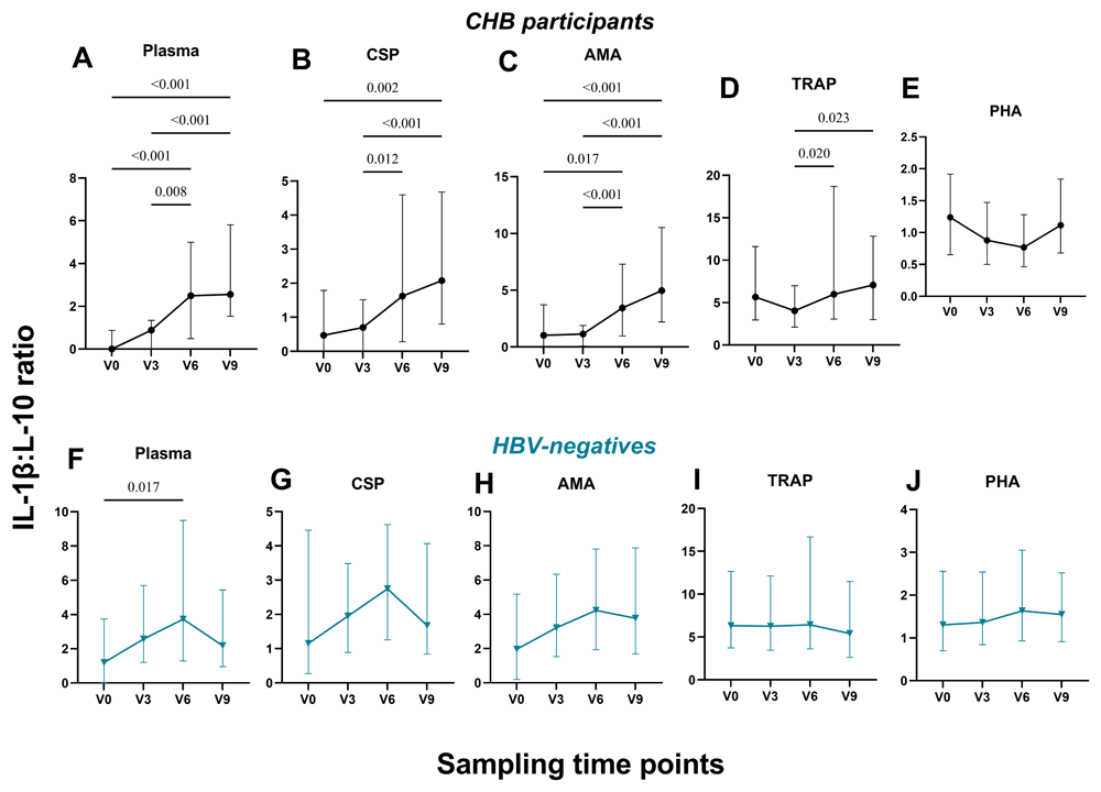
Figure 9. IL-1β/IL-10 ratios.
IL-1β /IL-10 ratios were computed and compared among chronic HBV (black) and HBV-negative (sea blue) individuals.
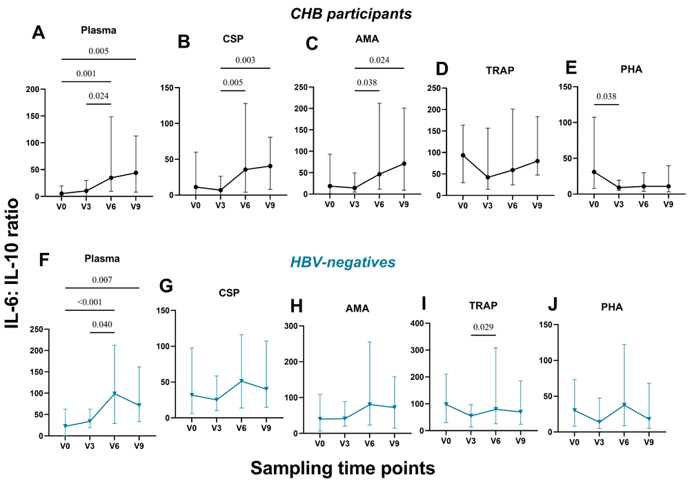
Figure 10. IL-6/IL-10 ratios.
IL-6/IL-10 ratios were computed and compared among chronic HBV (black) and HBV-negative (sea blue) individuals.
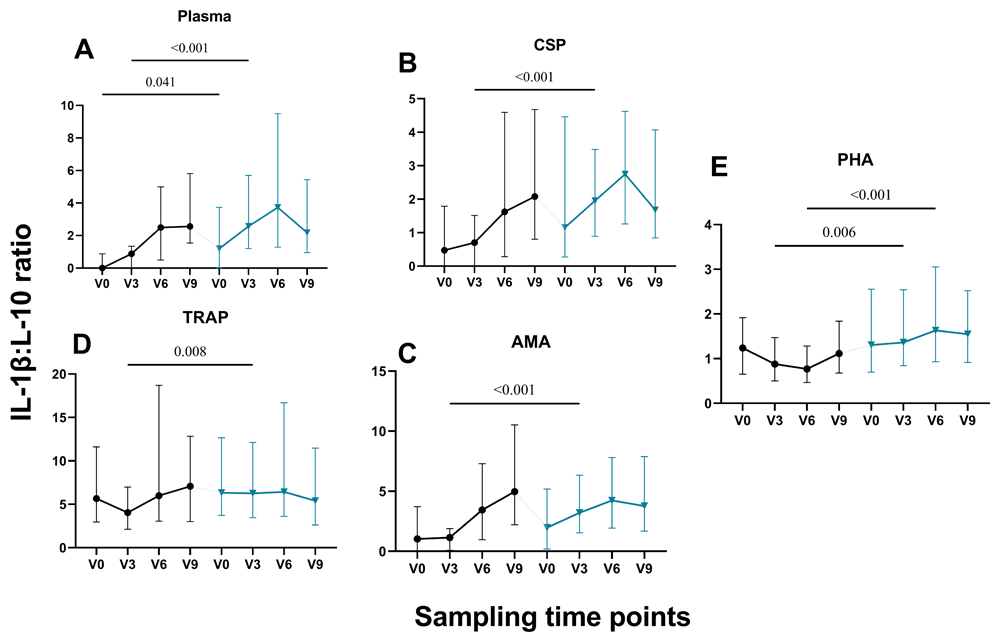
Figure 11. IL-1β/IL-10 ratios.
IL-1β /IL-10 ratios were computed and compared between chronic HBV (black) and HBV-negative (sea blue) individuals.
Discussion
Cytokines play vital roles in the regulation of HBV replication and robust responses promote the clearance of the virus in the acute stage31. However, cytokines have been implicated in the pathophysiology of HBV, providing evidence for the use of recombinant forms of some cytokines for treatment15,17. From our results, chronically infected HBV participants produced similar levels of IL-6 and IL-10. However, IL-6 levels was the highest of the expressed cytokines (Figure 1A, D & F). This observation may be as a result of the many functions of IL-6 in the human body32–34 requiring its constant production. The IL-6 levels may also account for the ability of the CHB group to control the HBV levels below the target levels for antiviral therapy. Additionally, similar levels of IL-1β, and TNF-α were produced by both groups (Figure 2B & C). Similar findings were reported by Ribeiro, et al.35 however, IL-6 levels were significantly different between chronically infected HBV participants (not on antiviral therapy) and healthy controls. This finding supports the practice at the liver clinics where the caregivers exclude these individuals from antiviral therapy as they exhibited similar immune functionality as controls as they may have the ability to control the virus.
To determine how early T cell exhaustion state establishes in CHB, we measured cytokines produced in response to whole blood stimulation every 3 month over a year. This was used as exhausted T cells will not be able to express cytokines in response to antigen stimulation. We found that our cases and controls showed similar ability in the production of TNF-α, IFN-α, IFN-γ, IL-17A, IL-1β, IL-6, IL-2, and IL-10 following stimulation with malaria antigens (Figure 2–Figure 4). TRAP induced the highest level of TNF-α, IL-1β, IL-6, and IL-10 suggesting that a TRAP-based vaccine may work better in these individuals (Figure 4). Additionally, when TRAP-induced cytokines levels were compared to those of unstimulated plasma, CSP and AMA-1 samples, both groups expessed higher levels of IL-1β (Figure 5A–G), IL-6 (Figure 6A–G), IL-10 (Figure 7A–C, E–F), and TNF-α (Figure 8B, C, E, F &G). This suggests that the cells from these chronically infected HBV participants are exhibiting similar immunological characteristics as those of HBV-negative individuals. These results are similar to that of Kennedy, et al.36, where they also used the expression of cytokines as an outcome measure for T cell function using HBV peptides. Their results showed that immune-tolerant CHB participants had similar ability to express cytokines as healthy controls.
Increasing levels of TNF-α, IL-1β, IL-6, and IL-10 were observed in both groups across the time points and in both unstimulated and stimulated samples. This may be due to the exposure of the participants to infectious bites during the study (Figure 12). We also determine the regulation of immune response among the two groups by computing pro-inflammatory and T-regulatory cytokine ratios (L-6:IL-10, TNF-α:IL-10, and IL-1β:IL-10). Our results show a balance in pro-inflammatory and T-regulatory cytokine ratios as there was no significant difference between both groups at all time points. Of note, increasing IL-1β:IL-10 ratios were observed across the four time points among CHB group, however, HBV-negatives recorded significantly higher IL-1β:IL-10 ratios compared to CHB-positives at visit 1 and 3 (Figure 9 & Figure 11).
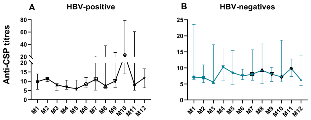
Figure 12. Anti-CSP titres among CHB and HBV-negatives over a twelve-month period.
Conclusion
A limitation to this study was that samples were obtained from CHB in the early phase of chronicity only and did not include CHB individuals with advanced disease, who are treatment-naïve and those on antiviral therapy, for comparisons and deductions to be made. This could have given as the opportunity to understand how advance CHB influences immune responses to P. falciparum and probably malaria vaccines. However, this study showed that there is a difference immune responses and clinical presentation between individuals with advanced CHB and those in early chronicity. There is no impairment in the ability to induce the needed pro-inflammatory response against Plasmodium falciparum among these individuals. These findings suggest that T cell exhaustion may not set in at the early stages of chronicity in CHB.
Ethics and consent statement
Ethical clearance was obtained from the Institutional Review Board (IRB) of the Noguchi Memorial Institute for Medical Research and the Scientific and Technical Committee (STC) of Noguchi Memorial Institute for Medical Research (NMIMR) on the 8th of January, 2020 and 13th of December, 2019 respectively The ethics and protocol review committee of the Korle-Bu Teaching Hospital (KBTH) also gave approval on the 9th of April, 2020. The NMIMR-IRB has a United States Government Federal-wide Assurance (FWAA00001824) from the Office for Human Research Protections and has approved thousands of biomedical research protocols. The approval numbers, FWAA00001824(NMIMR-IRB/046/19-20) and KBTH-IRB/00024/2020) were given by the above institutions respectively. Before recruitment, trained study personnel thoroughly explained the purpose of the study to potential participants and informed them about their rights as participants in the study. Participants completed and signed informed consent forms after the consenting process. The principles outlined in the Declaration of Helsinki were adhered to in every activity carried out in this study.
Data availability statement
The authors confirm that the data supporting the findings of this study have been deposited at the Open Science Framework’s website. Open Science Framework: HEPMAL Study. DOI: https://doi.org/10.17605/OSF.IO/YQJ5T37
This project contains the following underlying data:
• The concentrations of 13 cytokines (IFN-γ, IFN-α, IFN-β, TNF-α, IL-1β, IL-2, IL-6, IL12p70, IL-4, IL-13, IL-21, IL-17A, and IL-10) in pg/ml, age and sex.xlsx. (The dataset.)
Data are available under the terms of the Creative Commons Zero “No rights reserved” data waiver (CC0 1.0 Public domain dedication).
Faculty Opinions recommendedReferences
- 1.
WHO: Hepatitis B. 2021. Reference Source
- 2.
WHO: Global hepatitis report 2017. World Health Organization, 2017. Reference Source
- 3.
Abesig J, Chen Y, Wang H, et al.: Prevalence of viral hepatitis B in Ghana between 2015 and 2019: a systematic review and meta-analysis.
PLoS One.
2020; 15(6): e0234348. PubMed Abstract
| Publisher Full Text
| Free Full Text
- 4.
Sharpe AH, Wherry EJ, Ahmed R, et al.: The function of programmed cell death 1 and its ligands in regulating autoimmunity and infection.
Nat Immunol.
2007; 8(3): 239–245. PubMed Abstract
| Publisher Full Text
- 5.
Boni C, Fisicaro P, Valdatta C, et al.: Characterization of Hepatitis B Virus (HBV)-specific T-cell dysfunction in chronic HBV infection.
J Virol.
2007; 81(8): 4215–4225. PubMed Abstract
| Publisher Full Text
| Free Full Text
- 6.
Ye B, Liu X, Li X, et al.: T-cell exhaustion in chronic hepatitis B infection: current knowledge and clinical significance.
Cell Death Dis.
2015; 6(3): e1694. PubMed Abstract
| Publisher Full Text
| Free Full Text
- 7.
Salimzadeh L, Le Bert N, Dutertre CA, et al.: PD-1 blockade partially recovers dysfunctional virus–specific B cells in chronic hepatitis B infection.
J Clin Invest.
2018; 128(10): 4573–4587. PubMed Abstract
| Publisher Full Text
| Free Full Text
- 8.
Wang H, Luo H, Wan X, et al.: TNF-α/IFN-γ profile of HBV-specific CD4 T cells is associated with liver damage and viral clearance in chronic HBV infection.
J Hepatol.
2020; 72(1): 45–56. PubMed Abstract
| Publisher Full Text
- 9.
Wherry EJ: T cell exhaustion.
Nat Immunol.
2011; 12(6): 492–499. PubMed Abstract
| Publisher Full Text
- 10.
Wherry EJ, Kurachi M: Molecular and cellular insights into T cell exhaustion.
Nat Rev Immunol.
2015; 15(8): 486–499. PubMed Abstract
| Publisher Full Text
| Free Full Text
- 11.
Belloni L, Allweiss L, Guerrieri F, et al.: IFN-α inhibits HBV transcription and replication in cell culture and in humanized mice by targeting the epigenetic regulation of the nuclear cccDNA minichromosome.
J Clin Invest.
2012; 122(2): 529–537. PubMed Abstract
| Publisher Full Text
| Free Full Text
- 12.
McNab F, Mayer-Barber K, Sher A, et al.: Type I interferons in infectious disease.
Nat Rev Immunol.
2015; 15(2): 87–103. PubMed Abstract
| Publisher Full Text
| Free Full Text
- 13.
Hyodo N, Nakamura I, Imawari M: Hepatitis B core antigen stimulates interleukin-10 secretion by both T cells and monocytes from peripheral blood of patients with chronic Hepatitis B Virus infection.
Clin Exp Immunol.
2004; 135(3): 462–466. PubMed Abstract
| Publisher Full Text
| Free Full Text
- 14.
Stoop JN, van der Molen RG, Baan CC, et al.: Regulatory T cells contribute to the impaired immune response in patients with chronic Hepatitis B Virus infection.
Hepatology.
2005; 41(4): 771–778. PubMed Abstract
| Publisher Full Text
- 15.
Zhong S, Zhang T, Tang L, et al.: Cytokines and chemokines in HBV infection.
Front Mol Biosci.
2021; 8: 805625. PubMed Abstract
| Publisher Full Text
| Free Full Text
- 16.
Harker JA, Lewis GM, Mack L, et al.: Late interleukin-6 escalates T follicular helper cell responses and controls a chronic viral infection.
Science.
2011; 334(6057): 825–829. PubMed Abstract
| Publisher Full Text
| Free Full Text
- 17.
de Almeida Ribeiro CR, Martinelli KG, da Motta de Mello V, et al.: Cytokine, genotype, and viral load profile in the acute and chronic hepatitis B.
Viral Immunol.
2020; 33(10): 620–627. PubMed Abstract
| Publisher Full Text
- 18.
He Y, Hwang S, Ahmed YA, et al.: Immunopathobiology and therapeutic targets related to cytokines in liver diseases.
Cell Mol Immunol.
2021; 18(1): 18–37. PubMed Abstract
| Publisher Full Text
| Free Full Text
- 19.
Bouezzedine F, Fardel O, Gripon P: Interleukin 6 inhibits HBV entry through NTCP down regulation.
Virology.
2015; 481: 34–42. PubMed Abstract
| Publisher Full Text
- 20.
Park Y, Park JY, Han KH, et al.: Serum cytokine levels in chronic hepatitis B patients receiving peginterferon alpha-2a therapy.
Hepatobiliary Pancreat Dis Int.
2012; 11(5): 499–506. PubMed Abstract
| Publisher Full Text
- 21.
Huang L, Zhou L, Gan J, et al.: IL-21 is associated with virological relapse of HBeAg positive chronic hepatitis B after discontinuance of entecavir.
Turk J Gastroenterol.
2021; 32(2): 178–186. PubMed Abstract
| Publisher Full Text
| Free Full Text
- 22.
Yan T, He Y, Li Y, et al.: With cytometric bead assay, the interleukin-10/HBV DNA ratio is an early predictor for response to interferon-α treatment in chronic hepatitis B.
J Interferon Cytokine Res.
2015; 35(10): 779–784. PubMed Abstract
| Publisher Full Text
- 23.
Li MH, Lu HH, Chen QQ, et al.: Changes in the cytokine profiles of patients with chronic hepatitis B during antiviral therapy.
Biomed Environ Sci.
2021; 34(6): 443–453. PubMed Abstract
| Publisher Full Text
- 24.
Wang G, Liu Y, Huang R, et al.: Characteristics of regulatory B cells in patients with chronic Hepatitis B Virus infection in different immune phases.
Discov Med.
2017; 23(128): 295–304. PubMed Abstract
- 25.
Tian ZJ, Shen Y, Li XR, et al.: Increased interleukin-32, interleukin-1, and interferon-γ levels in serum from hepatitis B patients and in HBV-stimulated peripheral blood mononuclear cells from healthy volunteers.
J Infect Public Health.
2019; 12(1): 7–12. PubMed Abstract
| Publisher Full Text
- 26.
Wu JF, Wu TC, Chen CH, et al.:
Serum levels of interleukin-10 and interleukin-12 predict early, spontaneous Hepatitis B Virus e Antigen seroconversion.
Gastroenterology.
2010; 138(1): 165–172.e163. PubMed Abstract
| Publisher Full Text
- 27.
WHO: World malaria report 2023. Geneva: World Health Organization, 2023. Reference Source
- 28.
WHO: World malaria report 2022. 2022. Reference Source
- 29.
Segbefia SP, Asandem DA, Pobee A, et al.:
Expression patterns of immune checkpoint proteins and Plasmodium falciparum-induced cytokines in chronic Hepatitis B Virus-infected and uninfected individuals: a cross-sectional study.
Health Sci Rep.
2024; 7(8): e2280. PubMed Abstract
| Publisher Full Text
| Free Full Text
- 30.
Kusi KA, Bosomprah S, Dodoo D, et al.:
Anti-sporozoite antibodies as alternative markers for malaria transmission intensity estimation.
Malar J.
2014; 13: 103. PubMed Abstract
| Publisher Full Text
| Free Full Text
- 31.
Xia Y, Protzer U:
Control of Hepatitis B Virus by cytokines.
Viruses.
2017; 9(1): 18. PubMed Abstract
| Publisher Full Text
| Free Full Text
- 32.
Tanaka T, Narazaki M, Kishimoto T:
IL-6 in inflammation, immunity, and disease.
Cold Spring Harb Perspect Biol.
2014; 6(10): a016295. PubMed Abstract
| Publisher Full Text
| Free Full Text
- 33.
Sebina I, Fogg LG, James KR, et al.:
IL-6 promotes CD4+ T-cell and B-cell activation during Plasmodium infection.
Parasite Immunol.
2017; 39(10): e12455. PubMed Abstract
| Publisher Full Text
- 34.
Wilairatana P, Mala W, De Jesus Milanez G, et al.:
Increased interleukin-6 levels associated with malaria infection and disease severity: a systematic review and meta-analysis.
Sci Rep.
2022; 12(1): 1–24, 5982. PubMed Abstract
| Publisher Full Text
| Free Full Text
- 35.
de Almeida Ribeiro CR, Beghini DG, Lemos AS, et al.:
Cytokines profile in patients with acute and chronic hepatitis B infection.
Microbiol Immunol.
2022; 66(1): 31–39. PubMed Abstract
| Publisher Full Text
- 36.
Kennedy PT, Sandalova E, Jo J, et al.:
Preserved T-cell function in children and young adults with immune-tolerant chronic hepatitis B.
Gastroenterology.
2012; 143(3): 637–645. PubMed Abstract
| Publisher Full Text
- 37.
Segbefia SP, Bonney K, Amoah LE, et al.:
Ex-vivo evaluation of Plasmodium falciparum-induced cytokine expression in chronic Hepatitis B Virus-infected and uninfected individuals. [dataset], 2024. http://www.doi.org/10.17605/OSF.IO/YQJ5T