Introduction
Environmental DNA (eDNA) has emerged as a revolutionary tool for marine biodiversity monitoring, offering a valuable complement to traditional methods based on visual surveys or species capture1. eDNA-based approaches often demonstrate superior sensitivity, particularly for detecting species that are rare, cryptic, small, nocturnal, or otherwise elusive, which are frequently missed by conventional techniques2. A significant bottleneck in current methodologies is the time-consuming water filtration process, typically restricted to surface waters due to the logistical challenges of sampling at depth. This limitation may result in the loss of valuable information on benthic biodiversity. While technological advances, such as submersible pumps and robotic sampling devices3,4, are being developed to enable deeper sampling, they often remain prohibitively expensive. Additionally, for divers using manual underwater pumps, filtration time is constrained by bottom time and decompression limits.
A promising, low-tech alternative has been proposed by using marine sponges as natural eDNA samplers5. These benthic organisms filter large volumes of water daily, possess a rapid regeneration capacity, and have been successfully applied for biodiversity assessments in diverse ecosystems, including the North Atlantic6, Arctic7, Southern Ocean8,9, and the Indo-Pacific10. These studies have shown that different sponge species exhibit varying eDNA yields, which have been attributed to differences in metabolism, pumping rates, and associated microbial communities. For instance, sponges are categorized into low microbial abundance (LMA) and high microbial abundance (HMA)11, a factor influencing eDNA recovery with LMA species showing superior recovery rates7,12. Identifying the species that maximize eDNA recovery under standardized conditions is thus essential before implementing large-scale monitoring campaigns. Such comparisons, to our knowledge, have only been conducted in controlled aquaria experiments12, as eDNA recovery in natural settings is subject to additional confounding factors due to actual differences in fish occupancy and environmental parameters at the time of sampling.
In the Mediterranean Sea, despite the seminal study using opportunistic samples from two local sponge species5, systematic assessments of their eDNA sampling capacity are missing. The Northwestern Mediterranean façade is particularly rich in sponges, with steep walls and coralligenous habitats providing optimal conditions for sponge growth13. This region also hosts several marine protected areas (MPAs)14, where ongoing biomonitoring efforts could greatly benefit from novel eDNA-based approaches for biodiversity assessment. Here, we evaluated seven cosmopolitan Mediterranean sponge species thriving at a single site (coralligenous wall) to compare their effectiveness for eDNA recovery. By sampling 19 specimens under uniform environmental conditions and using a standardized processing protocol, we identified marked differences in eDNA recovery across species. Our results highlight Axinella verrucosa as the most promising candidate for future eDNA-based biodiversity monitoring efforts. Conversely, our findings also suggest certain species may be less effective as natural samplers, underscoring the importance of species selection in sponge-based eDNA studies.
Methods
Field work
Sampling was conducted on May 14, 2024, at the southern tip of Frioul Island, Marseille (Cap Caveau; 43° 15.614'N / 5° 17.360'E) between 10 and 11 a.m. Three specimens were sampled for each sponge species, except for Aplysina cavernicola and Axinella verrucosa, for which only two individuals were found at the site. Biopsies were excised using a blunt-ended diving knife at a depth of 10 meters, in an area characterized by a rocky vertical wall colonized by benthic organisms, descending to a sandy plain at 14 meters (Figure 1). Water temperature at the time of sampling was 17°C. Samples of the same species were stored in Ziploc bags within a diving mesh-bag and were transferred upon surfacing to a cooler with seawater for transport. Within 1–1.5 hours, excess water was removed, and biopsies were dried on paper tissue before being transferred into 50 mL falcon tubes with absolute ethanol. Samples were stored at -20°C until further processing.
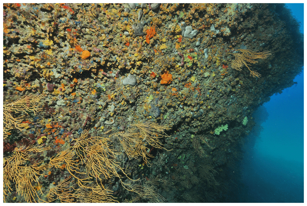
Figure 1. Sampling site.
Underwater photography of the coralligenous wall where sponge specimens were sampled.
Sample processing
Sponge tissues (0.5 cm3) were blotted dry with paper tissue, as outlined in Harper et al. (2023)15, before being chopped with a sterile scalpel on a petri dish. DNA extraction followed a modified Qiagen Blood & Tissue Kit protocol16. Briefly, sponge fragments were placed in 2 mL Eppendorf tubes and mixed with 720 µL Buffer ATL and 80 µL Proteinase K before incubation at 56°C overnight. An extraction control without starting material was included to detect potential contaminations. The next day, lysates (600 µL) were transferred to new tubes, combined sequentially with 600 µL Buffer AL and 600 µL 100% ethanol, and loaded onto DNeasy Mini spin columns in 2 mL collection tubes. After centrifugation at 6,000 × g for 1 min, the flowthroughs were discarded, and the process was repeated until all lysates had passed through the columns. The membranes were washed with 500 µL Buffer AW1 and centrifuged at 6,000 × g for 1 min, followed by a second wash with 500 µL Buffer AW2 and centrifugation at 20,000 × g for 3 min. Residual wash buffer was removed by an additional centrifugation at 20,000 × g for 1 min. DNA was eluted by adding 200 µL of preheated (56°C) AE buffer to the membranes, incubating for 1 min at room temperature, and centrifuging at 6,000 × g for 1 min. Following DNA extraction, qPCR was conducted using Fish16S primers17 to assess fish eDNA presence and optimize amplification conditions. This primer set was chosen because it was recently shown to recover a greater diversity of fish species than other primer pairs in a Mediterranean site18. The qPCR mix consisted of 10 µL of SsoAdvanced™ Universal SYBR® Green Supermix (BioRad), 2 µL of primers (5 µM each), 0.16 µL of BSA (Fisher), and 2 µL of DNA extract, with molecular-grade water making up a final volume of 20 µL. qPCRs were run on a CFX96 Connect Real-Time PCR Detection system (BioRad), the thermal cycling protocol included 10 minutes at 95°C, followed by 60 cycles of 30 seconds at 95°C, 30 seconds at 55°C, and 1 minute at 72°C. Samples were tested pure and diluted 10-fold and 100-fold. The 100-fold dilution performed best for all samples and was used for subsequent library preparation. Results indicated higher Fish16S detection per input DNA in the species Clathrina clathrus and Agelas oroides, which were therefore pinpointed as the best candidates and sequenced individually. The remaining samples were merged per species and sequenced as a pool. All samples were standardized to a concentration of 2.5 ng/µL to ensure uniform input for amplification. Eight technical replicates per sample were amplified using Fish16S primers tagged with unique 8-base identifiers, to allow sample dereplication during data processing. PCRs were run on a CFX96 Connect Real-Time PCR Detection system (BioRad), the reaction mix consisted of 10μL of Amplitaq Gold 360 mix (Fisher), 0.16μL of BSA (Fisher), 2μL of the primer F&R mix at 5μM each, 2μL of DNA extract and molecular grade water for a final volume of 20μL. The thermal cycling protocol included 10min at 95°C, followed by 48 cycles of 30s at 95°C, 30s at 55°C, 1min at 72°C, and a final elongation step of 7min at 72°C. Samples were purified with the MinElute kit (Qiagen), and products were controlled on a 2% agarose gel (E-Gel Power Snap®, Invitrogen). The library was then constructed and sequenced by FASTERIS (Geneva, Switzerland). It was prepared with the Metafast protocol and sequencing was performed on an Illumina MiSeq platform (2 × 150 bp).
Bioinformatic analyses
The OBITools suite19 and SumaClust20 were used for sequence processing and taxonomic assignment. Paired-end reads were merged (illuminapairedend), quality-filtered (obigrep), and dereplicated (obiuniq). Sequences were clustered at 97% similarity (SumaClust), with the most abundant sequence in each cluster retained as the representative cluster center. Clusters containing at least 10 reads in at least one replicate were selected for downstream analysis. Taxonomic assignment was performed with ecotag against a reference database constructed from GenBank entries using in silico PCR (ecoPCR) and filtered to retain sequences classified at the family level or higher (obigrep). Further filtering steps using the R package metabaR21 removed chimeras (best identity <95%), contaminants (contaslayer), low-abundance artefacts (<3% relative frequency, tagjumpslayer), and replicates with sequencing coverage below 100 reads. The remaining PCR replicates were aggregated by sample, and MOTUs observed fewer than 10 times per sample were excluded. The MOTU list was then manually cured and verified with BLASTn, and MOTUs Fish16S_00009 and Fish16S_00517 assigned only to genus level were renamed as Diplodus sp1 and Diplodus sp2, respectively.
Discussion
We observed marked differences in eDNA recovery across sponge species, with Axinella verrucosa outperforming the rest by recovering five of the six detected fish species, thus emerging as the most promising candidate for biomonitoring applications (Figure 2). Notably, it was the only tested species with a standing 3D structure protruding into the water column, a feature that may enhance eDNA capture. With previous studies arguing both in favor7 and against10 a correlation between sponge morphology and eDNA recovery, this aspect warrants further investigation. The World Porifera Database lists 100 accepted Axinella species, it will be valuable to determine whether others in this genus share similar eDNA retention capabilities. A compelling candidate is A. polypoides, an LMA species with a standing 3D structure similar to A. verrucosa., though its protected status necessitates careful handling. Our findings also align with previous work showing that LMA sponges recover more eDNA than HMA sponges7,12, reinforcing the need to focus on LMA species in future studies. Including other LMA species such as Crambe crambe – not included in this study but known for its antimicrobial compounds22,23 and possessing a flat body - will help disentangle the influence of microbial abundance and morphology on eDNA uptake.
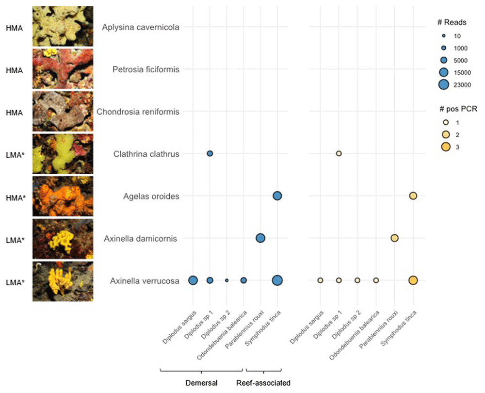
Figure 2. Metabarcoding results.
Bubble plot depicting the number of Fish16S reads in blue (#Reads) and positive PCR replicates in orange (#pos PCR). Tested sponge species are depicted on the Y-axis with a photography of a representative specimen and their HMA/LMA status (retrieved from refs 11,27. The * mark indicates a predicted status). Detected fish species are indicated on the X-axis with their corresponding habitat (retrieved from FishBase).
The detected fish community was composed of demersal and reef-associated species, highlighting the relevance of sponges as tools for benthic biodiversity monitoring. This is all the most relevant given recent findings suggesting that water eDNA is a poor proxy for benthic biodiversity24, emphasizing the potential of sponges for capturing eDNA from cryptic and substrate-associated taxa. Such ability makes them particularly valuable for monitoring habitats that are difficult to access through conventional methods, such as underwater caves, crevices, and vertical walls. Moreover, while we primarily detected bottom-dwelling species, pelagic fish may be recovered upon further methodological improvements. This is supported by Turon et al. (2020)10 who found no significant detection bias between pelagic and demersal fish when using sponge eDNA.
The overall number of detected fish species was low, and we identify several factors that may have contributed to this outcome. The sampling site lacked structural complexity and no visible fish schools were present during collection, potentially limiting the availability of eDNA in the water column. Additionally, environmental conditions at the time of sampling -such as water temperature- may have influenced DNA persistence and detection. Future campaigns targeting multiple sites and seasons, including high biodiversity MPA’s core areas, should overcome this issue. We also identify several methodological improvements that could enhance future studies. These include the immediate transfer of sponges to ethanol upon resurfacing to limit eDNA degradation, the use of multiple genetic markers to overcame reference database incompleteness and increase species detection probability18,25, and improved inhibitor removal during DNA extraction to limit sample inhibition26. Finally, a key priority is the completion of reference genetic databases. Expanding the current dataset is crucial for accurate species-level assignments, particularly for commercially important taxa such as Diplodus, given its importance for fisheries management and conservation.
Overall, A. verrucosa stands out as a promising candidate for future eDNA sampling campaigns, yet protocol optimization and further testing of other LMA sponges is warranted. As eDNA-based monitoring advances, sponge sampling offers a valuable complement to water eDNA surveys, especially for benthic communities and habitats where conventional sampling is challenging. Our results arise from a preliminary pilot study for a larger monitoring scheme, yet we share them promptly to benefit MPA practitioners and inform emerging biodiversity monitoring initiatives amid growing national and international demand.
Data availability statement
Zenodo, Evaluation of Mediterranean sponges as natural samplers for environmental DNA (eDNA) - Underlying DATA. https://doi.org/10.5281/zenodo.1489366228.
The project contains the following underlying data:
- [Evaluation of Mediterranean sponges as natural samplers for environmental DNA (eDNA) - Underlying DATA.xls’] (comprehensive information on sample treatment and analysis steps, from DNA extraction to metabarcoding analysis).
Data are available under the terms of the Creative Commons Attribution 4.0 International license (CC-BY 4.0).
Acknowledgments
We are grateful to the OSU-PYTHEAS diving service, the OMICS platform at MIO, and the Biogeochemistry team leaders, for their support and availability.
Faculty Opinions recommendedReferences
- 1.
Miya M:
Environmental DNA metabarcoding: a novel method for biodiversity monitoring of marine fish communities.
Ann Rev Mar Sci.
2022; 14: 161–185. PubMed Abstract
| Publisher Full Text
- 2.
Boulanger E, Loiseau N, Valentini A, et al.:
Environmental DNA metabarcoding reveals and unpacks a biodiversity conservation paradox in Mediterranean marine reserves.
Proc Biol Sci.
2021; 288(1949): 20210112. PubMed Abstract
| Publisher Full Text
| Free Full Text
- 3.
Preston C, Yamahara K, Pargett D, et al.:
Autonomous eDNA collection using an uncrewed surface vessel over a 4200–km transect of the Eastern Pacific Ocean.
Environ DNA.
2024; 6(1): e468. Publisher Full Text
- 4.
Hendricks A, Mackie CM, Luy E, et al.:
Compact and automated eDNA sampler for in situ monitoring of marine environments.
Sci Rep.
2023; 13(1): 5210. PubMed Abstract
| Publisher Full Text
| Free Full Text
- 5.
Mariani S, Baillie C, Colosimo G, et al.:
Sponges as natural environmental DNA samplers.
Curr Biol.
2019; 29(11): R401–R402. PubMed Abstract
| Publisher Full Text
- 6.
Neave EF, Cai W, Arias MB, et al.:
Trapped DNA fragments in marine sponge specimens unveil North Atlantic deep-sea fish diversity.
Proc Biol Sci.
2023; 290(2005): 20230771. PubMed Abstract
| Publisher Full Text
| Free Full Text
- 7.
Brodnicke OB, Meyer HK, Busch K, et al.:
Deep-sea sponge derived environmental DNA analysis reveals demersal fish biodiversity of a remote Arctic ecosystem.
Environ DNA.
2023; 5(6): 1405–1417. Publisher Full Text
- 8.
Jeunen GJ, Lamare M, Devine J, et al.:
Characterizing Antarctic fish assemblages using eDNA obtained from marine sponge bycatch specimens.
Rev Fish Biol Fish.
2024; 34: 221–238. Publisher Full Text
- 9.
Jeunen GJ, Lamare M, Cummings V, et al.:
Unveiling the hidden diversity of marine eukaryotes in the Ross Sea: a comparative analysis of seawater and sponge eDNA surveys.
Environ DNA.
2023; 5(6): 1780–1792. Publisher Full Text
- 10.
Turon M, Angulo-Preckler C, Antich A, et al.:
More than expected from old sponge samples: a natural sampler DNA metabarcoding assessment of marine fish diversity in Nha Trang Bay (Vietnam).
Front Mar Sci.
2020; 7: 605148. Publisher Full Text
- 11.
Moitinho-Silva L, Steinert G, Nielsen S, et al.:
Predicting the HMA-LMA status in marine sponges by machine learning.
Front Microbiol.
2017; 8: 752. PubMed Abstract
| Publisher Full Text
| Free Full Text
- 12.
Cai W, Harper LR, Neave EF, et al.:
Environmental DNA persistence and fish detection in captive sponges.
Mol Ecol Resour.
2022; 22(8): 2956–2966. PubMed Abstract
| Publisher Full Text
| Free Full Text
- 13.
Bertolino M, Cerrano C, Bavestrello G, et al.:
Diversity of Porifera in the Mediterranean coralligenous accretions, with description of a new species.
ZooKeys.
2013; (336): 1–37. PubMed Abstract
| Publisher Full Text
| Free Full Text
- 14.
MedPAN and SPA/RAC:
The 2016 status of marine protected areas in the Mediterranean. By Meola B. and Webster C. Ed SPA/RAC & MedPAN. Tunis 222 pages, 2019. Reference Source
- 15.
Harper LR, Neave EF, Sellers GS, et al.:
Optimized DNA isolation from marine sponges for natural sampler DNA metabarcoding.
Environ DNA.
2023; 5(3): 438–461. Publisher Full Text
- 16.
Jeunen GJ, Cane JS, Ferreira S, et al.:
Assessing the utility of marine filter feeders for environmental DNA (eDNA) biodiversity monitoring.
Mol Ecol Resour.
2023; 23(4): 771–786. PubMed Abstract
| Publisher Full Text
- 17.
Shaw JLA, Clarke LJ, Wedderburn SD, et al.:
Comparison of environmental DNA metabarcoding and conventional fish survey methods in a river system.
Biol Conserv.
2016; 197: 131–138. Publisher Full Text
- 18.
Roblet S, Priouzeau F, Gambini G, et al.:
Primer set evaluation and sampling method assessment for the monitoring of fish communities in the North-western part of the Mediterranean Sea through eDNA metabarcoding.
Environ DNA.
2024; 6(3): e554. Publisher Full Text
- 19.
Boyer F, Mercier C, Bonin A, et al.:
obitools: a unix-inspired software package for DNA metabarcoding.
Mol Ecol Resour.
2016; 16(1): 176–182. PubMed Abstract
| Publisher Full Text
- 20.
Mercier C, Boyer F, Kopylova E, et al.:
SUMATRA and SUMACLUST: fast and exact comparison and clustering of sequences. In: Programs and Abstracts of the SeqBio 2013 Workshop, 2013; 27–29.
- 21.
Zinger L, Lionnet C, Benoiston AS, et al.:
metabaR: an R package for the evaluation and improvement of DNA metabarcoding data quality.
Methods Ecol Evol.
2021; 12(4): 586–592. Publisher Full Text
- 22.
Jares-Erijman EA, Sakai R, Rinehart KL:
Crambescidins: new antiviral and cytotoxic compounds from the sponge Crambe crambe.
J Org Chem.
1991; 56(19): 5712–5715. Publisher Full Text
- 23.
Becerro MA, Lopez NI, Turon X, et al.:
Antimicrobial activity and surface bacterial film in marine sponges.
J Exp Mar Biol Ecol.
1994; 179(2): 195–205. Publisher Full Text
- 24.
Antich A, Palacín C, Cebrian E:
Marine biomonitoring with eDNA: can metabarcoding of water samples cut it as a tool for surveying benthic communities?
Mol Ecol.
2021; 30(13): 3175–3188. PubMed Abstract
| Publisher Full Text
- 25.
Roblet S, Priouzeau F, Gambini G, et al.:
From sight to sequence: Underwater Visual Census vs environmental DNA metabarcoding for the monitoring of taxonomic and functional fish diversity.
Sci Total Environ.
2024; 956: 177250. PubMed Abstract
| Publisher Full Text
- 26.
Dowell R, Craggs J, Head C, et al.:
DNA state influences the uptake and persistence of environmental DNA by marine sponge natural samplers. 2024. Publisher Full Text
- 27.
Gloeckner V, Wehrl M, Moitinho-Silva L, et al.:
The HMA-LMA dichotomy revisited: an electron microscopical survey of 56 sponge species.
Biol Bull.
2014; 227(1): 78–88. PubMed Abstract
| Publisher Full Text
- 28.
Garcia Seyda N:
Evaluation of Mediterranean sponges as natural samplers for environmental DNA (eDNA) - underlying DATA. [Data set]. Zenodo. 2025. http://www.doi.org/10.5281/zenodo.14893663









