Introduction
The increasing prevalence of antibiotic-resistant pathogens poses a major threat to human and animal health, underscoring the urgent need for novel antibiotics and alternative strategies to treat bacterial infections1–3. According to the 2024 WHO report on AMR, 50% of the fatal burden associated with antimicrobial resistance (AMR) is linked to two pathogens: S. aureus and E. coli4. With E. coli classified as a “critical” priority and methicillin-resistant S. aureus (MRSA) as “high” priority5,6, both pathogens remain central targets for antimicrobial research, where the development of both novel antibiotics and adjuvant therapies could help to overcome current treatment limitations.
Adjuvant therapies or “helper-drugs” are used in combination with antibiotics to enhance their efficacy. These compounds have typically limited or no antimicrobial activity on their own, but when used together with an antibiotic, they can significantly increase its ability to kill or inhibit bacterial growth7. An ideal helper compound can penetrate the bacterial cell envelope, has a low tendency for resistance development, and specifically targets bacterial components without harming the host3,8. This approach is especially valuable for challenging pathogens, such as Gram-negative bacteria, which possess a double membrane structure with embedded efflux pumps, explaining the low rate of novel antimicrobials for this group compared to Gram-positive bacteria9,10.
The class of aminoglycosides exhibits broad-spectrum activity against Gram-positive and Gram-negative bacteria. These antibiotics bind to the 16S subunit of the bacterial ribosome, causing error-prone protein biosynthesis11–13. Aminoglycosides, such as amikacin, gentamicin (GEN), and tobramycin, are used to treat life-threatening infections caused by multi-drug-resistant Gram-negative bacteria13,14. Additionally, streptomycin (STREP) and neomycin (NEO) are often the drugs of choice for treating infections caused by Yersinia pestis and Francisella tularensis, as well as E. coli-related infections in animals, for example post weaning diarrhea in pigs13,15–17.
Staphylococcus aureus, particularly MRSA, is a versatile pathogen capable of causing a wide range of infections, from skin infections to life-threatening conditions such as pneumonia and sepsis18. MRSA strains, due to their resistance to multiple antibiotics, present significant challenges in healthcare and community settings19,20. This study focuses on two MRSA strains with distinct genomic profiles: JE2, a highly virulent community-associated MRSA (CA-MRSA) strain21, and ST398, a livestock-associated MRSA (LA-MRSA) strain associated with zoonotic infections22.
To identify novel antimicrobial and helper-compounds, phenotypic screens can be used23,24. Whole-cell screening, first established in the 1920s by S. Waksman and his students, involved screening soil-dwelling Actinomycetes for antibacterials which led to the identification of antimicrobial compounds including streptothricin and STREP3,25,26. Whole cell screening approaches have the advantage that compounds, which are not able to reach their target in a sufficient concentration, are eliminated from the beginning, however after the identification of a hit compound, their mechanism of action must be determined23,24,27.
Actinomycetes, known for their prolific production of bioactive compounds, have historically been a rich source of antibiotics and remain promising for discovering novel antimicrobial agents and adjuvant compounds28. In the current study, we screened a library of 75 known antimicrobials and 880 extracts from Actinomycetes. To resensitize aminoglycoside-resistant E. coli to this class of antibiotics, we developed a helper-drug screen to identify compounds that enhance aminoglycoside activity. For MRSA strains, we screened a library of 75 known compounds and 160 Actinomycetes extracts for novel antimicrobials, followed by deconvolution of active molecules from promising hits.
Methods
Preparation of antimicrobial compound and extract plates
Two types of screening plates were prepared, a compound plate and extract plates from Actinomycetes. The antimicrobial compound plate contained 75 compounds with known antimicrobial activity. Compounds and their corresponding solvents appear in Table S1 and S2. One or two hundred μL of solved compounds (concentration 100 mg/L) were pipetted into a 96-well plate which was dried overnight using an oven. Until use, the dried compounds were stored at 4 °C and immediately before use, 100 μL of 10% DMSO was added, the compounds were resuspended and incubated for 30 min at room temperature. For compound testing, a final concentration of 20 mg/L was used for E. coli and 10 mg/L for MRSA.
For extract testing, plates with dried extracts from Actinomycetes strains from Naicons microbial collection29 were utilized. Full extracts were prepared from cultures (2 mL) by adding ethanol (4 mL) and shaking for 1 h at 30°C followed by centrifugation at 4000 rpm for 8 minutes. Fractionated extracts were obtained both from mycelium and cleared broth after centrifugation of 8 mL cultures at 4000 rpm for 8 minutes. Three mycelium extracts were obtained with 20%, 50%, and 100% ethanol (4 mL each, 1h shaking at 30°C followed by centrifuge at 4000 rpm for 8 minutes). Cleared broth was treated with HP20 (0,8 mL, shaking for 1h at 30°C). After centrifugation (8000 rpm 1 minute) the resin was extracted with increasing concentration of ethanol (5%, 30%, and 80%, shaking for 15 minutes at 30°C followed by centrifugation at 8000 rpm for 1 minute). All the extracts were dried overnight at room temperature and stored at 4°C. The extracts (originally 100 μL extract) were resuspended before use in 100 μL of 10% DMSO. Five to 20 μL of these Actinomycetes extracts were used for activity testing.
Bacterial strains and cultivation of bacterial strains
Four strains of E. coli30 were utilized in this study. The parent strain was E. coli K12 MG165531, and in addition, strains, which were resistant to selected aminoglycoside antibiotics due to the presence of plasmid encoding resistance to NEO (aph(3’)-Ia gene), GEN (aac(3)-IV gene) or STREP (strAB gene) were used (Table S3). Strains were grown in Luria Broth (LB) Lennox with shaking or on LB agar plates overnight at 37 °C. When appropriate, the media was supplemented with antibiotics including 50 mg/L NEO, 50 mg/L STREP, 20 mg/L GEN or 50 mg/L Chloramphenicol (CHL) (Sigma-Aldrich, Copenhagen, Denmark).
MRSA strains consisted of JE221 and ST39822. These were grown in Tryptic Soy Broth (TSB) with shaking overnight at 37 °C. When appropriate, 1 mL of the culture was added to 10 mL of Mueller-Hinton broth (CA-MHB) cation adjusted, mainly magnesium and calcium. TSB, LB, and MHB used are commercially available from BD (www.bd.com/en-us/).
Testing antimicrobial (helper-) activity of compounds
The minimum inhibitory concentration (MIC) of each antimicrobial compound was determined against both E. coli and MRSA strains using the broth micro-dilution method, following CLSI guidelines32. For the assay, we prepared each well with 40 μL of MHB media, 10 μL of the compound (final concentration: 20 mg/L for E. coli and 10 mg/L for MRSA, Table S1, S2), and 50 μL of bacterial culture (E. coli or MRSA), as shown in (Figure 1A). For testing aminoglycoside helper-compounds, a final concentration of 1/8 MIC of the relevant aminoglycoside (NEO, STREP, or GEN) was added to the MHB, along with 40 μL of MHB media, as shown in (Figure 1B). The bacterial suspensions were prepared by adding 100 μL of aminoglycoside-resistant E. coli/MRSA of an optical density (OD)600 of 0.08-0.1 (corresponding to a McFarland standard of 0.5) to 10 mL MHB media. Afterwards, the 96-well plate was incubated at 37 °C for 24 h followed by an OD measurement using a plate reader.
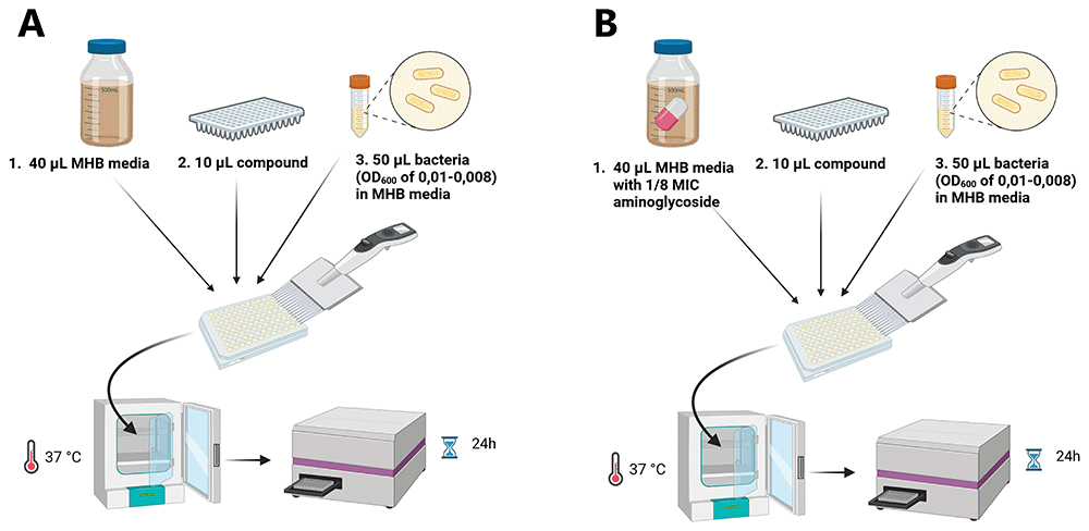
Figure 1.
Schematic representation of the screening approach for antimicrobials (A) and aminoglycoside helper-compounds (B). In a 96-well plate in each well, 40 μL MHB media, 10 μL compound and 50 μL bacterial suspension were mixed, incubated at 37 °C and the OD600 was measured after 24h using a plate reader. This figure was created using BioRender.com.
Screening of extracts and deconvolution of active molecules
A total of 880 extracts derived from Actinomycetes were initially screened against E. coli, while only 160 extracts were tested against MRSA due to the discovery of a hit compound, which warranted proceeding with purification steps.
On the testing day, each extract was prepared by suspending 100 µL in 10% DMSO. Then, 10 µL of each extract was added to wells containing 40 µL of bacterial solution and 50 µL of MHB, as outlined above. Extracts showing 70% or higher inhibition against MRSA compared to growth without the extract (calculations detailed below) were selected for verification, fractionation, and purification to identify the active compound. For E. coli, extracts demonstrating potential helper-drug activity, follow-up experiments were conducted by adding 5–20 μL of the extract, 45–30 μL of MHB, and 50 μL of bacterial suspension to a 96-well plate (final volume: 100 μL). Incubation and OD measurements were performed as described above.
Upon activity confirmation, six fractionated extracts (three from mycelium and three from the cleared broth) were also tested in serial dilutions. At first, the fractions were suspended in 125 µL of 10% DMSO and tested as for the full extracts. Active fractions were further sub-fractionated into 24 fractions (one fraction per minute) using High-Performance Liquid Chromatography (HPLC). We used a Shimadzu HPLC system LC 2010A-HT (Kyoto, Japan) equipped with a Merck LiChrospher RP-18 reverse-phase column (5 μm, I.D. × L 4.6 × 125 mm, Merck, Darmstadt, Germany) at 50°C. The mobile phases consisted of 0.05% formic acid (HCOOH) for phase A and acetonitrile (ACN) for phase B. The flow rate was set at 1 mL/min. Samples were fractionated using a linear gradient: ACN was increased from 10% to 95 in 18 minutes, followed by 5 minutes at 95%. UV detection was carried out at 230 and 270 nm. The 24 subfractions were dried using a speed vacuum concentrator and resuspended later in 10% DMSO for further screening, using the same screening technique as above. Upon activity confirmation, the active fraction was analyzed using high-resolution mass spectrometry (HRMS) to identify the specific compound responsible for the activity. For each active fraction detected, the adjacent inactive fractions (preceding and subsequent) were also analyzed to monitor the elution peak of the active fraction. The analysis was performed using a Vanquish UHPLC system connected to an Orbitrap Exploris 120 mass spectrometer, utilizing a YMC-Triart ODS column. The mobile phase included 0.1% formic acid in water (A), LCMS grade acetonitrile (B), and LCMS grade isopropyl alcohol (C), with a gradient flow program over a 23-minute cycle, as described in 33. The flow rate was set at 0.8 mL/min with a sample injection volume of 8 μL at 40°C. During the separation, 75% of the flow was directed to a diode array detector and 25% to the mass spectrometer, which was equipped with a heated electrospray ionization source operating in both positive and negative modes. Finally, MS/MS and UV-Vis spectra were analyzed and matched against both internal34 and external databases35–38 to identify the active compounds. Finally, each sub-fraction was then suspended in the appropriate volume, and their activity against MRSA was further tested.
Assessing bioactivity of identified molecules
To evaluate the bioactivity of the identified molecules against MRSA strains, we obtained purified elaiophylin (Santa Cruz Biotechnology, Inc.) and nigericin sodium salt (MedChemExpress.com). Each molecule was initially dissolved in DMSO to prepare a 1 mg/mL stock solution. MIC testing and checkerboard assays were then conducted in combination with ampicillin (AMP) to assess potential synergistic effects.
Checkerboard assay
The antimicrobial activity of the molecules in combination a) with AMP against MRSA strains b) with NEO against NEO-resistant E. coli was tested using the method described by Zhu et al.39 with some modifications. Briefly, overnight bacterial cultures were sub-cultured the following day and incubated until they reached the mid-exponential phase, corresponding to OD600 of 0.5–0.6. The bacterial suspension was then diluted to OD600 of 0.1 (equivalent to McFarland 0.5). Following this, 50 µL of the bacterial suspension was added to each well of a 96-well plate, containing 50 µL of MHB and the diluted molecules in combination with AMP or NEO in increasing concentrations. The fractional inhibitory concentration index (FICI) was calculated using the formula: FICI=FICA+FICB, where FICA= MICA in combination / MICA alone and FICB= MICB in combination / MICB alone. The FICI was used to categorize the interaction as synergistic (FICI ≤ 0.5), additive (FICI > 0.5 and ≤ 1.0), indifferent (FICI > 1.0 and ≤ 4.0) or as antagonistic (FICI > 4.0).
Data analysis and growth inhibition calculation
To calculate the growth percentage of the bacteria at a certain condition, the OD600 values obtained from the plate reader were first blank-corrected using the average blank value of the MHB media (without bacteria/compounds), which was recorded in column 12 of the 96-well plate (plate set-up in Figure S1). After blank correction, the average full growth (with and without 1/8 MIC of the corresponding aminoglycoside) of E. coli and MRSA in the absence of validation compounds or bacterial extracts was calculated by averaging the values from column 11. This value represented 100% growth for each pathogen under specific conditions. The percentage of growth for each well was then calculated by dividing the OD600 of each well by the average full growth and multiplying by 100 to determine the growth percentage in the presence of the different antimicrobial compounds or extracts. For E. coli, the percentage of growth was calculated with and without 1/8 MIC of the corresponding aminoglycoside. To assess the helper-compound activity of each extract, the difference between the growth percentages with and without 1/8 MIC of the aminoglycoside was calculated. A higher positive difference indicated more pronounced helper-drug activity. Chemical structures were drawn using ChemDraw Cloud (PerkinElmer Informatics, Waltham, MA, USA; https://chemdraw.perkinelmer.cloud).
Results
Growth of aminoglycoside-resistant E. coli in presence of the corresponding aminoglycoside
The MICs of NEO, STREP and GEN against aminoglycoside-resistant E. coli MG1655 strains were 2000, 1400 and 100 mg/L, respectively. To determine a baseline for further experiment, the average growth of the aminoglycoside-resistant E. coli without and with 1/8 MIC of the corresponding antibiotic was determined (Table 1). 1/8 MIC of the corresponding aminoglycoside inhibited the growth of each aminoglycoside-resistant strains slightly, resulting in a growth of 72 – 90% compared to the growth without aminoglycosides.
Table 1. Growth of aminoglycoside-resistant strains with and without 1/8 MIC of the corresponding antibiotic.
| Aminoglycoside
concentration
(in mg/L) | OD600 control | OD600 with
1/8 MIC
aminoglycoside | Growth with
aminoglycosides
in % (vs. control) |
|---|
| MG1655 aph(3’)-Ia | NEO: 250 | 1,16 (+/- 0,048) | 0,84 (+/- 0,113) | 72 % (+/- 8,2) |
| MG1655 strAB | STREP: 175 | 1,16 (+/- 0,032) | 1,04 (+/- 0,081) | 90 % (+/- 4,8) |
| MG1655 aac(3)-IV | GEN: 12,5 | 1,23 (+/- 0,027) | 0,94 (+/- 0,096) | 77 % (+/- 8,06) |
Assessing known antimicrobial compounds against E. coli and MRSA strains
To assess antimicrobial activity, we screened a library of 75 compounds with known antimicrobial properties against E. coli and MRSA strains (Table S1, S4 and Figure 2). Compounds that inhibited the growth of at least two aminoglycoside-resistant E. coli by more than 90 % included tetracyclines, polymyxins, the aminoglycosides GEN, spectinomycin and apramycin, fluoroquinolones, streptothricin and gargantulide (Figure 2A). Furthermore, purpuromycin, clindamycin, phosphomycin and the aminoglycosides NEO and STREP partly inhibited the growth (>50% inhibition) of at least two of the aminoglycoside-resistant E. coli (Figure 2A). The full list of compounds and the corresponding percentage of growth is listed in Table S1.
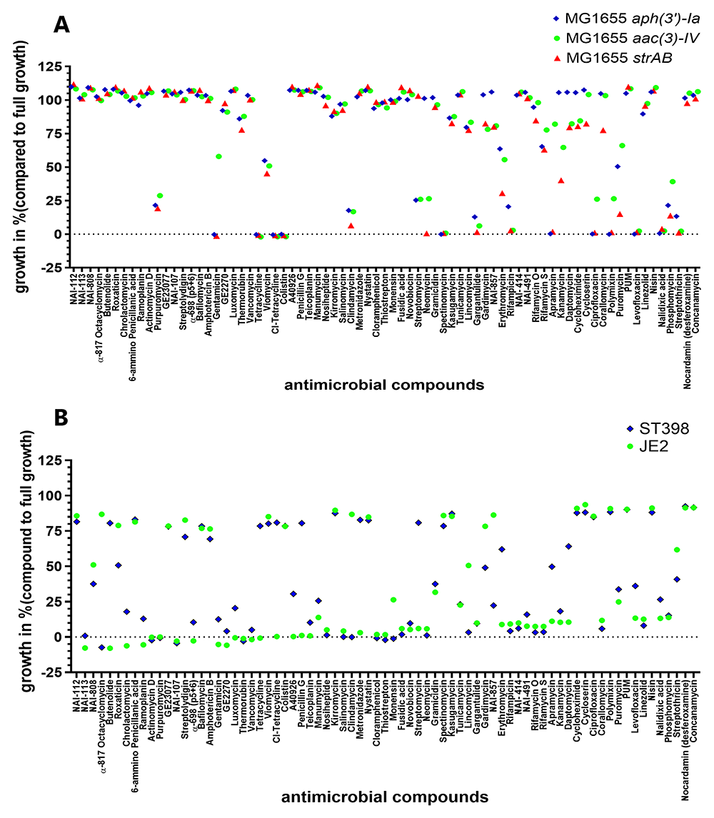
Figure 2. Growth inhibition of 75 compounds with antimicrobial activity on aminoglycoside-resistant E. coli and MRSA.
NEO-resistant E. coli, STREP-resistant E. coli, and GEN-resistant E. coli (A), MRSA ST398 and JE2 (B). The bacterial strains were exposed to 75 different antimicrobial compounds. The antimicrobial compounds were present in a 96-well plate (Table S1 and S4) The average percentage of growth in the presence of the antimicrobial (compared to average growth without antimicrobials) was assessed in four biological replicates, calculated using Excel and depicted with GraphPad Prism 10 (GraphPad Software, San Diego, USA).
When screened against MRSA, most compounds demonstrated similar inhibition profiles for both ST398 and JE2 strains, though some notable differences were observed. As shown in Figure 2B, 25 out of the 75 compounds achieved at least 90% growth inhibition in ST398, while 34 compounds had the same level of inhibition against JE2. However, 12 compounds exhibited significant differential activity (≥50% difference) between the two strains.
The polyethers α-817 octacyclomycin, the lincosamides clindamycin and lincomycin, and NAI-857 were all more effective against ST398 than against JE2. Similarly, tetracycline and chlorotetracycline, butanolide, the b-lactam penicillin G, metronidazole, the aminoglycoside STREP, the macrolide erythromycin, and daptomycin all exhibited stronger activity against JE2 than against ST398. Of note, these effects were not class-specific: for example, streptomycin exhibited much stronger activity against JE2 (~6% growth) compared to ST398 (~80.8% growth), while neomycin showed minimal difference, with around 1.1% growth for ST398 and 5.77% for JE2. Full profiles of each compound against both strains (growth percentage) are presented in Table S4.
Assessing helper-compound activity of compounds with antimicrobial activity for treatment of aminoglycoside-resistant E. coli MG1655
After investigating which compounds inhibited the growth of aminoglycoside-resistant E. coli on their own, we tested which antimicrobials could act as aminoglycoside helper-compounds, i.e., they inhibited the growth of resistant E. coli in the presence of 1/8 MIC of the corresponding aminoglycoside. The difference in growth between the control (with 20 mg/L antimicrobial compounds) and treatment (with 20 mg/L antimicrobial compound and 1/8 MIC of the corresponding aminoglycoside) is presented in Figure 3. A good helper-compound would show a big difference between the two groups, since the inhibition by the compound alone would be small while the inhibition by the compound in combination with 1/8 MIC of the corresponding aminoglycoside would be considerable. This identified two antibiotics that caused an average difference in growth of more than 80% in four biological replicates in the presence of 1/8 MIC of NEO (Figure 3). On their own, rifamycin O (20 mg/L) and thermorubin (20 mg/L) have no antimicrobial activity against NEO-resistant E. coli with an average OD600 of around 1 similar to full growth without the corresponding antimicobials. Combined with 1/8 MIC of NEO, the growth was considerably reduced with an OD600 of 0,02 for rifamycin O and 0,045 for thermorubin (Figure 3). The synergist interaction between NEO and thermorubin (FICI: 0.25-0.375) as well as NEO and rifamycin O (FICI: 0.188-0.375) was confirmed by checkerboard assay. In combination with 1/8 MIC of NEO, 4-8 mg/L thermorubin (the MIC of thermorubin alone was 32-64 mg/L) and 16-32 mg/L rifamycin O (the MIC of rifamycin O alone was 128 mg/L) inhibited the growth of the NEO-resistant E. coli.
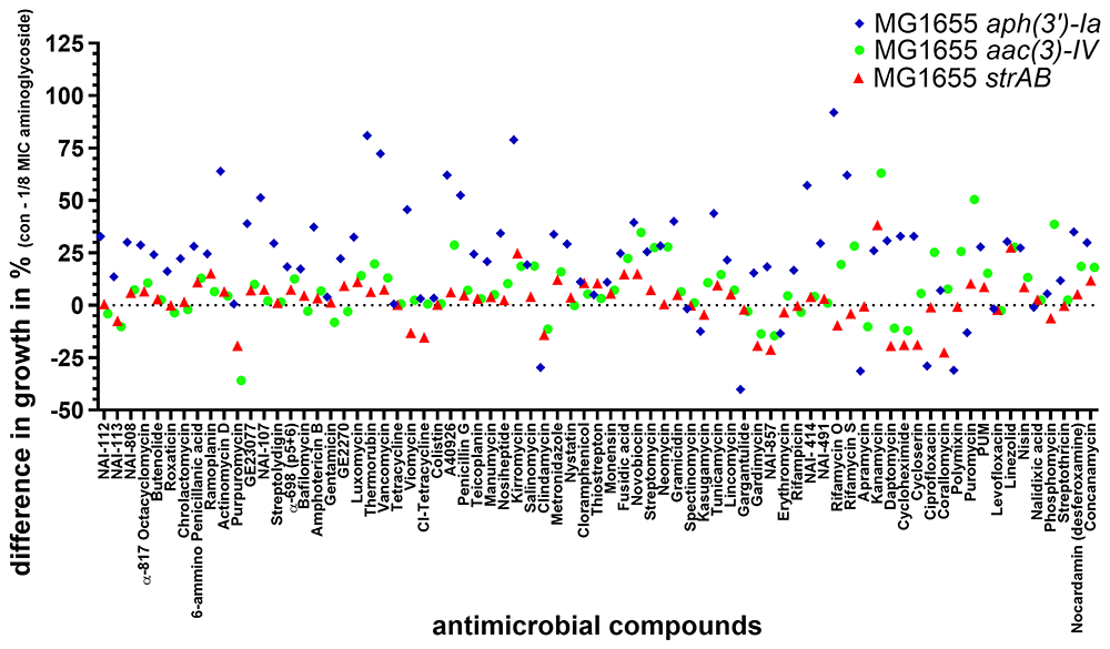
Figure 3. Testing of 75 compounds with known antimicrobial activity for potential aminoglycoside helper-compound activity NEO-resistant (blue), GEN-resistant (green) STREP-resistant (red) MG1655 strains were exposed to 75 different antimicrobial compounds (final concentration: 20 mg/L).
The antimicrobial compounds were present in a 96-well plate (Table S2). The difference in the percentage of growth with and without the aminoglycoside at 1/8 MIC was calculated by Excel (average of 4 biological replicates) and depicted using GraphPad Prism 10 (GraphPad Software, San Diego, USA).
For GEN and STREP, no promising helper-compounds were identified; the helper-drug candidate for GEN and STREP with the highest synergistic activity was the aminoglycoside antibiotic kanamycin (Figure 3) with an average difference of 63% and 38%, compared to growth in the presence of kanamycin alone.
Assessing antimicrobial and helper-compound activity of Actinomycetes extracts
We screened 880 Actinomycetes extracts from the NAICONS library as described previously by Simone et al.40. To identify those with antimicrobial or helper-compound activity against MRSA and GEN-resistant E. coli. Given GEN’s critical role in human health, we prioritized its use for helper-compound analysis. No extract inhibited the growth on their own nor in the presence of 1/8 MIC of GEN by more than 50 % (Figure 4). However, one extract with moderate antimicrobial and one with moderate helper-compound activity were identified. One extract from a Streptomyces strain41, showed a partial growth inhibition with and without 1/8 MIC of GEN (C3 in Figure 4 A, B), indicating that this extract contains antimicrobial compound(s). Mass spectrometry analysis of this extract revealed that molecules belonging to the class of streptothricin were present in this extract41 and streptothricin was shown to inhibit growth of aminoglycoside-resistant E. coli in the screen of compounds with known antimicrobial activity reported above (Figure 4, H3). In addition, one extract from Actinomadura41 showed no general antibacterial activity but helper-drug activity in combination with 1/8 MIC of GEN against the GEN-resistant E. coli (C3 in Figure 4, C, D). A follow-up investigation revealed that this extract showed moderate concentration-dependent GEN helper-compound activity, since this extract on its own did not inhibit growth and the largest effect in growth inhibition was observed with 20 μL of extract (Figure S2). Mass spectrometry analysis for this extract ID 57183 did not show the presence of any known molecule, presenting the need for further experiments.
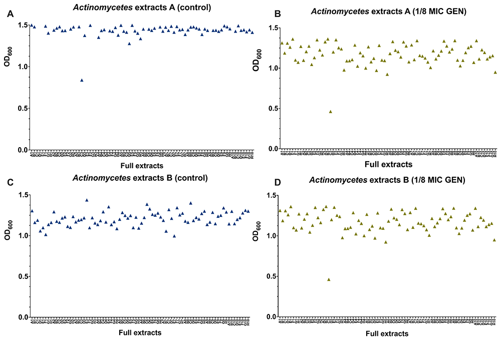
Figure 4. Screening of Actinomycetes extracts for antibacterial and helper-compound activity.
880 Actinomycetes extracts (shown are 160 extracts) were tested and for their antimicrobial activity (A, C) and as helper-compounds in combination with 1/8 MIC of GEN (B, D) against GEN-resistant E. coli by measuring their OD600 and depicted with GraphPad Prism 10 (GraphPad Software, San Diego, USA). The Actinomycetes extracts were present in a 96-well plate and the letter (A–H) combined with the number (1–10) referred to the position of each extract.
Additionally, we tested 80 Streptomyces extracts and 80 extracts from other Actinomycetes genera against ST398 and JE2 MRSA strains (Figures 5A, and 5B respectively). Among the Streptomyces extracts, we found 10 hits that inhibited the growth of both strains by 90%. Further analysis via serial dilution confirmed the antimicrobial efficacy of two extracts, (D6) and (H6), which demonstrated consistent 100% inhibition in at least one or two dilutions. From the other Actinomycetes extracts, we found four hits that inhibited both strains equally, with one (D8) displaying potent antimicrobial activity over three successive dilutions. Based on these findings, extracts D6 (Extract ID 44876), H6 (Extract ID 38424), and D8 (Extract ID 59998) were prioritized for further investigation.
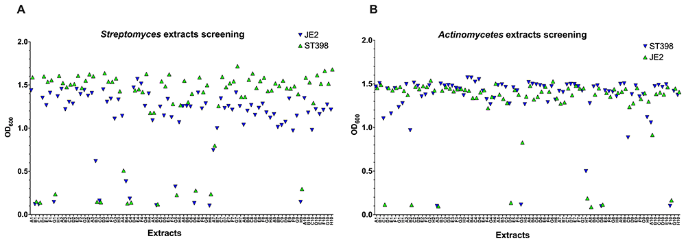
Figure 5. Screening of Actinomycetes extracts for antibacterial activity.
160 Actinomycetes extracts (80 from Actinomycetes rare extracts and 80 from Streptomyces strain) were tested for their antimicrobial activity. 10 hits were found in common from Streptomyces extracts (A) while 4 hits were found in common from Actinomyces rare extracts (B).
To understand better the activity profile of each extract, we generated six fractionated extracts (three from mycelium and three from cleared broth). The (D6) extract showed notable activity in fraction 4 of the cleared broth across four serial dilutions, while (H6) exhibited activity in fraction 5 of the cleared broth against both strains. Likewise, fraction 5 of the (D8) extract displayed strong antimicrobial effects. These active fractions were subsequently analyzed by HPLC, generating sub-fractions for detailed testing against MRSA strains. The results of screening the 72 sub-fractions are shown in Figure 6 for both strains. Among the sub-fractions, D6 (sub-fraction containing A1) inhibited ST398 by 40% and JE2 by 10.4%. H6 fractions 14 and 22 showed high activity, inhibiting ST398 by 96% and JE2 by 65% and 41%, respectively. In contrast, sub-fractions of D8 showed no significant activity.
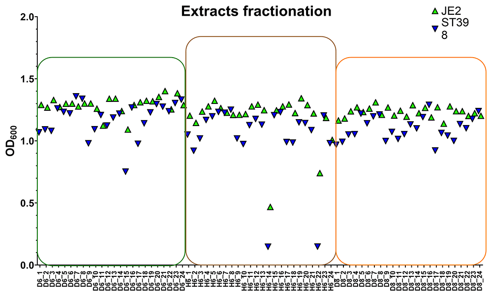
Figure 6. Scatter plots showing the measurement of OD600 of strains ST398 and JE2 exposed to 3 x 24 subfractions of each extract (D6), (H6), and (D8) obtained through fractionation in the HPLC system (each extract was fractioned into 24 fractions).
D6 subfractions are shown in green box, H6 in brown, and D8 in orange.
Identification of compounds by mass spectrometry analysis
After detecting the active fractions with antimicrobial activity, we proceeded with characterization using mass spectrometry. The first fraction analyzed, designated as A1, exhibited signals at 446.3112 in positive ion mode and 444.2968 in negative ion mode in the full scan. Upon fragmentation, two primary fragment ions were detected at m/z 272.1855 and m/z 106.0499. These MS signals did not correspond to any compounds in the ChemNet or MassBANK databases, which contain extensive collections of known chemical structures and mass spectral data. The second fraction, labeled B1, exhibited signals at m/z 742.5098 and m/z 747.4648 in the full scan, corresponding to the [M+NH4]+ and [M+Na]+ adducts, respectively. MS/MS analysis revealed fragmentation patterns consistent with the molecule nigericin, as previously described by Harvey et al.42. The fragmentation data are shown in Figure S3A, and the 2D structure of nigericin is provided in Figure S3B.
For the third fraction, B2, a precursor ion was detected at m/z 1047.5850 in the full scan. MS/MS analysis revealed fragment ions at m/z 729.3819, m/z 711.3713, and m/z 734.3972, which are characteristic of elaiophylin, as previously reported by Boya et al.43. The fragmentation spectra are displayed in Figure S4A, and the 2D structure of elaiophylin is shown in Figure S4B.
After confirming the activity of the molecules, by literature search, we found that both elaiophylin and nigericin are hexaene antibiotics produced by the microorganism Streptomyces hygroscopicus CH-744 with reported activity against MRSA (Gui et al., 2019; Zhu et al., 2022).
Antimicrobial susceptibility testing
The MIC of elaiophylin was 0.75 µg/mL against ST398 and 1 µg/mL against JE2. For nigericin, the MIC values were slightly lower at 0.5 µg/mL and 0.75 µg/mL, against ST398 and JE2, respectively. In contrast, AMP (B) exhibited much higher MICs of 12 µg/mL for ST398 and 6 µg/mL for JE2.
When elaiophylin was tested in combination with AMP, the FICI obtained was 2.22 for ST398 and 2.46 for JE2, indicating no synergistic effect between the two antimicrobials. Similarly, combining nigericin with AMP resulted in a FICI value of 2.33 for ST398 and 3.33 for JE2, showing, even in this case, no synergistic interaction between these compounds as shown in Table 2.
Table 2. The MICs of elaiophylin and nigericin alone and in the presence of AMP.
| Strain | Molecule A | MIC A (µg/mL) | MIC B (µg/mL) | FICA | FICB | FICI | Effect |
|---|
| ST398 | Elaiophylin | 0.75 | 12.00 | 1.22 | 1.00 | 2.22 | Indifferent |
| Nigericin | 0.5 | 12.00 | 1.33 | 1.00 | 2.33 | Indifferent |
| JE2 | Elaiophylin | 1.00 | 6.00 | 1.13 | 1.33 | 2.46 | Indifferent |
| Nigericin | 0.75 | 6.00 | 2.00 | 1.33 | 3.33 | Indifferent |
MIC A: Minimum inhibition concentration of the tested molecule (elaiophylin/nigericin)
MIC B: Minimum inhibition concentration of AMP
FICI: fractional inhibitory concentration index of molecule A together with molecule B
Discussion
Helper drug screening against E. coli
In this study, we have established an in vitro screening approach that can be utilized for antimicrobial as well as antibiotic helper-compound identification. At first, we used a library of 75 known antibiotics against S. aureus and aminoglycoside-resistant E. coli. The classes of antibiotics that showed antimicrobial activity against E. coli include tetracyclines, aminoglycosides, fluoroquinolones, polymyxins and streptothricin, all already known for their activity against Gram-negative bacteria45–47. In addition to compounds with antibiotic activity, we set out to identify aminoglycoside helper-compounds. We selected the class of aminoglycosides since these antimicrobials are highly potent against Gram-negative bacteria, and they show good penetration through the Gram-negative double membrane structure13,46. However, due to an increasing level of resistance and toxicity concerns, aminoglycosides are less frequently used nowadays12,13. The application of helper-drugs could address both problems: a helper-drug administered in combination with aminoglycosides could re-sensitize aminoglycoside-resistant bacteria to treatment, and the combination would allow a lower concentration of aminoglycosides decreasing the risk of toxic side effects48,49. We performed the helper-drug screen at a concentration of 1/8 MIC of the corresponding aminoglycoside antibiotic as this concentration showed a growth inhibition of 10-28 %. A lower concentration of the aminoglycoside would decrease the likelihood of finding a helper-compound and a higher concentration might be toxic enough to preclude further developments.
The aminoglycoside helper-drug screen with known antimicrobials, identified two antibiotics acting synergistically with NEO, which was confirmed by checkerboard assay. Among these, rifamycin inhibits the bacterial DNA-dependent RNA polymerase, showing limited activity against Gram-negatives50,51. Thermorubin interferes with protein biosynthesis by binding to the ribosomal 70S initiation complex and possesses activity against Gram-positives and selected Gram-negative bacteria including E. coli52,53. Rifamycin O and thermorubin did not show antimicrobial activity against E. coli, at the concentration used (20 mg/L), which is in line with previous studies where the MIC of rifamycin O was 75 mg/L50. However, for thermorubin activity against E. coli SKF 12140 has been reported at this concentration53. This discrepancy could be due to strain differences, and further studies are needed given the fact that resistance to thermorubin has not been well studied52,53. To the best of our knowledge, a synergistic combination of aminoglycosides and rifamycin O or thermorubin has not been reported previously. The reason for the observed synergy is currently unknown. A possible explanation is that rifamycin O and thermorubin increase the uptake of NEO or vice versa, requiring further investigations.
In addition to the screen with known antibiotics, we investigated the antimicrobial as well as GEN helper-compound activity of extracts from Actinomycetes. The choice of Actinomycetes extracts was based on their record as valuable resources in natural product discovery26,47,54–56. However, so far Actinomycetes extracts, including the extracts prepared by Naicons41, have mostly been screened for direct antimicrobial activity56, leaving room for discoveries of molecules with helper-compound activity23,57,58.
One Streptomyces extract with antimicrobial activity was identified (extract ID 36463), most likely resulting from the presence of streptothricin, which has been reported to be the most common class of molecules responsible for antimicobial activity against Gram-negative bacteria identified in previous NAICONS screens56. Additionally, one extract from Actinomadura (extract ID 57183) showed a moderate, concentration-dependent helper-compound activity, however, the responsible molecule needs to be identified. Nevertheless, the identification of helper-drugs and helper-extracts, despite the limited number of tested samples, confirms the applicability of our helper-drug screening approach.
Compound and extract screening against S. aureus
Screening of 75 compounds against S. aureus revealed that the strains we tested were susceptible to paramagnetoquinones. The compound showed great efficacy with a 95% growth inhibition against JE2 and 88% against ST398. This compound, isolated from Actinoallomurus, has previously shown activity against with a low MIC of 0.015 μg/mL against Gram-positive pathogens59. Here we confirm the activity of the molecule against MRSA strains relevant for humans and livestock. Another highly active compound was NAI-107, which inhibited ST398 and JE2 with a 94% inhibition rate. NAI-107, a lantibiotic known for its effectiveness against Gram-positive bacteria, including vancomycin-resistant S. aureus, disrupts cell wall formation, leading to the accumulation of the soluble peptidoglycan precursor UDP-N-acetylmuramic acid-pentapeptide (UDP-MurNAc-pentapeptide) within the cytoplasm60. Two additional compounds, enduracyclinones and NAI-414, demonstrated notable activity. Enduracyclinones exhibited 73 and 85% growth inhibition of ST398 and JE2, respectively. These compounds are supposed to inhibit both DNA and peptidoglycan biosynthesis, providing a dual mechanism of action against Gram-positive pathogens61. NAI-414, with an 83% inhibition rate, belongs to the spirotetronate class and has demonstrated both anti-staphylococcal and anti-tumor activities62. Generally, there were good similarities in the screening of anti-antimicrobial compounds between the two MRSA strains. However, we observed different activity by some compounds, with some being ineffective against one of them. This highlights the importance of strain selection before conducting screening.
Furthermore, two molecules, elaiophylin and nigericin, with activity against MRSA strains were identified. Elaiophylin, a macrodiolide, showed antimicrobial activity by inhibiting the NorA efflux pump in S. aureus, which appears to be overexpressed in S. aureus during biofilm growth63. This mechanism causes a reduction and/or inhibition of bacterial growth, as well as creating pores in the lipid bilayer of bacterial membranes64. Elaiophylin also has anti-cancer properties, including autophagy inhibition and antiangiogenic effects63. Numerous studies have also confirmed that it could be used in combination with other molecules, such as norfloxacin and ethidium bromide, to enhance the effect of pharmacological treatments65. The second molecule, nigericin, which reduces ATP production by interfering with the electron transport chain, is also known to have activity against Gram-positive bacteria39. The purified molecules were tested alone and for synergistic effects with the β-lactam antibiotic AMP, to determine if they could be used as helper molecules in combination therapies. AMP is known to bind to penicillin-binding proteins, which play a role in the final steps of peptidoglycan synthesis in the bacterial cell wall66. We hypothesized that disrupting the cell wall would enhance the penetration of these molecules, potentially reducing MIC values. However, no synergistic effect was observed, indicating that elaiophylin and nigericin do not have an additive effect with AMP. Although elaiophylin does not show an additive effect with β-lactams, it has been demonstrated to exhibit a synergistic effect with fluoroquinolones, such as norfloxacin, which inhibit the DNA gyrase enzyme and impede DNA replication65,67.
In conclusion, despite the limited scope of our screening, which included 75 purified compounds and 160 (for MRSA) / 880 (for E. coli) Actinomycetes extracts, we were able to identify several promising compounds with potential antimicrobial and antibiotic-helper activity that warrant further investigation. These findings highlight the importance and value of screening natural products for drug-like molecules. By utilizing a diverse library of natural compounds, we demonstrate the capacity to discover novel synergistic interactions and helper-drug candidates, further supporting the potential of Actinomycetes as a rich resource for future therapeutic development. The results from this initial screening highlight the advantage of expanding the search for new antibiotics and helper compounds beyond traditional synthetic libraries, which may lead to more effective combination therapies for treating resistant bacterial infections. However, the screening also confirms that more antibiotics are effective against Gram-positive bacteria than Gram-negative bacteria, as previously highlighted68.
Ethics and consent
Ethical approval and consent were not required.
Data availability
Underlying data
Repository name: Open Science Framework:
Screening Actinomycetes extracts for antimicrobial compounds against methicillin-resistant Staphylococcus aureus and helper-compounds against aminoglycoside-resistant E. coli. https://doi.org/10.17605/OSF.IO/SQ6GZ69.
The project contains the following underlying data:
Extended data
Repository name: Open Science Framework:
Screening Actinomycetes extracts for antimicrobial compounds against methicillin-resistant Staphylococcus aureus and helper-compounds against aminoglycoside-resistant E. coli. https://doi.org/10.17605/OSF.IO/SQ6GZ69.
The project contains the following underlying data:
Data are available under the terms of the Creative Commons Attribution 4.0 International license (CC-BY 4.0).
Funding
This research has received funding from the European Union’s Horizon 2020 research and innovation program under the Marie Sklodowska-Curie grant agreement number 956154.
Acknowledgements
We would like to thank the team at Naicons (Italy), especially Stefania Serina and Cristina Brunati for productive discussions, suggestions for protocol optimization and for providing Actinomycetes extracts. Also, Marianna Iorio and Matteo Simone for helping with compounds identification.
Faculty Opinions recommendedReferences
- 1.
European Union: A European one health action plan against Antimicrobial Resistance (AMR) contents. 2017. Reference Source
- 2.
World Health Organization: WHO: global action plan on antimicrobial resistance. ISBN 9789241509763, 2015. Reference Source
- 3.
Lewis K:
Platforms for antibiotic discovery.
Nat Rev Drug Discov.
2013; 12(5): 371–387. PubMed Abstract
| Publisher Full Text
- 4.
Murray CJ, Ikuta KS, Sharara F, et al.:
Global burden of bacterial Antimicrobial Resistance in 2019: a systematic analysis.
Lancet.
2022; 399(10325): 629–655. PubMed Abstract
| Publisher Full Text
| Free Full Text
- 5.
World Health Organization: WHO publishes list of bacteria for which new antibiotics are urgently needed. (Accessed Last: 19.7.2023). Reference Source
- 6.
World Health Organization: WHO bacterial priority pathogens list, 2024: bacterial pathogens of public health importance to guide research, development and strategies to prevent and control antimicrobial resistance. 2024. Reference Source
- 7.
González-Bello C:
Antibiotic adjuvants – a strategy to unlock bacterial resistance to antibiotics.
Bioorg Med Chem Lett.
2017; 27(18): 4221–4228. PubMed Abstract
| Publisher Full Text
- 8.
Silver LL:
Appropriate targets for antibacterial drugs.
Cold Spring Harb Perspect Med.
2016; 6(12): a030239. PubMed Abstract
| Publisher Full Text
| Free Full Text
- 9.
Nikaido H:
Molecular basis of bacterial outer membrane permeability revisited.
Microbiol Mol Biol Rev.
2003; 67(4): 593–656. PubMed Abstract
| Publisher Full Text
| Free Full Text
- 10.
Muñoz KA, Hergenrother PJ:
Facilitating compound entry as a means to discover antibiotics for Gram-negative bacteria.
Acc Chem Res.
2021; 54(6): 1322–1333. PubMed Abstract
| Publisher Full Text
| Free Full Text
- 11.
Magnet S, Blanchard JS:
Molecular insights into aminoglycoside action and resistance.
Chem Rev.
2005; 105(2): 477–498. PubMed Abstract
| Publisher Full Text
- 12.
Krause KM, Serio AW, Kane TR, et al.:
Aminoglycosides: an overview.
Cold Spring Harb Perspect Med.
2016; 6(6): a027029. PubMed Abstract
| Publisher Full Text
| Free Full Text
- 13.
Serio AW, Keepers T, Andrews L, et al.:
Aminoglycoside revival: review of a historically important class of antimicrobials undergoing rejuvenation.
EcoSal Plus.
2018; 8(1). PubMed Abstract
| Publisher Full Text
| Free Full Text
- 14.
Chaves BJ, Tadi P:
Gentamicin. StatPearls Publishing, Treasure Island (FL), 2020. PubMed Abstract
- 15.
Morsing MK, Larsen I, Pedersen KS, et al.:
Efficacy of neomycin dosing regimens for treating Enterotoxigenic Escherichia coli-related Post-Weaning Diarrhoea in a Danish nursery pig herd not using medicinal zinc oxide.
Porcine Health Manag.
2022; 8(1): 46. PubMed Abstract
| Publisher Full Text
| Free Full Text
- 16.
Godfred-Cato S, Cooley KM, Fleck-Derderian S, et al.:
Treatment of human plague: a systematic review of published aggregate data on antimicrobial efficacy, 1939–2019.
Clin Infect Dis.
2020; 70(70 Suppl 1): S11–S19. PubMed Abstract
| Publisher Full Text
- 17.
Hepburn MJ, Simpson AJ:
Tularemia: current diagnosis and treatment options.
Expert Rev Anti Infect Ther.
2008; 6(2): 231–240. PubMed Abstract
| Publisher Full Text
- 18.
Humphreys H:
Staphylococcus aureus: the enduring pathogen in surgery.
Surgeon.
2012; 10(6): 357–360. PubMed Abstract
| Publisher Full Text
- 19.
Otto M:
MRSA virulence and spread.
Cell Microbiol.
2012; 14(10): 1513–1521. PubMed Abstract
| Publisher Full Text
| Free Full Text
- 20.
DeLeo FR, Otto M, Kreiswirth BN, et al.:
Community-associated Meticillin-Resistant Staphylococcus Aureus.
Lancet.
2010; 375(9725): 1557–1568. PubMed Abstract
| Publisher Full Text
| Free Full Text
- 21.
Kennedy AD, Otto M, Braughton KR, et al.:
Epidemic community-associated Methicillin-Resistant Staphylococcus Aureus: recent clonal expansion and diversification.
Proc Natl Acad Sci U S A.
2008; 105(4): 1327–32. PubMed Abstract
| Publisher Full Text
| Free Full Text
- 22.
Guardabassi L, O’Donoghue M, Moodley A, et al.:
Novel lineage of Methicillin-Resistant Staphylococcus Aureus, Hong Kong.
Emerg Infect Dis.
2009; 15(12): 1998–2000. PubMed Abstract
| Publisher Full Text
| Free Full Text
- 23.
Melander RJ, Mattingly AE, Melander C:
Phenotypic screening of compound libraries as a platform for the identification of antibiotic adjuvants: identification of colistin adjuvants from a natural product library.
Methods Enzymol.
Academic Press Inc.,
2022; 665: 153–176. PubMed Abstract
| Publisher Full Text
| Free Full Text
- 24.
Park SW, Casalena DE, Wilson DJ, et al.:
Target-based identification of whole-cell active inhibitors of biotin biosynthesis in Mycobacterium tuberculosis.
Chem Biol.
2015; 22(1): 76–86. PubMed Abstract
| Publisher Full Text
| Free Full Text
- 25.
Woodruff HB:
Selman A. Waksman, winner of the 1952 Nobel Prize for Physiology or Medicine.
Appl Environ Microbiol.
2014; 80(1): 2–8. PubMed Abstract
| Publisher Full Text
| Free Full Text
- 26.
Schatz A, Bugle E, Waksman SA:
Streptomycin, a substance exhibiting Antibiotic activity against gram-positive and gram-negative bacteria.
Exp Biol Med.
1944; 55(1): 66–69. Publisher Full Text
- 27.
Payne DJ, Gwynn MN, Holmes DJ, et al.:
Drugs for bad bugs: confronting the challenges of Antibacterial discovery.
Nat Rev Drug Discov.
2007; 6(1): 29–40. PubMed Abstract
| Publisher Full Text
- 28.
Barka EA, Vatsa P, Sanchez L, et al.:
Taxonomy, physiology, and natural products of Actinobacteria.
Microbiol Mol Biol Rev.
2015; 80(1): 1–43. PubMed Abstract
| Publisher Full Text
| Free Full Text
- 29.
NAICONS Naicons Laboratories. (accessed on 6 January 2024). Reference Source
- 30.
Wellner SM, Alobaidallah MSA, Fei X, et al.:
Genome-wide identification of fitness-genes in aminoglycoside-resistant Escherichia coli during antibiotic stress.
Sci Rep.
2024; 14(1): 4163. PubMed Abstract
| Publisher Full Text
| Free Full Text
- 31.
Blattner FR, Plunkett G, Bloch CA, et al.:
The complete genome sequence of Escherichia coli K-12.
Science.
1997; 277(5331): 1453–1462. PubMed Abstract
| Publisher Full Text
- 32.
Patel, Cockerill, Bradford:
M100–S25 Performance Standards for Antimicrobial Susceptibility Testing; Twenty-Fifth Informational Supplement An Informational Supplement for Global Application Developed through the Clinical and Laboratory Standards Institute Consensus Process. Berwyn, PA USA, 2015.
- 33.
Vind K, Brunati C, Simone M, et al.:
Megalochelin, a Tridecapeptide Siderophore from a Talented Streptomycete.
ACS Chem Biol.
2023; 18(4): 861–874. PubMed Abstract
| Publisher Full Text
| Free Full Text
- 34.
Simone M, Monciardini P, Gaspari E, et al.:
Isolation and characterization of NAI-802, a new Lantibiotic produced by two different Actinoplanes Strains.
J Antibiot (Tokyo).
2013; 66(2): 73–78. PubMed Abstract
| Publisher Full Text
- 35.
van Santen JA, Poynton EF, Iskakova D, et al.:
The natural products Atlas 2.0: a database of microbially-derived natural products.
Nucleic Acids Res.
2022; 50(D1): D1317–D1323. PubMed Abstract
| Publisher Full Text
| Free Full Text
- 36.
Blunt J, Munro M, Upjohn M:
The role of databases in marine natural products research. In: Handbook of Marine Natural Products, Springer Netherlands: Dordrecht, 2012; 389–421. Publisher Full Text
- 37.
Sorokina M, Merseburger P, Rajan K, et al.:
COCONUT online: collection of open natural products database.
J Cheminform.
2021; 13(1): 2. PubMed Abstract
| Publisher Full Text
| Free Full Text
- 38.
Rutz A, Sorokina M, Galgonek J, et al.:
The LOTUS initiative for open knowledge management in natural products research.
eLife.
2022; 11: e70780. PubMed Abstract
| Publisher Full Text
| Free Full Text
- 39.
Zhu X, Hong A, Sun X, et al.:
Nigericin is effective against multidrug resistant gram-positive bacteria, persisters, and biofilms.
Front Cell Infect Microbiol.
2022; 12: 1055929. PubMed Abstract
| Publisher Full Text
| Free Full Text
- 40.
Simone M, Iorio M, Monciardini P, et al.:
The molecules gateway: a homogeneous, searchable database of 150k annotated molecules from Actinomycetes.
J Nat Prod.
2024; 87(11): 2615–2628. PubMed Abstract
| Publisher Full Text
- 41.
NAICONS Micro4all.
- 42.
Harvey BM, Mironenko T, Sun Y, et al.:
Insights into polyether biosynthesis from analysis of the nigericin biosynthetic gene cluster in Streptomyces sp. DSM4137.
Chem Biol.
2007; 14(6): 703–714. PubMed Abstract
| Publisher Full Text
- 43.
Boya CA, Fernández-Marín H, Mejiá LC, et al.:
Imaging mass spectrometry and ms/ms molecular networking reveals chemical interactions among cuticular bacteria and pathogenic fungi associated with fungus-growing ants.
Sci Rep.
2017; 7(1): 5604. PubMed Abstract
| Publisher Full Text
| Free Full Text
- 44.
Ilic S, Konstantinovic S, Ciric J, et al.:
Crude glycerol and whey as carbon and nitrogen sources for the production of antibiotics.
Advanced Technologies.
2016; 5(1): 5–9. Publisher Full Text
- 45.
EUCAST The European Committee on Antimicrobial Susceptibility Testing:
Breakpoint Tables for Interpretation of MICs and Zone Diameters. Breakpoint Tables for Interpretation of MICs and Zone Diameters. (accessed on 26 February 2024). Reference Source
- 46.
Kohanski MA, Dwyer DJ, Collins JJ:
How antibiotics kill bacteria: from targets to networks.
Nat Rev Microbiol.
2010; 8(6): 423–435. PubMed Abstract
| Publisher Full Text
| Free Full Text
- 47.
Waksman SA, Woodruff HB:
Streptothricin, a new selective bacteriostatic and bactericidal agent, particularly active against gram-negative bacteria.
Exp Biol Med.
1942; 49(2): 207–210. Publisher Full Text
- 48.
Rosenberg CR, Fang X, Allison KR:
Potentiating aminoglycoside antibiotics to reduce their toxic side effects.
PLoS One.
2020; 15(9): e0237948. PubMed Abstract
| Publisher Full Text
| Free Full Text
- 49.
Wright GD:
Antibiotic adjuvants: rescuing antibiotics from resistance.
Trends Microbiol.
2016; 24(11): 862–871. PubMed Abstract
| Publisher Full Text
- 50.
Bacchi A, Pelizzi G, Nebuloni M, et al.:
Comprehensive study on structure–activity relationships of rifamycins: discussion of molecular and crystal structure and spectroscopic and thermochemical properties of rifamycin O.
J Med Chem.
1998; 41(13): 2319–2332. PubMed Abstract
| Publisher Full Text
- 51.
Floss HG, Yu TW:
Rifamycinmode of action, resistance, and biosynthesis.
Chem Rev.
2005; 105(2): 621–632. PubMed Abstract
| Publisher Full Text
- 52.
Parajuli NP, Emmerich A, Mandava CS, et al.:
Antibiotic thermorubin tethers ribosomal subunits and impedes a-site interactions to perturb protein synthesis in bacteria.
Nat Commun.
2023;
14(1): 918. PubMed Abstract
| Publisher Full Text
| Free Full Text
- 53.
Cavalleri, B, Turconi M, Pallanza R:
Synthesis and antibacterial activity of some derivatives of the antibiotic Thermorubin.
J Antibiot (Tokyo).
1985; 38(12): 1752–1760. PubMed Abstract
| Publisher Full Text
- 54.
De Simeis, D, Serra S:
Actinomycetes: a never-ending source of bioactive compounds—an overview on antibiotics production.
Antibiotics (Basel).
2021; 10(5): 483. PubMed Abstract
| Publisher Full Text
| Free Full Text
- 55.
Monciardini, P, Iorio M, Maffioli S, et al.:
Discovering new bioactive molecules from microbial sources.
Microb Biotechnol.
2014; 7(3): 209–220. PubMed Abstract
| Publisher Full Text
| Free Full Text
- 56.
Zdouc MM, Iorio M, Vind K, et al.:
Effective approaches to discover new microbial metabolites in a large strain library.
J Ind Microbiol Biotechnol.
2021; 48(3–4): kuab017. PubMed Abstract
| Publisher Full Text
| Free Full Text
- 57.
Katz L, Baltz RH:
Natural product discovery: past, present, and future.
J Ind Microbiol Biotechnol.
2016; 43(2–3): 155–176. PubMed Abstract
| Publisher Full Text
- 58.
Hutchings M, Truman A, Wilkinson B:
Antibiotics: past, present and future.
Curr Opin Microbiol.
2019; 51: 72–80. PubMed Abstract
| Publisher Full Text
- 59.
Iorio M, Cruz J, Simone M, et al.:
Antibacterial paramagnetic quinones from Actinoallomurus.
J Nat Prod.
2017; 80(4): 819–827. PubMed Abstract
| Publisher Full Text
- 60.
Münch D, Müller A, Schneider T, et al.:
The lantibiotic NAI-107 binds to bactoprenol-bound cell wall precursors and impairs membrane functions.
J Biol Chem.
2014; 289(17): 12063–12076. PubMed Abstract
| Publisher Full Text
| Free Full Text
- 61.
Monciardini P, Bernasconi A, Iorio M, et al.:
Antibacterial aromatic polyketides incorporating the unusual amino acid enduracididine.
J Nat Prod.
2019; 82(1): 35–44. PubMed Abstract
| Publisher Full Text
- 62.
Mazzetti C, Ornaghi M, Gaspari E, et al.:
Halogenated spirotetronates from Actinoallomurus.
J Nat Prod.
2012; 75(6): 1044–1050. PubMed Abstract
| Publisher Full Text
- 63.
Gui M, Zhang MX, Wu WH, et al.:
Natural occurrence, bioactivity and biosynthesis of elaiophylin analogues.
Molecules.
2019; 24(21): 3840. PubMed Abstract
| Publisher Full Text
| Free Full Text
- 64.
Grigoriev PA, Schlegel R, Gräfe U:
Cation selective ion channels formed by macrodiolide antibiotic elaiophylin in lipid bilayer membranes.
Bioelectrochemistry.
2001; 54(11): 11–15. PubMed Abstract
| Publisher Full Text
- 65.
Rodrigues dos Santos Barbosa C, Feitosa Muniz D, Silvino Pereira P, et al.:
Evaluation of elaiophylin extracted from Streptomyces hygroscopicus as a potential inhibitor of the NorA efflux protein in Staphylococcus aureus: an in vitro and in silico approach.
Bioorg Med Chem Lett.
2021; 50: 128334. PubMed Abstract
| Publisher Full Text
- 66.
Tipper DJ:
Mode of action of beta-lactam antibiotics.
Pharmacol Ther.
1985; 27(1): 1–35. PubMed Abstract
| Publisher Full Text
- 67.
Goldstein EJC:
Norfloxacin, a fluoroquinolone antibacterial agent. Classification, mechanism of action, and in vitro activity.
Am J Med.
1987; 82(6B): 3–17. PubMed Abstract
| Publisher Full Text
- 68.
Bhujugade SR, Karande G, Patil S:
Bacteriological profile and antibiogram pattern of wound infection in a tertiary care hospital.
Cureus.
2024; 16(10): e72185. PubMed Abstract
| Publisher Full Text
| Free Full Text
- 69.
Al Nahhas D, Wellner SM, Sosio M, et al.:
Screening actinomycetes extracts for antimicrobial compounds against methicillin-resistant Staphylococcus Aureus and helper-compounds against aminoglycoside-resistant E. Coli. (accessed on 26 March 2025). https://osf.io/sq6gz/









