Introduction
Uveal melanoma (UM), which arises from melanocytes in the uvea, is the most common primary intraocular tumour in adults.1 UM most commonly occurs in the choroid (90% of cases), followed by the ciliary body (6% of cases) and finally the iris (4% of cases).2 First line treatment options for primary UM (pUM) include local resection, radiation therapy and enucleation.3 Metastatic spread occurs in ~50% of UM patients, the most common site of metastatic spread is the liver. Liver-directed local treatments, such as hepatic artery embolization, are often employed in mUM patients.4–6 Unfortunately, regardless of the treatment received the prognosis for mUM is poor. One study conducted a systemic review and meta-analysis of 78 articles containing information about UM-patient survival, totalling 2494 patients. The study reported the median overall survival across all treatments, to be 1.07 years.7
Tebentafusp-tebn (KIMMTRAK) became the first Food and Drug Administration (FDA) and European Medicines Agency (EMA) approved disease-specific treatment for mUM in January and April 2022, respectively.8,9 Tebentafusp-tebn is restricted to HLA-A*0201 haplotypes, whose prevalence is higher in European and North American Caucasian populations than other ethnic groups.9–13 In August 2023, the HEPZATO KIT, which consists of melphalan injection via the hepatic delivery system, was approved by the FDA for mUM based off the FOCUS study results.14,15 In this study, the median overall survival in the treated population was 20.5 months with 80% of patients surviving at least 1 year and 43% surviving at least 2 years. Of the 95 patients assessed for safety after treatment with melphalan all reported at least one treatment-emergent adverse effects (TEAE), with 17.9% discontinuing treatment due to TEAEs. However, eligibility for the melphalan clinical trial curtailed strict criteria including patients must have unresectable metastases in less than half of the liver and extrahepatic disease limited to the bone, lymph nodes, subcutaneous tissues or lung that is amenable to resection or radiation.15 These strict criteria would leave patients at advanced stages of disease unable to participate.
Due to the poor prognosis and inadequate therapeutic interventions for mUM, there is a need to identify alternative therapeutic approaches. GNAQ and GNA11 mutations are present in ~90% of UM cases.16,17 Consequential constitutive activation of downstream signalling pathways, such as the protein kinase C (PKC) pathway, leads to cell proliferation contributing to cancer progression.1 Darovasertib is a first-in-class oral, small molecule inhibitor of protein kinase C (PKC). Darovasertib potently inhibits the activity of the novel η, θ, δ, ε and classical α, β PKC isoforms, and was granted orphan drug designation for mUM by the FDA on 2 May 2022.18,19 However, a recent study reported that darovasertib cannot suppress multiple active pathways in UM tumours. This study involved the incubation of 11 different UM cell lines (92.1, MP46, MEL270, MP38, OMM1.3, OMM1.5, MP41, MEL285, MEL290 and MEL202) with 1 or 5 μM darovasertib.20 The UM cells had three distinct responses to darovasertib: cytotoxic, cytostatic and resistant. PKC inhibition was cytotoxic in only the GNAQ-mutant Mel270 and GNA11-mutant OMM1 cell lines. Cytostatic UM cell lines included the 92.1, Mel202, OMM1.3, OMM1.5, MP38, MP41, and MP46. Meanwhile the Mel290 and Mel285 cell lines were resistant to darovasertib.20
Since darovasertib decreases UM cell proliferation but does not directly produce cell death, it was considered that it could be more efficacious when used in combination.20 Indeed, darovasertib in combination with crizotinib, a c-Met receptor tyrosine signalling inhibitor, has gained traction as a potential therapeutic strategy. C-Met is a receptor tyrosine kinase, that when bound to its ligand hepatocyte growth factor (HGF), leads to activation of the Ras-ERK, STAT, and PI3K-AKT pathways, all implicated in oncogenic cell proliferation, survival, and motility.21 Crizotinib is a small-molecule inhibitor that is selective for c-Met as well as anaplastic lymphoma kinase (ALK) and ROS proto-oncogene 1, receptor tyrosine kinase.22 It inhibits cell proliferation, migration, and invasion of several tumour cell lines in vitro. Crizotinib also displayed significant antitumor activity in UM xenograft mouse models.23 It is approved for the treatment of ALK expressing advanced and metastatic non-small cell lung cancer.23 Darovasertib and crizotinib have been previously tested in vitro in the 92.1 and MEL202 (primary) and MM28 (metastatic) UM cell lines. Crizotinib synergised strongly with darovasertib in MEL202 and MM28 but not in the 92.1 cells.24 However, these results are only reported in an abstract and not yet been peer-reviewed.
Darovasertib and crizotinib both initially entered clinical trials as single agents. In the phase I clinical trial with darovasertib the median overall survival for mUM patients treated with darovasertib was 13.2 months.18 Crizotinib was evaluated in a single-arm, multicentre study in high-risk UM patients, who received treatment for pUM and had no evidence of metastasis. 34 patients enrolled and received at least one dose of crizotinib, but two patients withdrew early. Of these 32 patients left, 8 patients did not complete the trail due to disease recurrence or toxicity. After a median follow up duration of 47.1 months, 21 patients developed distant recurrent disease. The median recurrence-free survival was 34.9 months, with a 32-month recurrence rate of 50% (95% CI, 33–67%).25
Following the clinical trials as single agents, darovasertib and crizotinib entered clinical trials in combination. Positive interim phase 2 data was reported for darovasertib (300 mg twice daily) and crizotinib (200 mg twice daily) given to 20 evaluable first line and 63 evaluable any-line patients, 63% of these patients received at least 1 previous line of therapy and 43% received 2 or more prior lines of therapy in the metastatic setting (Clinical trial: NCT03947385). In the 20 evaluable first-line mUM patients, the confirmed overall response rate (ORR) was 45%, with 9 patients experiencing a partial response (PR). Furthermore, the disease control rate (DCR) was 90% and the median progression-free survival (PFS) was approximately 7 months. In evaluable patients who received the regimen in any line (n = 63), the confirmed ORR was 30%. The DCR was 87% and the median PFS was approximately 7 months. In the group of first- and any-line patients with hepatic-only disease (n = 20), the confirmed ORR with darovasertib plus crizotinib was 35%. The DCR was 100% and the median PFS was approximately 11 months. Darovasertib plus crizotinib was found to have a manageable toxicity profile.26 Currently, there is an ongoing phase 2/3, multi-arm, multi-stage, open-label study of human leukocyte antigen (HLA)-A*02: 01 negative participants with mUM who will be randomized to receive either darovasertib + crizotinib or investigator’s choice of treatment (pembrolizumab, ipilimumab + nivolumab, or dacarbazine) (Clinical trial number: NCT05987332). Overall, it appears that darovasertib and crizotinib in combination shows promise as therapy for mUM.
The aim of this research was to determine in vitro, if darovasertib and crizotinib in combination have a greater effect on mUM cell proliferation than either drug alone. To our knowledge there are no publications detailing the effect of darovasertib and crizotinib, in combination, on the OMM2.5 cell line. The OMM2.5 cell was utilised as it is derived from a liver metastasis, which is the most common metastatic site in UM patients. Darovasertib and crizotinib have been previously tested in combination in the 92.1 (female, primary) and MEL202 (female, primary) and MM28 (male, metastatic with a Bap1 and GNA11 mutation) UM cell lines. The OMM2.5 cell line, originates from a male patient and has a GNAQ mutation and is BAP1 wildtype. Thus, this specific cell line has a distinctly different genetic make-up compared to the metastatic MM28 cell line. Therefore, the use of this OMM2.5 cell line provides a unique insight into the effect of these drugs both alone and in combination on mUM. Also, the previous experiments using darovasertib and crizotinib in combination utilised cell-titer glo assay to analyse cell viability. While in this study the IncuCyte Live-Cell Analysis System was used which provides a novel insight into the effect of the drugs in real-time on OMM2.5 cell proliferation.27
Methods
Cell culture
OMM2.5 cells (kindly provided by Dr. Martine Jager, Leiden, The Netherlands) were cultured and passaged in complete media containing RPMI 160 with GlutaMAX ™ Supplement (Gibco; Waltham, MA, USA) + 10% foetal bovine serum (FBS; Gibco) + 2% penicillin-streptomycin (PEST) and incubated at 37°C with 5% CO2, unless otherwise stated.28 Cell passage number ranged from 23 to 77.
Drug preparation
0.08, 0.16, 0.31, 0.63, 1.25, 2.5, 5, 10 and 20 μM of darovasertib (#TA9H93CFC345-5MG, Merck) or crizotinib (#PZ0191-5MG, Merck) were prepared through a 1:2 serial dilution. For the drug combination experiments, a stock of 100 μM of crizotinib and 100 μM of darovasertib was prepared. 0.16, 0.31, 0.63 or 1.25 μM crizotinib were each tested in combination with 1.25, 2.5, 5 or 10 μM darovasertib. Total combination concentrations (TCC) of 1.88, 2.5, 2.81, 3.13, 3.75, 5.31, 5.63, 6.25, 10.63 and 11.25 μM crizotinib and darovasertib were prepared from the same 100 μM stock for the drug combination experiments. For the drug combination experiment, crizotinib and darovasertib were added to the plate at the same time point and cells were treated in duplicate.
MTT assay
Cell metabolism, an indirect measure of viability was determined using MTT (3-(4,5-dimethylthiazol-2-yl)-2,5-diphenyltetrazolium bromide) (#M5655, Sigma-Aldrich; St. Louis, MO USA) assay. 5000 cells per well were seeded into 96-well plates and allowed to adhere overnight. Cells were treated in either duplicate or triplicate with either 0.2% DMSO or 1% DMSO (#276855-100ML, Sigma-Aldrich) (vehicle control) or specific concentrations of darovasertib (#TA9H93CFC345-5MG, Merck) or crizotinib (#PZ0191-5MG, Merck) alone or in combination prepared in completed media for 96 hours. After 96 hours, the drug treatment was removed and 90 μL of prewarmed serum-free medium and 10 μl of 10% MTT solution, prepared by placing 5 mg of thiazolyl blue tetrazolium bromide in 1 ml of Phosphate-buffered saline (Gibco, Gaithersburg, MD, USA) in a fume hood, was added to each well. The plate was covered with tin foil and incubated for 2.5 hours at 37°C. After incubation, the MTT dye and media were removed and replaced with 100 μl of 100% DMSO to dissolve the formazan crystals. Cells with high metabolic activity stained purple, while cells with low metabolic activity were clear. Absorbance values were read at 570 nm using a SpectraMax M2 microplate reader (Molecular Devices Corporation, Sunnyvale, CA, USA). The overall % metabolic activity was calculated based on the vehicle control treated samples, following the formula: % metabolic activity = sample average absorbance/control average absorbance × 100. Statistical analysis was carried out using GraphPad Prism software (GraphPad, SAN Diego, CA, USA). Data is presented as mean +/− standard deviation of the mean. Differences were considered statistically significant at p < 0.05. Background was not subtracted from final absorbance values.28
IncuCyte S3 Live-Cell Analysis System proliferation assay set-pp
5000 cells/well were seeded into a 96 well plate, 100 μL of cell/media solution was pipetted into each well. The 96 well plate was then placed into 37°C incubator to allow the cells to adhere. After 24 hours, the plate was examined under the microscope to check cell adherence. An image of the plate was then taken using the IncuCyte S3 Live-Cell Analysis System (Sartorius, Germany) at 10 × magnification, this served as the time 0 time point. Cell media was then removed and replaced with the desired drug concentration. For darovasertib and crizotinib alone, cells were treated in triplicates with 0.2% DMSO (vehicle control) or specific concentrations of either darovasertib (Merck, TA9H93CFC345-5MG) or crizotinib (Merck, PZ0191-5MG). For drug combination experiments, cells were treated in duplicate, the drugs were added to the cells at the same time point, with 0.2% or 1% DMSO control. Cells were incubated for 96 hours with the drugs. The IncuCyte S3 Live-Cell Analysis System was set to take images of the cells every 3 hours at 10 × magnification throughout the 96-hour long experiment.
IncuCyte S3 Live-Cell Analysis
Following completion of the 96-hour treatment, an adherent cell by cell analysis was carried out on the obtained images using the IncuCyte S3 Live-Cell Analysis System. The cell boundary, seed and cell by cell parameters were adjusted based off the images obtained of the cells. Following completion of this analysis, a dead/alive analysis was carried out on the cells using the IncuCyte S3 Live-Cell Analysis Software. Dead cells were selected based off wells with the highest level of cell death, while alive cells were selected based off wells with the highest level of cell viability. Statistical analysis was carried using GraphPad Prism software. Data is presented as mean ± standard deviation of the mean. Differences were considered statistically significant at p < 0.05. The average confluence of each well was calculated and normalised against the baseline (time 0). Adherent cell by cell analysis, which performs label-free adherent cell counts and subsequent cell-by-cell classification based on cell shape, size or fluorescence intensity, was carried out using the IncuCyte S3 Live-Cell Analysis System software.
Additive or greater than additive analysis
For the combination data, the results are shown as the % reduction in cell metabolic activity or cell proliferation. This was calculated by subtracting the cell metabolic activity or cell proliferation at a given concentration from the cell metabolic activity or cell proliferation of the DMSO-treated cells at that time point. This allowed for easier comparison between the effects of the drugs alone and in combination. If a treatment showed a negative % reduction in comparison to DMSO, the reduction was set to 0%. If the calculated combined predicted effect of darovasertib and crizotinib was greater than 100%, it was adjusted to 100% for data analysis. A combination was deemed to be greater than additive if the drug combination was statistically different than crizotinib alone, darovasertib alone and the predicted % reduction of crizotinib and darovasertib. If a combination was statistically significant in comparison to crizotinib alone, darovasertib alone and equivalent to the predicted % additive reduction of darovasertib and crizotinib, it was concluded that the combination was additive. Conversely, a combination was indicated as antagonistic if it was statistically less effective than either crizotinib alone or darovasertib alone.
Statistical analysis
Statistical analysis was performed using GraphPad Prism v8.2.1 (GraphPad Software, San Diego, CA, USA). Normality test was not performed on the data. Either one-way or two-way ANOVA, with Dunnett’s post-hoc analysis, was used as indicated in figure legends. Error bars depict the standard deviation of the mean (St. Dev), indicating the variability among samples. The investigators were not blinded to allocation during experiments and outcome assessment. Significance was set at p-value less than 0.05.28 Prior to statistical testing, data were evaluated for normality and homoscedasticity using residual-based diagnostic tests. For datasets meeting the assumptions of normality and equal variances, group differences were assessed using a one-way ANOVA (F-test) or a two-way ANOVA. When normality assumptions were not met, a Kruskal-Wallis or Mixed-Effects Analysis was applied. All tests were two-sided and statistical significance was set at p < 0.05.
IC50 curves
IC50 curves were generated using GraphPad Prism v8.2.1. Concentrations were transformed to log values. DMSO control was represented as 1, every other concentration of crizotinib or darovasertib was increased by 1 in line with the DMSO control. Non-linear regression was performed, using log (inhibitor) vs. normalized response—Variable slope. 1 was subtracted from the generated IC50 value to account for the addition of 1 to all of the concentrations.
Combenefit analysis
Combenefit 2 Software was used to visually analyse and quantify the synergistic and/or antagonistic interactions between crizotinib and darovasertib in combination. IncuCyte S3 Live-Cell Analysis System (n = 2), duplicate results for the combination were formatted and saved as individual.xls files compatible with the Combenefit software i.e., one file per replicate. The Combenefit software was launched and all available reference models (Bliss, HSA and Loewe), and graphical outputs selected and the resulting figures exported.29
Results
Crizotinib or darovasertib alone reduce metastatic uveal melanoma cell metabolic activity
Darovasertib is a first-in-class oral, small molecule inhibitor of PKC (schematic in
Figure 1A). It potently inhibits the activity of the novel η, θ, δ, ε and classical α, β PKC isoforms.18 Meanwhile crizotinib is a small-molecule inhibitor that is selective for c-Met (schematic in
Figure 1A) as well as anaplastic lymphoma kinase (ALK) and ROS proto-oncogene 1, receptor tyrosine kinase22 (
Figure 1A). In this study, the effect of crizotinib and darovasertib alone, on metabolic activity was initially assessed using the MTT assay (
Figure 1B–E).
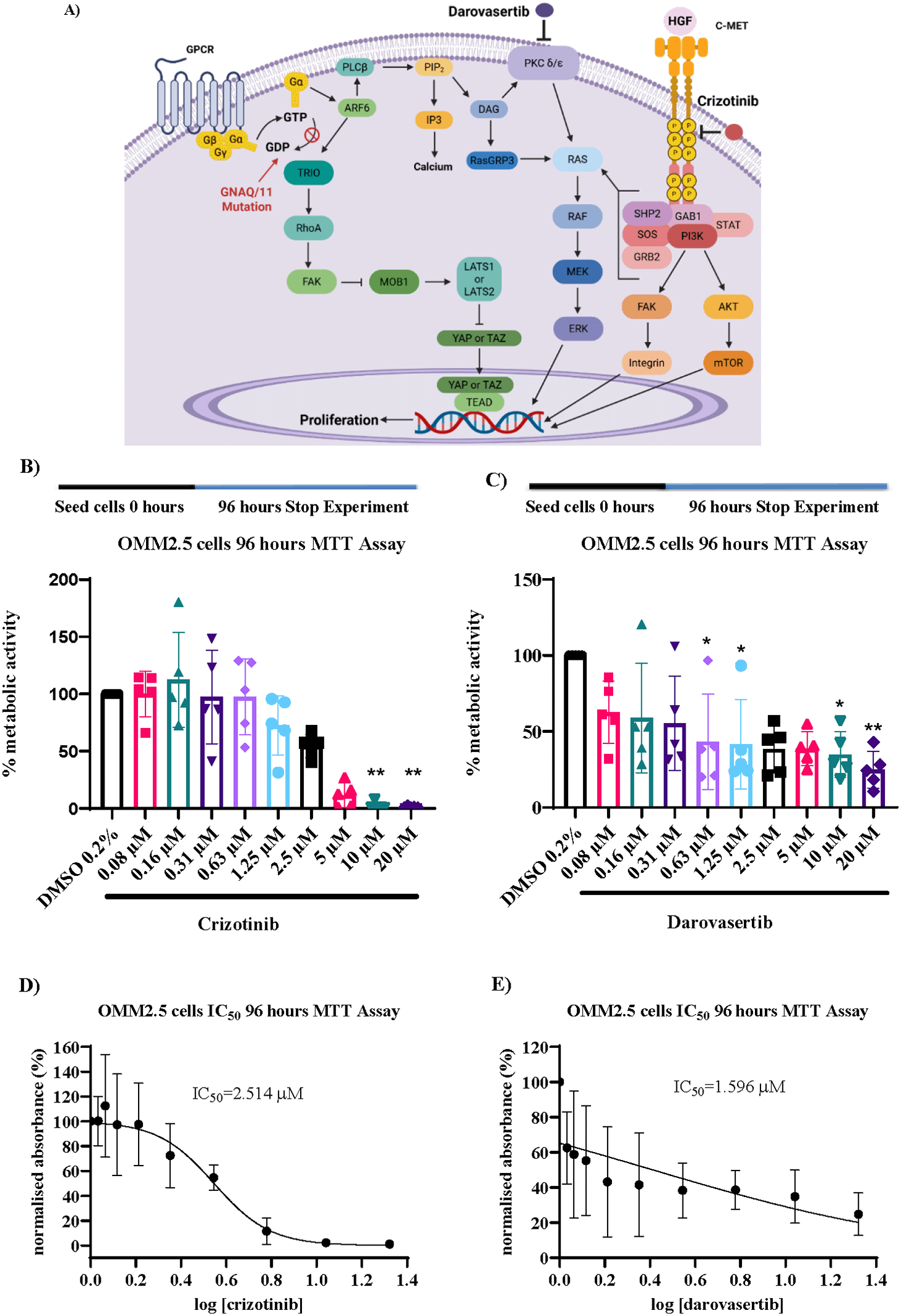
Figure 1. Crizotinib and darovasertib alone significantly reduce metastatic uveal melanoma cell metabolic activity.
A) Schematic of the cellular pathways targeted by darovasertib and crizotinib. Darovasertib inhibits protein kinase C signalling while crizotinib inhibits c-MET receptor tyrosine kinase signalling, which can attenuate cancer cell proliferation. This dual inhibition may suppress downstream activation of the MAPK and PI3K/AKT pathways, which are key drivers of uveal melanoma cell growth and survival [Adapted from 1,18, 28,30–32]. Created with BioRender.com. Subscription: Institution (University College Dublin). B) Graph showing the effect on OMM2.5 cellular metabolism of 96-hour treatment with crizotinib (n = 5). C) Graph showing the effect on OMM2.5 cellular metabolism of 96-hour treatment with darovasertib (n = 5). D) IC50 for crizotinib in OMM2.5 cells following a 96-hour treatment. E) IC50 for darovasertib in OMM2.5 cells following 96-hour treatment. Cell metabolic activity was determined using MTT (3-(4,5-dimethylthiazol-2-yl)-2,5-diphenyltetrazolium bromide) assay. 5000 cells were seeded and treated in triplicate for each individual experiment. Statistical analysis was performed by Kruskal-Wallis with Dunnett’s post hoc multiple comparison test. Error bars are mean ± S.D. *p < 0.05, **p < 0.01. 0.2% DMSO (vehicle control).
Table 1. Crizotinib and darovasertib in combination do have a greater effect on metastatic uveal melanoma cell proliferation than either drug alone.
Average % reduction in proliferation across four biological IncuCyte S3 Live-Cell replicates with crizotinib and darovasertib in combination (n = 2, 3 or 4). †Indicates that 1% DMSO was used to normalise the data, otherwise 0.2% DMSO was used to normalise the data. *Indicates that the concentration was prepared in 0.2% DMSO but compared to 1% DMSO. Data is shown ± Standard Deviation. Statistical analysis was performed by Kruskal-Wallis or One-Way Anova with either Tukey’s or Dunnett’s post hoc multiple comparison test, comparing the combination treatments (n = 3 or 4) to crizotinib alone, darovasertib alone and the % predicted additive effect. Statistical test chosen was based off normality test results. Only significant p values are shown. Data is normalized to number of cells present in each well at time 0 and presented as % alive cells divided by the number of total cells present in the well at that time point. TCC = Total Combination Concentration.
| IncuCyte S3 Live-Cell Data From 4 Biological Replicates 96 Hours |
|---|
Crizotinib and Darovasertib Combination
(μM) |
Crizotinib +Darovasertib % Reduction ± St.Dev |
Crizotinib Alone % Reduction ± St. Dev |
Darovasertib Alone % Reduction ± St.Dev |
Predicted (Crizotinib + Darovasertib) % Reduction ± St.Dev |
Crizotinib + Darovasertib % Reduction – Predicted % Reduction ± St.Dev |
TCC
(μM) |
Crizotinib at TCC % Reduction ± St.Dev |
Darovasertib at TCC % Reduction ± St.Dev |
|---|
| 0.63 Crizotinib + 1.25 Darovasertib
| 73.8 ± 9 | 1.8 ± 2.5 (p < 0.0001)
| 62 ± 11.5 | 63.9 +/− 9.4 | 9.9 ± 0.4 | 1.88 | 25.1 ± 29.6 | 67.9 ± 2.3 |
| 0.63 Crizotinib + 2.5 Darovasertib
| 74.4 ± 0.8 | 1.8 ± 2.5 (p < 0.0001)
| 65.6 ± 1.9
(p = 0.0010)
| 67.5 ± 2.5
(p = 0.0057)
| 7 ± 3.2 | 3.13 | 33.1 ± 21.9 | 74.6 ± 7.6 |
| 0.63 Crizotinib + 10 Darovasertib
| 40.5 ± 31.3† | 0†* | 34.6 ± 29.7† | 34.6 ± 29.7† | 5.9 ± 1.7† | 10.63† | 75.4 ± 36.8† | 43.1 ± 27.6† |
| 1.25 Crizotinib + 10 Darovasertib
| 48.6 ± 21.8† | 0†* (p = 0.0373)
| 42.8 ± 25.4† | 42.8 ± 25.4† | 5.8 ± 3.8† | 11.25† | 81.8 ± 30.8† | 44.6 ± 25.1† |
| 1.25 Crizotinib + 1.25 Darovasertib
| 80.5 ± 3 | 13.8 ± 18.3 (p = 0.0001)
| 62 ± 11.5 | 75.8 ± 10.2 | 4.7 ± 10.7 | 2.5 | 43 ± 28.8 | 65.8 ± 1.5 |
| 0.63 Crizotinib + 5 Darovasertib
| 75.8 ± 3.3 | 1.8 ± 2.5 | 71.6 ± 0.6 | 73.5 ± 2.1 | 2.3 ± 5.1 | 5.63 | 80.9 ± 16.2 | 72.3 ± 3.3 |
| 1.25 Crizotinib + 2.5 Darovasertib
| 79.7 ± 2.3 | 11.8 ± 15.4 (p < 0.0224)
| 65.8 ± 1.5 | 77.6 ± 14.8 | 2.1 ± 17 | 3.75 | 45.1 ± 22 | 70 ± 0.3 |
| 0.31 Crizotinib + 2.5 Darovasertib
| 67.7 ± 2 | 0.7 ± 1 | 65.9 ± 2.6 | 66.6 ± 1.5 | 1.1 ± 3.5 | 2.81 | 37.4 ± 27.2 | 67.9 ± 7 |
| 0.31 Crizotinib + 5 Darovasertib
| 72.3 ± 1.9 | 0.7 ± 1.0 | 71.9 ± 0.7 | 72.6 ± 0.3 | −0.3 ± 2.3 | 5.31 | 74.8 ± 2.1 | 69.9 ± 3.8 |
| 0.31 Crizotinib + 1.25 Darovasertib
| 68 ± 0.7 | 0.7 ± 1 | 68.3 ± 5.4 | 69 ± 6.4 | −1 ± 5.7 | 1.56 | Not calculated | Not calculated |
| 1.25 Crizotinib + 5 Darovasertib
| 78.6 ± 4.6 | 11.8 ± 15.4 (p = 0.0321)
| 71.7 ± 0.6 | 82.2 ± 12.2 | - 3.5 ± 16.2 | 6.25 | 98.4 ± 20.8 | 69.9 ± 6.2 |
| 0.16 Crizotinib + 1.25 Darovasertib
| 65.7 ± 3.5 | 3 ± 4.2 | 68.3 ± 5.4 | 71.2 ± 9.6 | −5.5 ± 13.1 | 1.41 | Not calculated | Not calculated |
96-hour crizotinib treatment resulted in a dose-dependent reduction in OMM2.5 cell metabolic activity, 10 or 20 μM crizotinib significantly reduced OMM2.5 metabolic activity on average to 2.2% (p = 0.0081) or 1.1% (p = 0.0069), respectively (
Figure 1B). As the data did not meet the assumptions of normality, group differences were analysed using a Kruskal-Wallis test. This analysis revealed a significant overall effect of treatment on the measured outcome (Kruskal-Wallis statistic = 37.82, p < 0.0001, 10 groups, n = 50), indicating that the medians differed significantly among treatment groups. The magnitude of this effect was large as indicated by the substantial separation in rank distributions across treatment levels. Post hoc analysis using Dunn’s multiple comparison test with correction for multiple testing was performed to identify pairwise differences relative to control (0.2% DMSO). While treatments from 0.08 to 5 μM shows small and non-significant effects relative to the control (adjusted p > 0.05), 10 and 20 μM treatments produced large shifts in mean ranks (mean rank differences = 30.6 and 31.0, respectively), corresponding to statistically significant differences (adjusted p = 0.0081 and p = 0.0089).
Similarly, 96-hour darovasertib treatment resulted in a dose dependent reduction in OMM2.5 cell metabolic activity. 0.63, 1.25, 10 or 20 μM darovasertib significantly reduced OMM2.5 metabolic activity on average to 43.3% (p = 0.0402), 41.6% (p = 0.0305), 34.8% (p = 0.0284) or 24.8% (p = 0.0013), respectively (
Figure 1C). The data did not pass the normality test, so the Kruskal-Wallis test was utilised. The results reveal that the treatment had a significant effect on cell metabolic activity (Kruskal-Wallis statistic = 21.30, p = 0.0114, 10 groups, n = 50), indicating that the medians differed significantly among treatment groups. Subsequently, post hoc analysis using Dunn’s multiple comparisons test with correction for multiple testing was performed to identify pairwise differences relative to control (0.2% DMSO). 0.63, 1.25, 10 and 20 μM treatments produced large shifts in mean ranks (mean rank differences = 26.20, 27, 27.20 and 35.0, respectively), corresponding to statistically significant differences (adjusted p = 0.0402, p = 0.035, p = 0.0284, p = 0.0013).
In the OMM2.5 MTT assays, the IC50 of crizotinib at 96 hours is 2.514 μM (95% CI: 1.926 μM to 3.279 μM), while the IC50 of darovasertib is 1.596 μM (95% CI, 0.578 μM to 3.529 μM) at 96 hours (
Figure 1D, E).
Crizotinib and darovasertib in combination do not reduce OMM2.5 cell metabolic activity to a greater extent than either drug alone
Consequently, the effect of crizotinib and darovasertib in combination, on metabolic activity was assessed using the MTT assay (schematic in
Figure 2A). For the combinations, crizotinib was tested at 0.16 μM, 0.31 μM, 0.63 μM or 1.25 μM. While darovasertib was tested at 1.25 μM, 2.5 μM, 5 μM or 10 μM. For the drug combinations, the MTT results are calculated as % reduction in cell metabolic activity compared to DMSO controls. In the tabulated data, the specific drug combination is compared to i) each of the drugs tested alone at the same concentration and ii) the predicted additive effect of the specific combination based on summing the activity from the individually tested drugs (
Figure 2B). A drug combination being greater than additive was defined as displaying activity statistically higher than: a) equimolar crizotinib alone, b) equimolar darovasertib alone and c) the predicted % reduction for additive effects. A drug combination was deemed additive if the activity was significantly greater to a) equimolar crizotinib alone and b) equimolar darovasertib alone but equivalent to the predicted % additive reduction. As the data did not meet the assumptions of normality, group differences were analysed using a Kruskal-Wallis with Dunn’s multiple comparisons test.
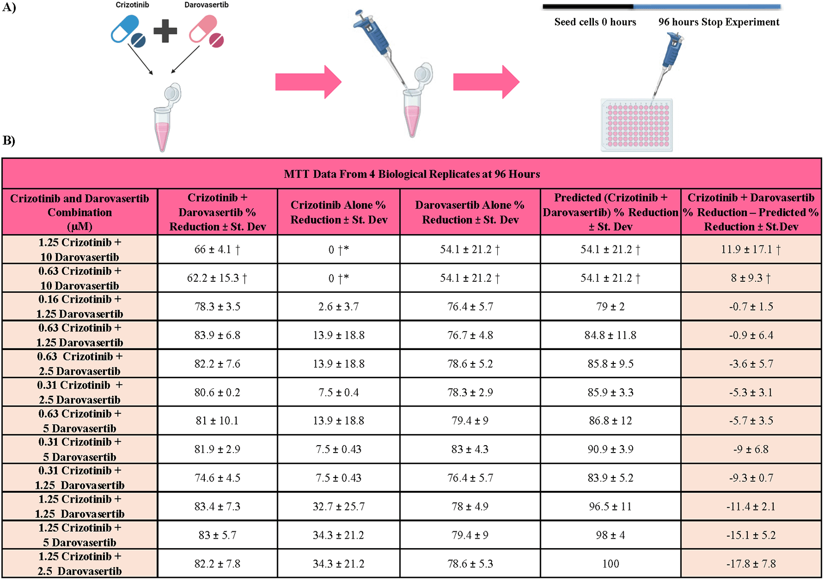
Figure 2. Crizotinib and darovasertib in combination do not have a greater effect on metastatic uveal melanoma cell metabolic activity than either drug alone.
A) Schematic showing how darovasertib and crizotinib were added in combination to the OMM2.5 cells. B) Average % reduction in metabolic activity across four biological MTT replicates with crizotinib and darovasertib in combination (n = 2, 3 or 4). The drug combination results for the MTT assay are ordered with the combination with the greatest difference between the observed and predicted % reduction at the top. †Indicates that 1% DMSO was used to normalise the data, otherwise 0.2% DMSO was used to normalise the data. *Indicates that the concentration was prepared in 0.2% DMSO but compared to 1% DMSO. Data is presented as mean ± Standard Deviation. Statistical analysis was performed by Kruskal-Wallis with Dunnett’s post hoc multiple comparison test, comparing the combination treatments (n = 3 or 4) to crizotinib alone, darovasertib alone and the % predicted additive effect. No significance was found for any combination.
The drug combination results for the MTT assay are ordered with the combination with the greatest difference between the observed and predicted % reduction at the top (
Figure 2B). The table shows the average % reduction for each treatment across the MTT replicates for the most promising drug combinations (
Figure 2B). Any combinations using 10 μM darovasertib were compared to 1% DMSO, to account for higher vehicle DMSO concentrations in those solutions. Based on these parameters, the 96-hour MTT results with crizotinib plus darovasertib drug combinations indicate that none of the combinations are additive or greater than additive (
Figure 2B). Kruskal-Wallis analysis revealed no significant difference between the groups. Therefore, these combinations did not meet the criteria for additive drug effects (
Figure 2B).
Crizotinib alone significantly reduces metastatic uveal melanoma live cell proliferation
To assess the effects of single and combinations drugs on the proliferation of OMM2.5 mUM cells, live cell proliferation was analysed from 12 to 96 hours post treatment using an IncuCyte S3 Live-Cell Analysis System (
Figure 3A). Time points of every 12 hours were used for data analysis, which revealed that crizotinib had a time-dependent as well as a dose-dependent effect on OMM2.5 cell proliferation (
Figure 3A). As the data did not meet the assumptions of normality, group differences were analysed using a Mixed-Effects Analysis. Post hoc analysis using Dunnett’s multiple comparisons with correct for multiple testing was performed to identify pairwise differences relative to the control (0.2% DMSO) (
Figure 3A). Cell images revealed a difference in OMM2.5 cell morphology with concentrations of crizotinib above 1.25 uM, compared to the 0.2% DMSO controls (
Figure 3B). The changes included fewer cells and cellular shrinkage, which is a characteristic of cell death.33 At 96 hours, 5, 10 or 20 μM crizotinib significantly reduced live cell proliferation to 19.4% (p < 0.0001), 11.24% (p < 0.0001), or 9.4% (p < 0.0001) respectively (
Figure 3C). As data meet the assumptions of normality, group differences were analysed using a One-Way Anova test. The analysis results show a significant effect of crizotinib treatment on cell proliferation (F statistic = 38.25, p < 0.0001, 10 groups, n = 30). Post hoc analysis using Dunnett’s multiple comparisons test with correction for multiple testing was performed to identify pairwise differences relative to control. 5, 10 and 20 μM produced large shifts in mean ranks (mean rank differences = 96.17, 104.3 and 106.2, respectively), corresponding to statistically significant differences (adjusted p < 0.0001, p < 0.0001 or p < 0.0001, respectively). The IC50 of crizotinib for live cell proliferation was 4.122 μM at 96 hours, GraphPad prism software could not generate a confidence interval (
Figure 3D).
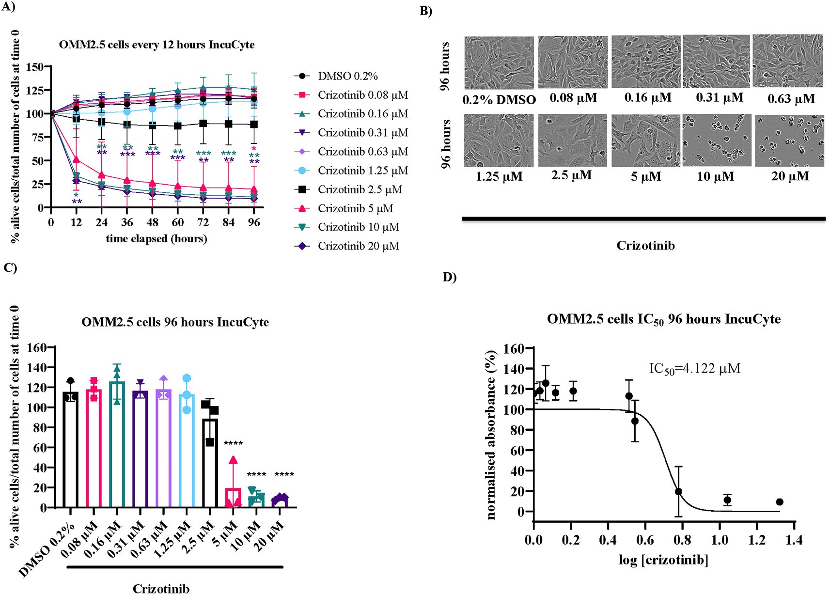
Figure 3. Crizotinib alone significantly reduces metastatic uveal melanoma cell proliferation.
A) Graph showing the effect of 96-hour treatment with crizotinib on OMM2.5 cell line proliferation (n = 3). Statistical analysis was performed by Mixed Effect Analysis with Dunnett’s post hoc multiple comparison test. *p < 0.05, **p < 0.01, ***p < 0.001. The pink * indicates significance for 5 μM crizotinib, the green * indicates significance for 10 μM crizotinib and the purple * indicates significance for 20 μM crizotinib, compared to 0.2% DMSO. B) IncuCyte S3 Live-Cell images of OMM2.5 cells, taken at 10 x magnification, treated with DMSO and crizotinib at 96 hours. C) The effect of 96-hour crizotinib treatment on OMM2.5 cell line proliferation (n = 3). Statistical analysis was performed by One-Way Anova with Dunnett’s post hoc multiple comparison test. ****p < 0.0001. D) IC50 of crizotinib in OMM2.5 cells following a 96-h treatment (n = 3). Data is normalized to number of cells present in each well at time 0 and presented as % alive cells divided by the number of total cells present in the well at that time point. Error bars are mean ± standard deviation. 0.2% DMSO (vehicle control).
Darovasertib alone significantly reduces metastatic uveal melanoma live cell proliferation
For darovasertib, the effects of single drug concentrations on the proliferation of OMM2.5 mUM cells, was likewise analysed from 12 to 96 hours post treatment using IncuCyte S3 Live-Cell Analysis (
Figure 4A). Time points of every 12 hours were used for data analysis, which revealed that darovasertib had a time- and dose-dependent effect on OMM2.5 cell proliferation (
Figure 4A). As the data did not meet the assumptions of normality, group differences were analysed using a Mixed-Effects Analysis. Post hoc analysis using Dunnett’s multiple comparisons with correction for multiple testing was performed to identify pairwise differences relative to the control (0.2% DMSO) (
Figure 4A). Cell images revealed a difference in OMM2.5 cell morphology for concentrations of darovasertib above 1.25 μM, compared to the 0.2% DMSO control (
Figure 4B). The changes again are consistent with cell death, including fewer cells and cellular shrinkage.33
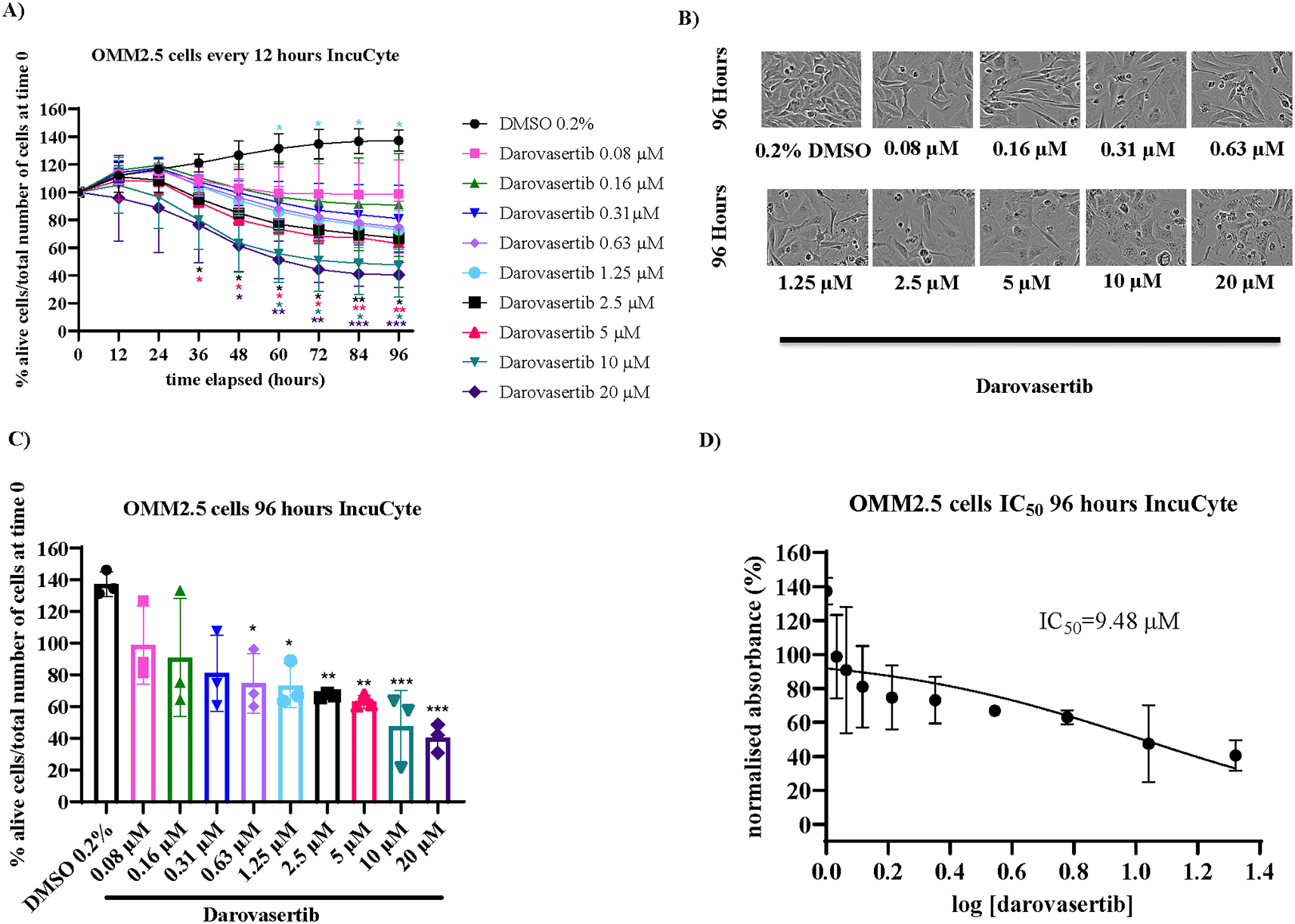
Figure 4. Darovasertib alone significantly reduces metastatic uveal melanoma cell proliferation.
A) Graph shows the effect of 96-hour treatment with darovasertib on OMM2.5 cell line proliferation (n = 3). Statistical analysis was performed by Mixed Effects Analysis with Dunnett’s post hoc multiple comparison test. *p < 0.05, **p < 0.01, ***p < 0.001. The light blue * indicates significance for 1.25 μM darovasertib, the black * indicates significance for 2.5 μM darovasertib, the pink * indicates significance for 5 μM crizotinib, the light green * indicates significance for 10 μM crizotinib and the dark purple * indicates significance for 20 μM crizotinib compared with 0.2% DMSO. B) IncuCyte S3 Live-Cell images of OMM2.5 cells treated with DMSO and darovasertib at 10x magnification at 96 hours. C) The effect of 96-hour crizotinib treatment on OMM2.5 cell line proliferation (n = 3). Statistical analysis was performed by One-Way Anova with Tukey’s post hoc multiple comparison test. *p < 0.05, **p < 0.01, ***p < 0.001. D) IC50 of darovasertib in OMM2.5 cells following a 96-h treatment (n = 3). Data is normalized to number of cells in each well at time 0 and presented as % alive cells divided by the number of total cells in the well at that time point. Error bars are mean ± standard deviation.
At 96 hours, 0.631.25, 2.5, 5,10 or 20 μM darovasertib significantly reduced cell proliferation to 81% (p = 0.0141), 74.7% (p = 0.0219), 73% (p = 0.0175), 66.9% (p = 0.0076), 63% (p = 0.0045), 47.5% (p = 0.0005) or 40.5% (p = 0.0002), respectively (
Figure 4C). As data met the assumptions of normality, group differences were analysed using a One-Way Anova test. This analysis revealed a significant overall effect of darovasertib on cell metabolic activity (F statistic = 6.015, p = 0.0004, 10 groups, n = 30). Post hoc analysis using Tukey’s multiple comparisons test with correction for multiple testing was performed to identify pairwise differences relative to control. 0.63, 1.25, 2.5, 5, 10 and 20 μM produced large shifts in mean ranks (mean rank differences = 62.56, 64.22, 70.41, 74.26, 89.76 and 96.76 respectively), corresponding to statistically significant differences (adjusted p = 0.0219, p = 0.0175, p = 0.0076, p = 0.0045. p = 0.0005 or p = 0.0002, respectively).
The IC50 of darovasertib for live cell proliferation at 96 hours was 9.48 μM (95% CI: 5.046 to 25.61) (
Figure 4D).
Crizotinib and darovasertib combinations exhibit additive or greater effects on metastatic uveal melanoma live cell proliferation
For the IncuCyte S3 Live-Cell analysis of pharmacological effects on cell proliferation of crizotinib plus darovasertib drug combinations (Table 1), either 0.16 μM, 0.31 μM, 0.63 μM or 1.25 μM crizotinib were tested in combination with either 1.25 μM, 2.5 μM, 5 μM or 10 μM darovasertib. The IncuCyte results were calculated as % reduction in cell proliferation compared to DMSO controls. This data is tabulated, and the specific drug combination is compared to; i)
each of the drugs tested alone at the same concentration and ii) the predicted additive effect of the specific combination based on summing the activity from the individually tested drugs (Table 1).
The data is ordered from highest difference between the observed % reduction of darovasertib plus crizotinib in combination, minus the predicted % reduction of darovasertib and crizotinib in combination. Any combinations using 10 μM darovasertib were compared to 1% DMSO, to account for higher DMSO in the solution. Based on these parameters, the 96-hour live cell proliferation results with crizotinib plus darovasertib drug combinations identifies the four-highest rank combinations (0.63 uM crizotinib +1.25 uM darovasertib; 0.63 uM crizotinib +2.5 uM darovasertib 0.63 uM crizotinib +10 uM darovasertib and 1.25 uM crizotinib +10 uM darovasertib) where the observed activity of the combinations appears greater (~5.9 to ~9.9%) than the calculated additive effect of the individual drugs (Table 1). For statistical analysis either Kruskal-Wallis with Dunn’s post hoc test or One-Way Anova with either Tukey or Dunnett’s post hoc test were utilised. The test used was dependent on if the data passed the assumption of normality.
Based on this ranking, selective drug combinations were analysed in greater detail. A combination being greater than additive was defined as a drug combination being statistically greater than a) equimolar crizotinib alone, b) equimolar darovasertib alone and c) the predicted % reduction in proliferation for the additive effects of darovasertib and crizotinib. A drug combination was deemed additive if it were significantly greater to a) equimolar crizotinib alone and b) equimolar darovasertib alone but equivalent to the predicted % additive reduction. We focused on the 48-hour and 96-hour timepoints as OMM2.5 cells have a doubling time of 50 hours.34 Applying these parameters the combination of 0.63 μM crizotinib and 1.25 μM darovasertib was identified as being greater than additive (Table 1).
1.25 μM crizotinib and 2.5 μM darovasertib in combination have an additive effect on metastatic uveal melanoma cell proliferation at 48 hours
1.25 μM crizotinib and 2.5 μM darovasertib in combination had a time dependent effect on OMM2.5 cell proliferation. (
Figure 5A). As the data did not meet the assumptions of normality, group differences were analysed using a Mixed-Effects Analysis. Post hoc analysis using Sidak multiple comparisons with correction for multiple testing was performed to identify pairwise differences relative to the control (0.2% DMSO) (
Figure 5A).
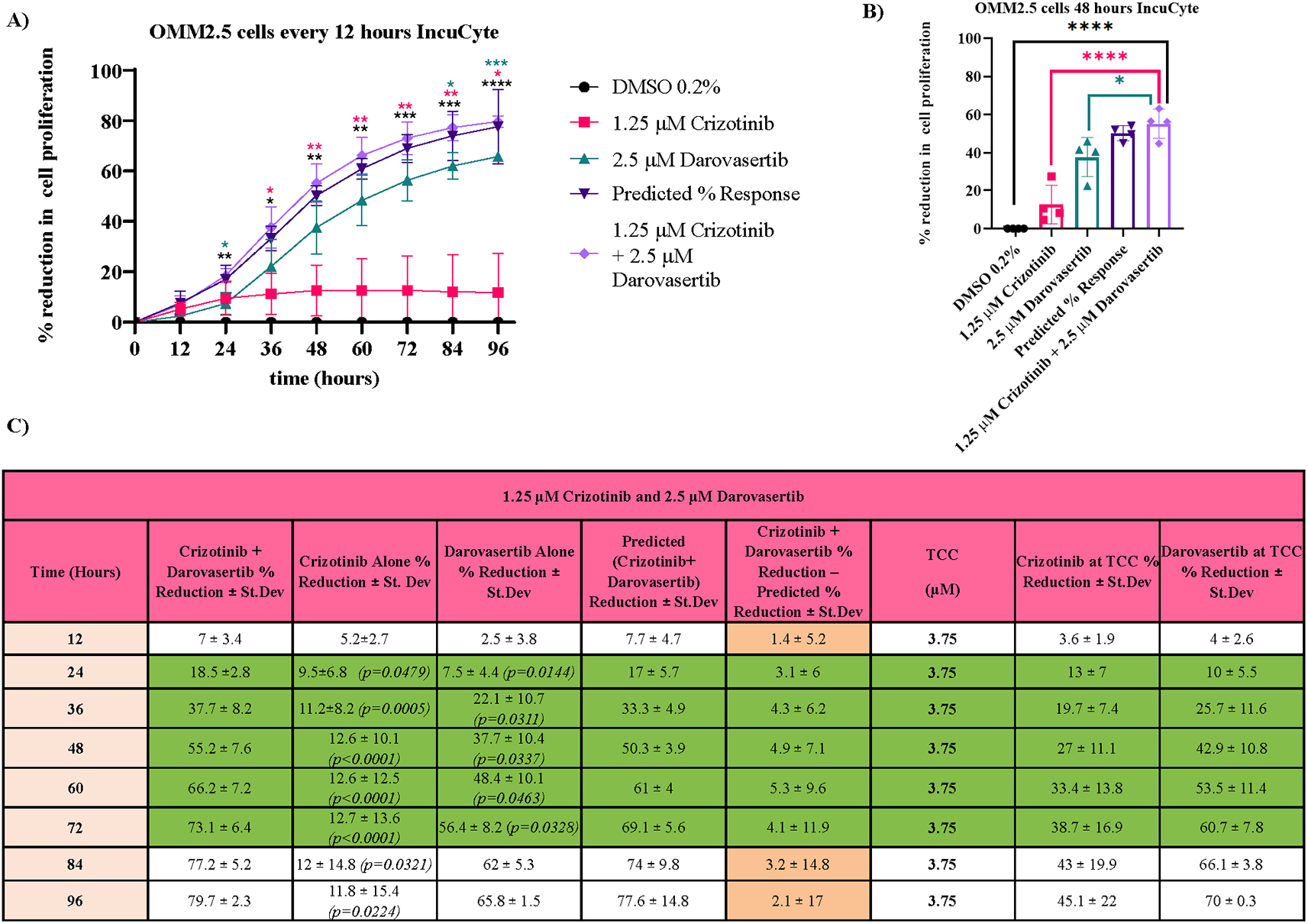
Figure 5. 1.25 μM crizotinib and 2.5 μM darovasertib in combination have an additive effect on metastatic uveal melanoma cell proliferation at 48 hours.
A) The effect of 96-hour treatment with 1.25 μM crizotinib and 2.5 μM darovasertib in combination on cell proliferation (n = 4). Statistical analysis was performed by Mixed-Effects Analysis with Sidak’s post hoc multiple comparison test, comparing 1.25 μM crizotinib and 2.5 μM darovasertib to all the other conditions, *p < 0.05, **p < 0.01,***p < 0.001 ****p < 0.0001. The black * indicates significance of 1.25 μM crizotinib and 2.5 μM darovasertib in combination compared to 0.2% DMSO, the pink * indicates significance of 1.25 μM crizotinib and 2.5 μM darovasertib in combination compared to 1.25 μM crizotinib and the green * indicates significance of 1.25 μM crizotinib and 2.5 μM darovasertib in combination compared to 2.5 μM darovasertib. B) The effect of 48-hour treatment with 1.25 μM crizotinib and 2.5 μM darovasertib in combination on cell proliferation (n = 4). Statistical analysis was performed by One-Way Anova with Tukey’s post hoc multiple comparison test, comparing 1.25 μM crizotinib and 2.5 μM darovasertib to all the other conditions, *p < 0.05, ****p < 0.0001. C) The average % reduction of IncuCyte S3 Live-Cell replicates every 12 hours across the 96-hour treatment (n = 4). TCC = Total Combination Concentration. Statistical analysis was performed by Kruskal-Wallis or One-Way Anova with either Tukey’s or Dunnett’s post hoc multiple comparison test, comparing 1.25 μM crizotinib and 2.5 μM darovasertib (n = 4) to crizotinib alone, darovasertib alone and the % predicted additive effect. Only significant p
values are shown. Green indicates additive, white indicates that the combination is neither antagonistic, additive or greater than additive at that timepoint. Data is normalized to number of cells present in each well at time 0 and presented as % reduction in cell viability. Error bars are mean ± standard deviation.
At 48 hours, One-Way Anova showed that the decreased cell proliferation caused by this combination is significantly greater than the decrease seen with DMSO controls (p < 0.0001), crizotinib alone (p < 0.0001) or darovasertib alone (p = 0.0337), but this combination was not significantly different in comparison to the predicted % additive effect (p = 0.8835). Thus, this combination appears to be additive, not greater than additive (
Figure 5B,C). As data met the assumptions of normality, group differences were analysed using a One-Way Anova test. This analysis revealed a significant overall effect of treatment on cell proliferation (F statistic = 40.62, p < 0.0001, 5 groups, n = 20), indicating that the medians difference significantly among treatment groups. Post hoc analysis using Tukey’s multiple comparisons test with correction for multiple testing was performed to identify pairwise differences relative to the combination. 0.2% DMSO, 1.25 μM crizotinib and 2.5 μM darovasertib produced large shifts in mean ranks (mean rank differences = −55.20, −42.57 and − 17.55 respectively), corresponding to statistically significant differences (adjusted p < 0.0001, p < 0.0001 or p = 0.0337, respectively).
The combination of 1.25 μM crizotinib and 2.5 μM darovasertib was found to be additive at 24, 36, 60 and 72 hours also (
Figure 5C). For statistical analysis either Kruskal-Wallis with Dunn’s post hoc test or One-Way Anova with either Tukey or Dunnett’s post hoc test were utilised. The test used was dependent on if the data passed the assumption of normality.
0.63 μM crizotinib and 2.5 μM darovasertib in combination have a greater than additive effect on metastatic uvea melanoma cell proliferation at 96 hours
In contrast, the combination of 0.63 μM crizotinib and 2.5 μM darovasertib in combination had a time dependent, greater than additive effect on OMM2.5 cell proliferation. (
Figure 6A). As the data did not meet the assumptions of normality, group differences were analysed using a Mixed-Effects Analysis. Post hoc analysis using Sidak multiple comparisons with correction for multiple testing was performed to identify pairwise differences relative to the control (0.2% DMSO) (
Figure 6A).
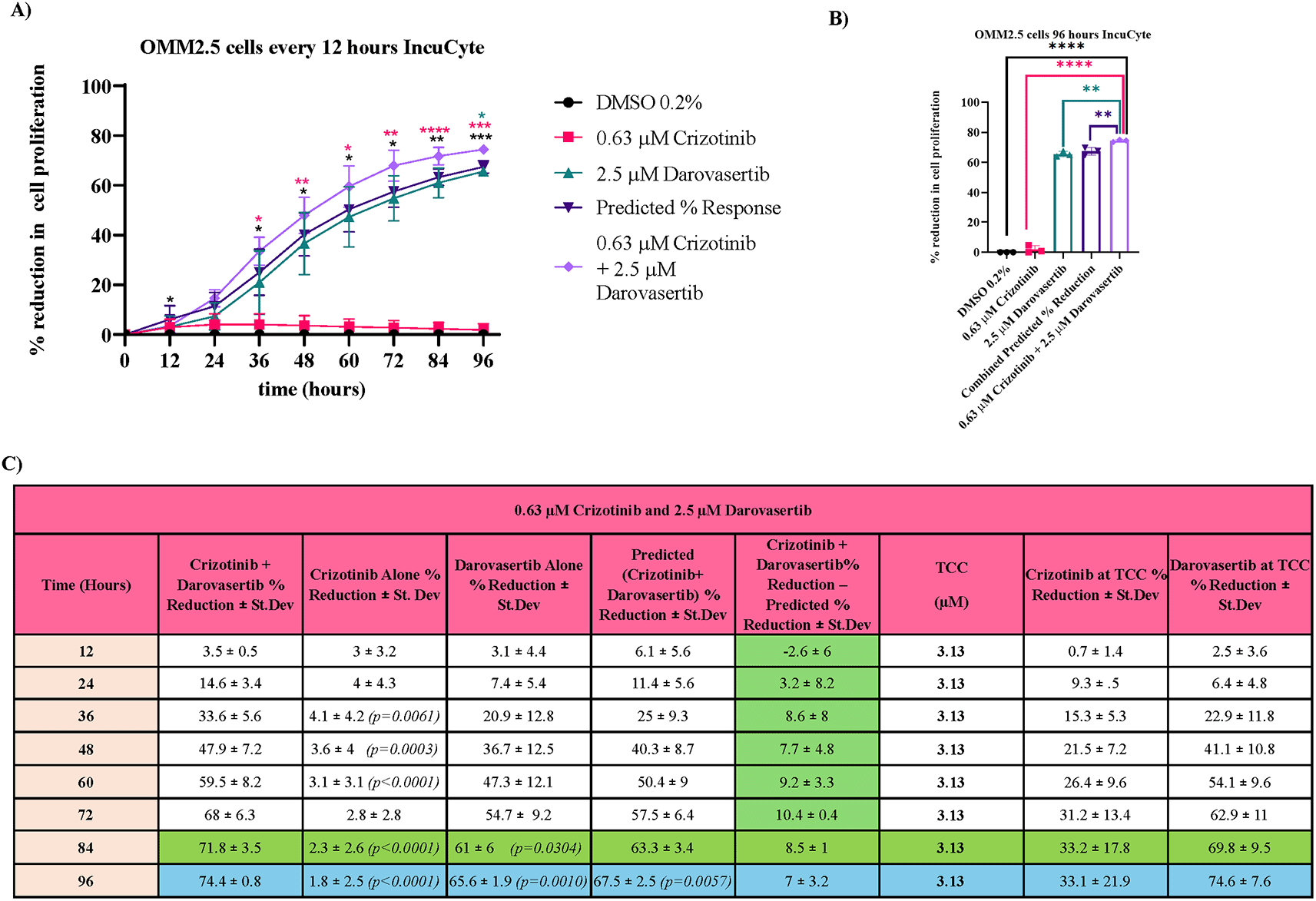
Figure 6. 0.63 μM crizotinib and 2.5 μM darovasertib in combination have a greater than additive effect on metastatic uveal melanoma cell proliferation at 96 Hours.
A) The effect of 96-hour treatment with 0.63 μM crizotinib and 2.5 μM darovasertib in combination on OMM2.5 cell proliferation (n = 3). Statistical analysis was performed by Two-Way Anova with Sidak’s post hoc multiple comparison test, comparing 0.63 μM crizotinib and 2.5 μM darovasertib to all the other conditions. *p < 0.05, **p < 0.01,***p < 0.001, ****p < 0.0001. The black * indicates significance of 0.63 μM crizotinib and 2.5 μM darovasertib in combination compared to 0.2% DMSO, the pink * indicates significance of 0.63 μM crizotinib and 2.5 μM darovasertib in combination compared to 0.63 μM crizotinib and the green * indicates significance of 0.63 μM crizotinib and 2.5 μM darovasertib in combination compared to 2.5 μM darovasertib. B) The effect of 96-hour treatment with 0.63 μM crizotinib and 2.5 μM darovasertib in combination on OMM2.5 cell proliferation (n = 3). Statistical analysis was performed by One-Way Anova with Tukey’s post hoc multiple comparison test, comparing 0.63 μM crizotinib and 2.5 μM darovasertib to all the other conditions. **p < 0.01, ****p < 0.0001. C) The average % reduction of IncuCyte S3 Live-Cell replicates every 12 hours across the 96-hour treatment (n = 3). TCC = Total Combination Concentration. Statistical analysis was performed by Kruskal-Wallis or One-Way Anova with either Tukey’s or Dunnett’s post hoc multiple comparison test, comparing 0.63 μM crizotinib and 2.5 μM darovasertib (n = 3) to crizotinib alone, darovasertib alone and the % predicted additive effect. Only significant p values are shown. Blue indicates greater than additive, green indicates additive, white indicates that the combination is neither additive, antagonistic nor greater than additive. Data is normalized to number of cells present in each well at time 0 and presented as % reduction in cell viability. Cell metabolic activity was determined using MTT assay. Error bars are mean ± standard deviation.
At the 96-hour timepoint, 0.63 μM crizotinib plus 2.5 μM darovasertib in combination is statistically significantly different in comparison to DMSO controls (p < 0.0001),
0.63 μM crizotinib (p < 0.0001), 2.5 μM darovasertib (p = 0.001) and the predicted % reduction of the pair (p = 0.0057) (
Figure 6B). This indicates this combination is greater than additive. As data met the assumptions of normality, group differences were analysed using a One-Way Anova test. This analysis revealed a significant overall effect of treatment on OMM2.5 cell proliferation (F statistic = 1288, p < 0.0001, 5 groups, n = 15). Post hoc analysis using Tukey’s multiple comparisons test with correction for multiple testing was performed to identify pairwise differences relative to the combination. 0.2% DMSO, 1.25 μM crizotinib, 2.5 μM darovasertib and the predicted % reduction produced large shifts in mean ranks (mean rank differences = −74.43, −72.60, −8.819 and − 6.980 respectively), corresponding to statistically significant differences (adjusted p < 0.0001, p < 0.0001, p = 0.001 or p = 0.0057).
Analysis of the 96-hour time-course of this combination indicates that the difference between the observed and calculated additive effects on live cell proliferation maximised (10.4% ± 0.4%) at ~72 hours (
Figure 6C). The combination of 0.63 μM crizotinib and 2.5 μM darovasertib was found to be additive at the 84 hours timepoint, with the combination being statistically significant in comparison to 0.63 μM crizotinib
(p < 0.0001),
2.5 μM darovasertib (p = 0.0304) but not the predicted % reduction (
Figure 6C). For statistical analysis either Kruskal-Wallis with Dunn’s post hoc test or One-Way Anova with either Tukey or Dunnett’s post hoc test were utilised. The test used was dependent on if the data passed the assumption of normality.
Crizotinib and darovasertib combinations exhibit synergistic effects on metastatic uveal melanoma live cell proliferation
To further analyse the nature of the interactions between crizotinib and darovasertib, Combenefit, a drug interaction analysis tool, was used. Combenefit was loaded with data from the IncuCyte S3 Live-Cell Analysis System following crizotinib and darovasertib combination treatment, and a colour-coded synergy distribution matrix of synergy scores was generated using the Bliss synergy model. The matrix was coloured according to the synergy scores and statistical significance. Predominantly additive interactions were observed between crizotinib and darovasertib across the synergy distribution matrix, with statistically significant synergistic interactions observed at intermediate concentrations of the combination. Synergy scores indicate statistically significant interactions between crizotinib and darovasertib at: crizotinib 0.63 μM and darovasertib 2.5 μM, and crizotinib 1.25 μM and darovasertib 2.5 μM (
Figure 7).
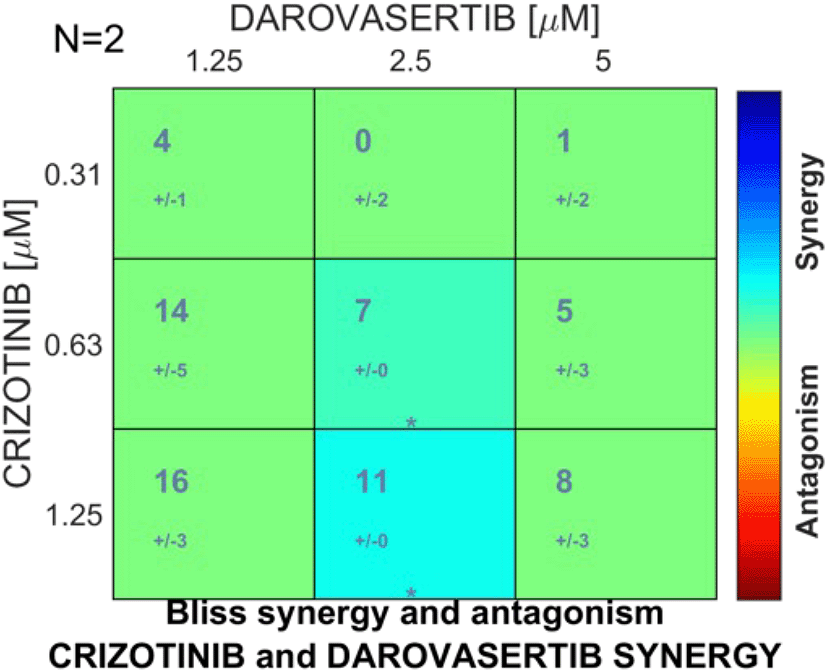
Figure 7. Synergy distribution matrix of crizotinib and darovasertib combination drug treatment using Bliss synergy model.
Synergy levels for crizotinib and darovasertib at different concentrations in combination. Synergy scores indicate statistically significant interactions between crizotinib and darovasertib at: crizotinib 0.63 μM and darovasertib 2.5 μM, and crizotinib 1.25 μM and darovasertib 2.5 μM. The levels of synergy and/or antagonism are colour-coded based on the calculated score, provided the result was statistically significant as determined by a one-sample t-test (*p < 0.05). A darker blue colour indicates more synergy between the drugs and a darker red colour indicates more antagonism between the drugs. Synergy scores were calculated using the Combenefit reference modelled dose-response matrix data and the IncuCyte S3 Live-Cell Analysis data following 96-h treatment with different concentrations of crizotinib and darovasertib alone and in combination (n = 2), data were expressed as percentage of the control (0.2% DMSO).
Discussion
mUM carries a poor prognosis, with very few treatments available. This study provides pre-clinical evidence that crizotinib and darovasertib, both alone and in combination, are efficacious in conferring anti-cancer effects in a specific mUM cell line.
Darovasertib received approval as an orphan drug by the U. S FDA on 2 May 2022, for treating mUM.18 It is currently being evaluated in clinical trials in combination with crizotinib.18,26 To date, darovasertib and crizotinib in combination has been tested in the OMM2.5 cell line. Furthermore, the effect of darovasertib and crizotinib either alone or in combination on mUM cells has not previously been monitored using the IncuCyte S3 Live-Cell Analysis System.
Firstly, the effect of crizotinib and darovasertib alone on OMM2.5 cell metabolic activity and live cell proliferation was analysed using MTT and IncuCyte S3 Live-Cell Analysis System, respectively. Crizotinib and darovasertib alone significantly reduced OMM2.5 cell metabolic activity, with darovasertib having higher potency while crizotinib had higher efficacy (
Figure 1). However, this is not seen with the IncuCyte S3 Live-Cell Analysis Software results, with darovasertib having a higher IC50 value than crizotinib. These results indicate that darovasertib has a greater effect on cell metabolic activity than proliferation. A recent study found that darovasertib cannot suppress multiple active pathways in UM tumours. The incubation of 11 different UM cell lines with 1 or 5 μM darovasertib, produced cell cycle inhibition but not cell death in most of the GNAQ/GNA11-mutant cell lines. Since OMM2.5 is a GNAQ Q209P mutant cell line,20,35 darovasertib’s mechanism of action may explain its differing effects on cell metabolic activity and proliferation.
Darovasertib and crizotinib combinations have been previously tested in 92.1 and MEL202 (primary) and in MM28 (metastatic) UM cells. Crizotinib was reported to synergize strongly with darovasertib at clinically relevant doses (80 nM) in MEL202 and MM28 in contrast to the 92.1 cells, where mild or no synergy was observed.24 However, these results are only reported in an abstract and not yet been peer-reviewed. For the drug combination experiments here, 0.16 μM, 0.31 μM, 0.63 μM or 1.25 μM crizotinib were tested in combination with either 1.25 μM, 2.5 μM, 5 μM or 10 μM darovasertib.
Based on MTT assay data, none of the combinations were found to be additive or greater than additive. However, the combination of 0.63 μM crizotinib plus 2.5 μM darovasertib (1:4 ratio) appears to hold the most promise as a greater than additive combination, according to the IncuCyte S3 Live-Cell Analysis System data. In Table 1, combinations such as 0.63 μM crizotinib +1.25 μM darovasertib and 1.25 μM crizotinib +10 μM darovasertib ranked among the most effective. However, statistical analysis revealed that neither combination is statistically greater than darovasertib alone or the predicted % reduction. In the phase 2 clinical trials, patients were given 200 mg of crizotinib and 300 mg of darovasertib, therefore a ratio of 1:1.5. The findings suggest that darovasertib and crizotinib can produce a greater than additive effect on mUM cell proliferation, but at a different ratio than the one used in clinical trials.26 The reduction in viability compared to DMSO for 0.63 μM crizotinib +2.5 μM darovasertib in combination is circa 10% and statistically significant to either drug alone. Therefore, the drugs in combination at these concentrations are biologically relevant regarding the effect it has on the proliferation of cancer cell lines. However, it is difficult to conclude at this point if this result is biologically relevant in vivo. To determine this further research would need to be carried out on UM patient samples or patient xenograft models.
Differences were seen between the MTT and IncuCyte S3 Live-Cell Analysis System results. Recently issues with over/underestimation of cell viability with the MTT assay have emerged. This over/underestimation may be due to adaptive metabolic and mitochondrial reprogramming in cells caused by inhibitor off-target effects or pharmacological treatment-mediated stress. In a study comparing the MTT assay versus direct cell counting of TMZ-treated glioblastoma cells, the MTT assay results showed either over- or under estimation of cell death in several glioblastoma cell lines. Thus, it is recommended to supplement the tetrazolium salt-based assays with other non-metabolic methods.36 In our opinion, the IncuCyte S3 Live-Cell Analysis provides a more comprehensive and robust analysis than the MTT assay. However, there is potential for error with the IncuCyte S3 Live-Cell Analysis as user-based bias can occur in regard to deciding if a cell is dead or alive. Furthermore, darovasertib has been found to decrease UM cell proliferation but not directly produce cell death. This likely had an impact on the MTT and IncuCyte results.20 Ultimately, these assays interrogate distinct biological endpoints and divergence between them is expected. With the MTT measuring metabolic activity, hence the results reflect cell metabolism and not direct cell number. While the IncuCyte results reflect cell number and cell morphology.
A limitation of this study is that all in vitro experiments were carried out using the OMM2.5 cell line, which a BAP1 negative cell line. However, 84% of mUMs are BAP1 positive.37 This means that the results collated here are only applicable to 16% of mUM cases. So, a future direction of this research is to repeat these experiments in a BAP1 positive cell line. Another limitation is that for the IncuCyte analysis the user was not blinded to drug location on the plate which may have led to potential bias when analysing the IncuCyte images. A further limitation is the low number of replicates for certain combinations enabled only N = 2 biological replicates to be evaluated using the Bliss Independence Analysis Model.
Conclusion
This study provides novel preclinical evidence supporting the anti-cancer potential of darovasertib and crizotinib, both individually and in combination, against mUM in the OMM2.5 cell line. Discrepancies between MTT and IncuCyte S3 Live-Cell assay results highlight the importance of using complementary methods to assess drug efficacy and underscore the limitations inherent in metabolic-based viability assays. Additionally, the reliance on a single, BAP1-negative mUM cell line restricts the application of these findings to the broader UM patient population, the majority of whom are BAP1-positive. Nonetheless, this work lays the foundation for more refined combination strategies and highlights the need for further investigation in diverse mUM models. Future studies incorporating BAP1-positive lines, combination software analysis, additional biological replicates, and enhanced assay techniques will be critical to validating and optimizing this potentially synergistic therapeutic combination.
Ultimately, the translational relevance of in vitro drug concentrations must be interpreted with caution, as effective doses observed in monolayer cell culture systems do not directly translate to achievable or tolerable exposures in patients. Both darovasertib and crizotinib are under clinical investigation, and their pharmacokinetic profiles in humans are increasingly well characterised. While the absolute concentrations associated with activity in this study may not be directly achievable in vivo, the observation that a particular ratio of two agents resulted in enhanced anti-tumour effects suggests that relative dosing relationships, rather than absolute dose escalation, may be an important determinant of efficacy. These findings are therefore best viewed as hypothesis-generating and supportive of future in vivo and pharmacokinetics/pharmacodynamics studies to assess pharmacological feasibility and therapeutic potential in a physiological context.
Ethics and consent
Ethical approval and consent were not required.
Data availability
Underlying data
Zenodo: Crizotinib and Darovasertib Significantly Reduce Metastatic Uveal Melanoma Cell Proliferation, Alone and In Combination. https://doi.org/10.5281/zenodo.16530866.38
This project contains the following underlying data:
•
Figure 1 B-E-Crizotinib and darovasertib alone significantly reduce metastatic uveal melanoma cell metabolic activity … xlsx
•
Figure 1 B-Crizotinib and darovasertib alone significantly reduce metastatic uveal melanoma cell metabolic activity … xlsx
•
Figure 1 C-Crizotinib and darovasertib alone significantly reduce metastatic uveal melanoma cell metabolic activity … xlsx
•
Figure 1 D-Crizotinib and darovasertib alone significantly reduce metastatic uveal melanoma cell metabolic activity...xlsx
•
Figure 1 E-Crizotinib and darovasertib alone significantly reduce metastatic uveal melanoma cell metabolic activity...xlsx
•
Figure 2 B-Crizotinib and darovasertib in combination do not have a greater effect on metastatic uveal melanoma cell metabolic activity than either drug alone...xlsx
•
Figure 3 A-Crizotinib alone significantly reduces metastatic uveal melanoma cell proliferation … xlsx
•
Figure 3 B-Crizotinib alone significantly reduces metastatic uveal melanoma cell proliferation … tiff
•
Figure 3 C-Crizotinib alone significantly reduces metastatic uveal melanoma cell proliferation … xlsx
•
Figure 3 D-Crizotinib alone significantly reduces metastatic uveal melanoma cell proliferation … ..xlsx
•
Figure 4 A-Darovasertib alone significantly reduces metastatic uveal melanoma cell proliferation … xlsx
•
Figure 4 B-Darovasertib alone significantly reduces metastatic uveal melanoma cell proliferation … tiff
•
Figure 4 C-Darovasertib alone significantly reduces metastatic uveal melanoma cell proliferation … xlsx
•
Figure 4 D-Darovasertib alone significantly reduces metastatic uveal melanoma cell proliferation … .xlsx
•
Table 1-Crizotinib and darovasertib in combination do have a greater effect on metastatic uveal melanoma cell proliferation than either drug alone … xlsx
•
Figure 5 A-1.25 μM crizotinib and 2.5 μM darovasertib in combination have an additive effect on metastatic uveal cell proliferation at 48 hours … xlsx
•
Figure 5 B-1.25 μM crizotinib and 2.5 μM darovasertib in combination have an additive effect on metastatic uveal cell proliferation at 48 hours … xlsx
•
Figure 5 C-1.25 μM crizotinib and 2.5 μM darovasertib in combination have an additive effect on metastatic uveal cell proliferation at 48 hours … xlsx
•
Figure 6 A-0.63 μM crizotinib and 2.5 μM darovasertib in combination have a greater than additive effect on metastatic uveal melanoma cell proliferation at 96 hours … xlsx
•
Figure 6 B-0.63 μM crizotinib and 2.5 μM darovasertib in combination have a greater than additive effect on metastatic uveal melanoma cell proliferation at 96 hours … xlsx
•
Figure 6 C-0.63 μM crizotinib and 2.5 μM darovasertib in combination have a greater than additive effect on metastatic uveal melanoma cell proliferation at 96 hours … xlsx
•
Figure 7 Synergy distribution matrix of crizotinib and darovasertib combination drug treatment using Bliss synergy model rep 1 … xlsx
•
Figure 7 Synergy distribution matrix of crizotinib and darovasertib combination drug treatment using Bliss synergy model rep 2 … xlsx
Data are available under the terms of the Creative Commons Attribution 4.0 International.









