Introduction
Distinguishing between wild and cultivated olive trees based on their pollen morphology is an important consideration for reconstructing the domestication history of this species. However, current methods do not adequately address this distinction and subsequently, tracing the long-term process of olive domestication remains an elusive task (Beug, 2004; Messora et al., 2017; Ribeiro et al., 2012). Therefore, new research on characterizing Olea europaea (both wild and cultivated) pollen is needed to advance how we measure and describe olive pollen to more effectively discriminate between cultivars or varieties based on their morphology. Additionally, we recognize that morphological studies of modern olive pollen should be applicable to past pollen reconstructions, which will aid in characterizing olive cultivation history and identifying potential “historical” varieties.
The olive tree (Olea europaea L.) is a major feature of current circum-Mediterranean landscapes and an essential element of modern Mediterranean agriculture. It is one of the most economically important trees in the Mediterranean basin and had significant cultural and symbolic value through history. Wild olive (Olea europaea L. subsp. europaea var. sylvestris) is also common in macchia and garrigues across the Mediterranean climate region. The early management of (wild) olive trees has often been related to village development and the neolithization process in the eastern Mediterranean (Langgut et al., 2019). However, there is an increasing consensus that the spread of olive orchards was likely driven by different factors in different areas, including human activities and climate (Carrión et al., 2010; Mercuri et al., 2013; Terral et al., 2004). The integration data from within and outside of archaeological sites is essential to shed light on the cultivation and large-scale management of this tree. The study of olive tree domestication in the Mediterranean remains a challenging question despite recent advances from several disciplines, including DNA (Besnard et al., 2013) and macro-botanical analysis (Terral et al., 2021). Pollen records are also providing new lines of evidence to understand long-term olive tree cultivation history and large-scale olive management (Langgut et al., 2019).
In this framework, the EU-funded OLEA-project (G.A. 895735) aims to focus on the drivers and timing of the spread of Olea shrubland environments as a central feature of the current Balearic landscape by combining past (from archaeological sites and non-archaeological sites) and modern records (modern analogues and high-resolution morphopalynological analysis). Among the latter, this project has a special focus on olive tree pollen grain morphology and studying pollen grains from different varieties of both cultivated and wild olive trees from Mallorca. The goal of these efforts is to propose morphotypes that can be applied to fossil pollen records and consequently trace back olive cultivation through time.
There are numerous and distinctive morphological attributes within pollen grains which are important taxonomic descriptors. These include pollen size, shape, number, position and character of apertures, and cell wall structure (Erdtman, 1945; Erdtman, 1952; Kremp, 1965; Stanley & Linskens, 1974). The olive tree generally produces small or medium-sized pollen grains with a predominantly oblate-spheroidal shape and three apertures (colpi) along the equatorial zone. The outer layer of the pollen wall (exine) ranges from thick (1.75 µm) to very thick (3.5 µm) and gives rise to a reticular structure with cavities (lumina) that characterizes the surface of the granule (De Leonardis et al., 1995; Punt et al., 2007).
These morphological descriptors are defined through specific parameters measurable in the two distinct views of the granule (Figure 1). In the equatorial view, the measurable parameters are: polar axis (P), equatorial diameter (E), P / E ratio, exine thickness (EV-Ex), maximum distance between colpi in mesocolpium (MES), maximum length of lumina in mesocolpium (Lumina M) and reticulum thickness (Muri). In the polar view the following measurements can be observed: distance between the apices of two adjacent colpi in apocolpium (DAC), exine thickness (PV-Ex), reticulum thickness (Muri) and maximum length of lumina in apocolpium (Lumina A).
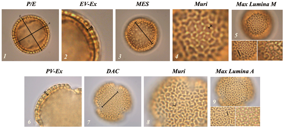
Figure 1. Representation of the morphological parameters in equatorial (1–5) and polar (6–9) views.
(1) P / E: polar axis and equatorial diameter ratio; (2) EV-Ex: exine thickness in equatorial view; (3) MES: maximum distance between two colpi in mesocolpium; (4 and 8) Muri: reticulus thickness; (5) Lumina M: maximum length of lumina in mesocolpium; (6) PV-Ex: exine thickness in polar vision; (7) DAC: distance between the apices of two adjacent colpi; (9) Lumina A: maximum length of lumina in apocolpium.
Measuring these parameters in thousands of granules is a slow and laborious process. Consequently, researchers are beginning to adopt automated techniques or digital methods meant to increase the speed and accuracy of measuring pollen grain morphology. However, studies using these techniques rarely publish information on the software configurations used to measure pollen grains, or they rely on proprietary software protected by paywalls. All these factors greatly inhibit the standardization, comparability, and reproducibility of the data and analyses such studies produce (see Daood et al., 2016; Khanzhina et al., 2018; Sevillano & Aznarte, 2018 for examples).
In response to these limitations, there is a growing trend in which funding sources and institutional research standards expect compliance with the FAIR principles (Findable, Accessible, Interoperable, and Reusable). This framework is also linked to the development of Open Science, in which the analytical procedures, data management plans and software configurations are presented transparently and available to the entire scientific community to encourage access, equity and research reproducibility (Bartling & Friesike, 2014; Marwick et al., 2017). Using open-access and open-source software, in addition to publishing code and detailed user guides, supports these Open Science principles.
In this context, we present OLEAtool, an open-access macro extension for ImageJ, which provides standardized tools for digital data collection of Olea pollen morphological metrics. ImageJ is a free, open-source image and video analysis program originally developed in the 1990’s by the United States National Institutes of Health for processing medical imagery (Schneider et al., 2012). In the years since, ImageJ has been adapted to multidisciplinary scientific applications across a diverse community of open-access researchers and practitioners. OLEAtool continues to expand the range of applications for ImageJ by including tools specifically tailored for morphopalynological data collection and analyses. By creating OLEAtool within the larger ImageJ open-science ecosystem, we are ensuring our data collection procedures are open and accessible to all. OLEAtool is updated regularly to fix bugs and add features via its source code on GitHub.
Methods
Implementation
The schematic workflow for OLEAtool is shown in Figure 2. OLEAtool is built using the ImageJ Macro language (version 1.53k;) to customize and implement a series of standardized tools for measuring pollen grain morphology from high-magnification images collected from camera-mounted light microscopes. Since it operates within ImageJ, OLEAtool is platform independent and is compatible with most common image file formats (e.g., TIFF, JPEG, GIF, BMP, PGM, PNG). OLEAtool includes two modules for measuring pollen morphology: 1) a general module for collecting manual measurements of all morphological parameters ("Manual Measurements Module"), and 2) a semi-automated module for measuring pollen lumina size, shape, and spatial distribution ("Lumina Module"). The operation of each module is described in detail below. Additionally, OLEAtool includes workflows for creating systematic metadata which documents important information about each sample and the data generated through the analysis (e.g., sample number, collection code, polar or equatorial view, analyst, date of analysis). This information is vital for archiving, sharing, and replicating pollen morphology datasets generated through OLEAtool.
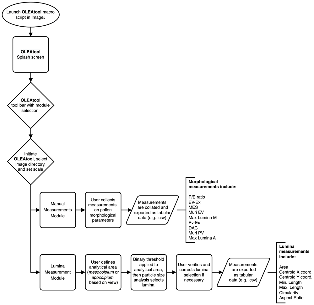
Figure 2. Schematic workflow for OLEAtool.
P / E: polar axis and equatorial diameter ratio; EV-Ex: exine thickness in equatorial view; MES: maximum distance between two colpi in mesocolpium; Muri: reticulus thickness; Lumina M: maximum length of lumina in mesocolpium; PV-Ex: exine thickness in polar vision; DAC: distance between the apices of two adjacent colpi; Lumina A: maximum length of lumina in apocolpium.
Manual Measurements Module description. Basic operations in OLEAtool are performed using the Manual Measurements Module, which includes linear measurements pollen morphological features in both polar and equatorial views. A user opens an image sequence of a pollen grain and selects the image (or images) that highlights the specific morphological parameter of interest (e.g., P, E, EV-Ex, etc.). For example, if measuring the equatorial diameter (E) of the pollen grain, the user will select an image where the exine is clearly visible and focused. They will draw a line to make the measurement, then click the “E” metric button on the toolbar. This measurement is then collected, and the result is automatically populated within the results table. This process is repeated for all desired measurements in the Manual Measurements Module. See Table 1 for descriptions of each morphological parameter measured in the OLEAtool Manual Measurements Module.
Table 1. Description of the parameters measured through the manual and lumina modules.
| Module | Metric | Description |
|---|
| Manual | P | Polar axis diameter |
| E | Equatorial axis diameter |
| P/E ratio | Polar axis/equatorial axis ratio |
| EV-Ex | Exine thickness in equatorial view |
| MES | Maximal distance between colpus in the mesocolpium |
| Muri EV | Exine reticulum thickness in mesocolpium |
| Max Lumina M | Maximal lumina length in the mesocolpium |
| Pv-Ex | Exine thickness in polar view |
| DAC | Distance between the apices of two colpi |
| Muri PV | Exine reticulum thickness in apocolpium |
| Max Lumina A | Maximal lumina length in the apoccolpium |
| Lumina | Area | Area of selected pixels in calibrated units |
| Centroid X | The x coordinate of the centroid derived from the center point of the selected lumen |
| Centroid Y | The y coordinate of the centroid derived from the center point of the selected lumen |
| Min Length | The shortest distance between any two points along the selected lumen |
| Max Length | The longest distance between any two points along the selected lumen |
| Circularity | Calculated as 4π × area ÷ perimeter2; A value of 1.0 indicates the selected pixels are a perfect circle;
As values approach 0.0, the selected pixels are increasingly elongated |
| Aspect ratio | Aspect ratio of selected pixels; calculated as the major (primary) axis / minor (secondary) axis of the
best fitting ellipse |
Lumina Module description. The Lumina Module is an enhance data collection workflow designed to measure a pollen grain’s lumina lengths, locations, and shapes as they are viewed within both the apocolpium and the mesocolpium. See Table 1 for descriptions of each lumen parameter measured in the OLEAtool Lumina Module. This module functions by using a user-defined area of interest within the image of a pollen grain to quantify lumina attributes by selecting and measuring them through image thresholding and a particle size analysis procedure. Image thresholds are automatically set but can be fine-tuned by the analyst to highlight as many lumina as possible within the field of view. Particle size analysis is then performed to identify and select all lumina identified through the procedure. Manual adjustment can be made to add or delete misidentified or unidentified lumina. Once the final selection is made, the length, location, and shape of each lumina is measured and compiled into a results table.
This approach offers several key advantages over traditional protocols for measuring pollen lumina in the apocolpium and the mesocolpium. Importantly, this protocol identifies all lumina within the area of interest and measures their maximum and minimum length, allowing a user to quickly identify the largest lumina in each pollen grain, which is commonly used as a distinctive morphological attribute in differentiating between pollen taxa. However, the OLEAtool Lumina Module expands the use of lumina attributes for differentiating pollen morphologies by: 1) constructing the distribution of all lumina maximum lengths within the pollen grain, and 2) collecting additional metrics for each lumen, including shape descriptors and relative centroid coordinates. Eliminating the need to manually measure each lumen within a pollen grain presents new pathways for using multiple metrics to describe all lumina to differentiate between pollen taxa or cultivars.
Operation
System requirements. OLEAtool is operated using customized macro extensions in ImageJ, and consequently has the same system requirements. ImageJ runs on operating systems that have Java 8 (or later) runtime installed. See installation instructions and examples of system configurations on the ImageJ website (here).
Download and start-up instructions
1. Download OLEAtool .zip folder from GitHub - see Software availability (Snitker et al., 2022)
2. Unzip the folder and place in an easily accessible location, such as the desktop, applications folder, or the home drive.
3. The file structure for photos of pollen used in OLEAtool can be arranged in any manner that is convenient to the user. However, we suggest creating a main folder with an identifying sample number (e.g., sample type, location, or depth) that contains subfolders containing photo sequences for each individual pollen grain that is analyzed. Please note that the main folder name and image file names are extracted and used to create a sample identifier in OLEAtool results table.
4. Open ImageJ and click the “Launch OLEAtool” option from the “More Tools” menu, which can be accessed by clicking the double arrows on the righthand side of the ImageJ toolbar.
5. Click the OLEAtool icon to initiate the program’s start window. A splash screen with the OLEAtool logo will appear on the screen and close. The main menu will then appear, providing the user with a choice to run the manual measurement module or the lumina module in OLEAtool.
6. Detailed and up to date tutorials, demonstration videos, and additional instructions can be found in the OLEAtool folder in GitHub and Zenodo repositories - see Software availability (Snitker et al., 2022).
Operating the Manual Measurements Module. This module has been created to measure the standard parameters used in Olea pollen morphology studies (e.g., Javady & Arzani, 2001; Lanza et al., 1996; Messora et al., 2017; Ribeiro et al., 2012). The measurements are collected by using a measurement toolbar, which is divided in equatorial (EV) and polar view (PV) parameters. The operational workflow for the Manual Measurements Module is highlighted in Figure 3 and described below:
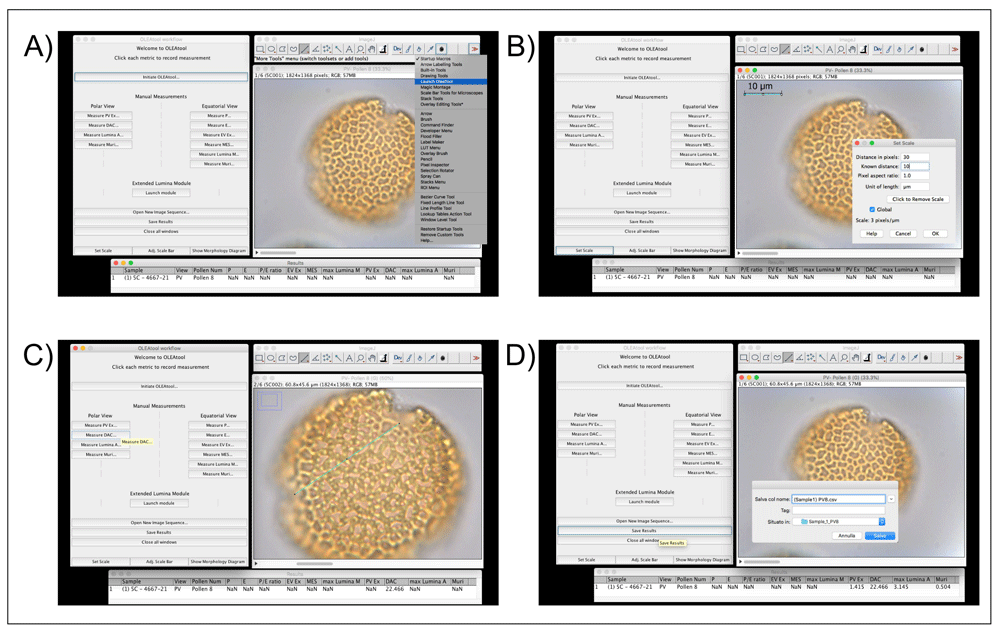
Figure 3. Example of OLEAtool operation in the Manual Measurements Module.
Operational steps include A) launching OLEAtool from the ImageJ menu; B) setting the scale for all measuring operations; C) collecting manual measurements by drawing on the image with the line tool and selecting the appropriate measurement to collect; and D) saving the output and clearing the images.
1. Open the images containing the photos of pollen grains that need to be measured. OLEAtool will extract the metadata from the image and displays them in a results table. All images will be loaded into an image stack. Individual images can be viewed by scrolling left or right using the scrollbar at the bottom of the viewing window.
2. Before proceeding with the measurements, it is essential to set the measurement scale and units using the Set Scale workflow within OLEAtool. To do so, the software will prompt the user to draw a line of a known length on the current image. Once the line is draw, click [OK], then enter in the known distance of the line in microns (µm). Click [OK]. OLEAtool’s scale is now calibrated.
3. To proceed with the measurements, the user draws a segment on the image and specifies the type of morphological parameter it is measuring, after which the measured value will be immediately reported in the results table.
4. Once all desired measurements are completed, the analyst saves the results table as a tabular dataset (Comma separated values [.csv] or Microsoft Excel file [.xlsx] formats) and can quit the session or open another image sequence to analyze.
Operating the Lumina Module. Exine structure and decoration patterns are key parameters in distinguishing between wild and cultivated Olea pollen types. Consequently, the semi-automated components of the Lumina module can efficiently collect multiple measurements of the lumina in the meso- and apocolpium areas. As mentioned previously, this module is expanding the ability for an analyst to evaluate different parameters for each lumen that would otherwise be logistically difficult and very time-consuming if assessed using traditional manual and optical microscopy techniques. The operational workflow for the Lumina Module is highlighted in Figure 4 and described below:
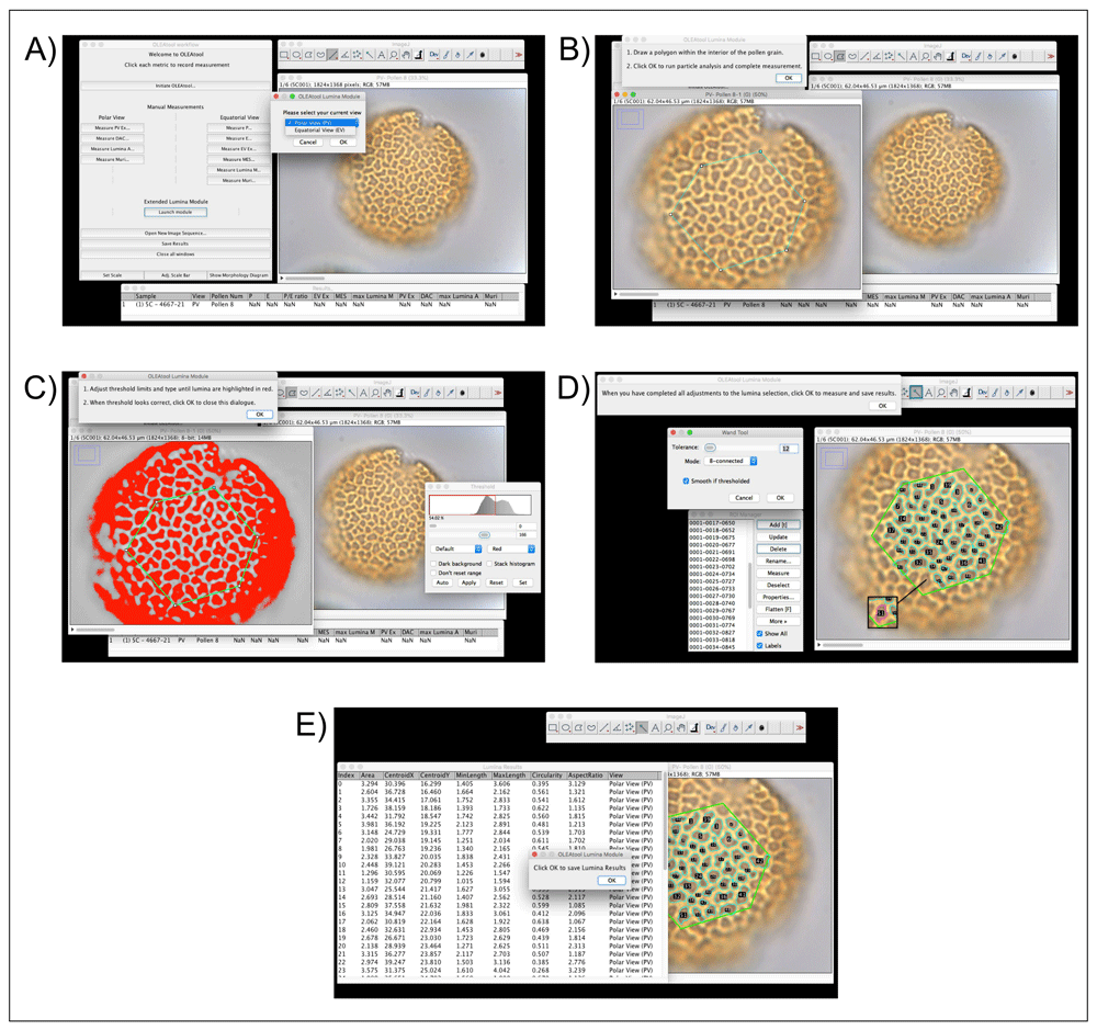
Figure 4. Example of OLEAtool operation in the Lumina Module.
Operational steps include A) starting the Lumina module and selecting the current view of the pollen grain; B) drawing a polygon in the center of the pollen grain to indicate the area of interest; C) apply the automated threshold and making any necessary manual adjustments; D) running the particle analysis procedure and making any manual adjustments to the selected lumina; and E) measuring all selected lumina and saving the results.
1. After setting the scale, the analyst launches the Lumina Module and specifies the type of view used in the analysis (equatorial or polar view).
2. OLEAtool opens a new window where is possible to choose the image in which the lumina are most visible. The analyst then uses the polygon tool to outline the boundaries of the mesocolpium (in equatorial view) or the apocolpium (in polar view). All lumina measurements will be take place in area of interest, which is defined by the polygon.
3. OLEAtool then thresholds the image using a black to white color scale. The analyst must either accept the automated threshold or adjust it using the sliders to highlight the most lumina possible. Following this selection, OLEAtool runs a particle size analysis procedure on the threshold image to select the lumina.
4. The lumina identified through the particle analysis are overlayed on the original image and their corresponding selection polygon are listed in the ImageJ region of interest (ROI) viewer. The analyst then verifies that the lumina selection is correct and can add or remove lumina using the ROI viewer. Once the final selections are made, all of the lumina are quantified (i.e., max length, shape, or location).
5. The lumina results can then be exported as tabular data (Common Separated Values [.csv] or Microsoft Excel file [.xlsx] formats), along with the image with the final overlay of selected lumina, which is saved as a JPEG or other supported image format.
Use Case: Quantifying Olea pollen morphology in the Balearic Islands
The main motivation to develop OLEAtool was to increase the number of measurable parameters we could collect, as well as improve the quality and accuracy of Olea pollen morphology studies. In this section we present a use case developed from research conducted during the EU-funded OLEA-project (G.A. 895735). The larger study design analysed the similarity/dissimilarity of cultivated and wild olive pollen grains from the Balearic Islands and proposed potential morphotypes that could also be applied to fossil palynological records (Mercuri et al., 2022; Ricucci, 2022). Here, we highlight a small portion of this work to illustrate the types of data required for using OLEAtool and the process of collecting morphological measurements using the Manual Measurements Module. The data associated with the Use case is available in Underlying data (Ricucci et al., 2022).
Input for OLEAtool began by collecting Olea pollen from olive tree flowers representing different individuals from throughout the Balearic Islands. Samples were then processed using standardized laboratory procedures (Erdtman, 1969). This includes treating each sample with glacial acetic acid to facilitate the release and dehydration of pollen from the anthers. The solution is then filtered, treated with the acetolytic mixture (acetic anhydride and sulfuric acid 9: 1) and boiled (90°C for 3–5 minutes). This procedure removes the cytoplasm and the lower layer of the granule wall, so that the outer layer (with diagnostic characters) is more visible. Pollen was mounted in a solution of 1:1 glycerol and water. Individual Olea pollen were photographed using a light-microscope at 1000x magnification (Mercuri et al., 2022; Ricucci, 2022). The resulting images were saved as JPGs with sample numbers in their file names in preparation for analysis in OLEAtool.
In the larger OLEA-project, all possible measurements of Olea morphological parameters (see Table 1 and Figure 1) were collected using both the Manual Measurements and Lumina Modules. Here we highlight the results of a subset of measurements made in the Manual Measurements Module and that include the Polar axis (P) and Equatorial diameter (E) collected for wild and cultivated varieties of Olea (Figure 5). Example images of the pollen used to collect these measurements (inputs) and the resulting measurements (outputs), are available via the data archives linked the Data availably section below (Ricucci et al., 2022). The mean polar axis measurements for wild varieties were 23.68 ± 1.71µm, while the mean measurements of cultivated varieties slightly higher, with values of 29.41 ± 1.80µm. Similarly, mean equatorial diameters for wild varieties were 23.88 ± 1.48µm and 28.67 ± 2.05µm in pollen corresponding to olive tree cultivars. Basic pollen dimensions accompanied by complementary measurements collected in the larger research project have proven to be key parameters in discriminating group of cultivars (Messora et al., 2017). In this sense, OLEAtool is an important step forward in collecting multiple pollen morphological parameters that when applied, will advance efforts to further distinguish between domestic and wild olive, and among agronomic varieties, though morphological pollen analysis.
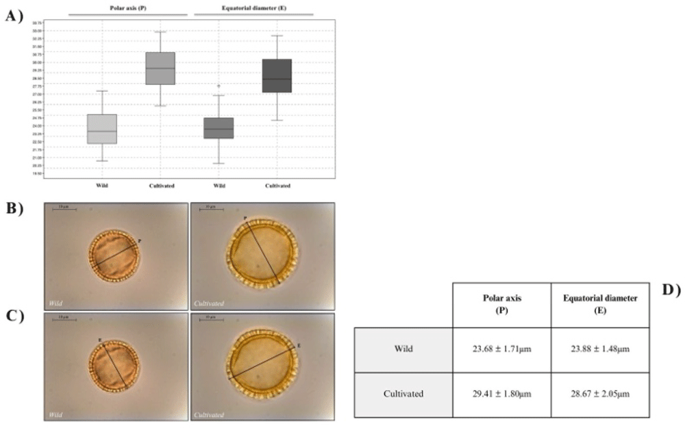
Figure 5. Results from the OLEA-project use study.
A) Boxplot obtained from the measurements of the polar axis (P) and equatorial diameter (E) parameters in wild and cultivated olive tree samples; B) examples of the polar axis (P) measurement in wild and cultivated Olea pollen; C) examples of the equatorial diameter (E) measurement in wild and cultivated Olea pollen; and D) results summary table of the OLEAtool use case.
Conclusions
OLEAtool has proven to be a powerful software tool for morphopalynological analysis on Olea pollen by increasing the quality, quantity, and speed at which these data can be collected. Specifically, it improves the data collection in the following ways:
1) OLEAtool enhances morphological data collection by increasing the speed, efficiency, replicability of the process by migrating all analyses to a digital platform.
2) The data produced by OLEAtool are standardized, comparable, and reproducible, which can facilitate collaboration and data sharing within the palynological research community.
3) The Lumina Module collects information on maximum lumen length, as well as a suite of other metrics that describe lumina shapes, sizes, and locations within the pollen grain. These new parameters will undoubtably advance the characterization of pollen exine pattern and provide new avenues for morphological analysis of Olea pollen.
Forthcoming research will include new statistical approaches to more definitively discriminate between wild and cultivated olive tree pollen, as well as expand the applicability of this work to other sectors, such as identifying modern agronomic varieties of olive trees. The morphotypes generated through this project aims to be applied to fossil pollen datasets obtained in natural and archaeological site sequences from the Balearic Islands to understand the timing and process of olive cultivation over the last several millennia. Despite Olea pollen grains have a highly resistance exine, taphonomical processes potentially affecting the exine structure of fossil pollen grains should be considered when trying to apply modern morphotypes to fossil palaeoenvironmental records. Moreove, Olea pollen grains are recurrent and abundant both Mediterranean palynological literature (Mercuri et al., 2013; Mercuri et al., 2019), including the Balearic Islands (Burjachs et al., 2017; Servera-Vives et al., 2018). Finally, we envision OLEAtool as an initial step in creating more collaboration between palynologists working in the Mediterranean by offering standardized methods for collecting morphological data. Furthemore, OLEAtool may be used in its current form in other pollen types such as the Brassicaceae family and other reticulate pollen grains where the lumina module can help in distinguishing between pollen types.
Ethics and consent
Ethical approval and consent were not required.
Data availability
Underlying data
Zenodo: Pollen Images for OLEAtool Manual Measurements Demonstration. https://doi.org/10.5281/zenodo.7412014 (Ricucci et al., 2022).
This project contains the following underlying data:
- 120 pollen images [JPEG format] of wild and cultivated pollen used as input data in demonstration case presented in the text.
- OLEAtool_example_dataset_manual_measurements.xlsx (all morphological measurements that can be collected in the Manual Measurements Module - see Table 1 - generated as output data from the example pollen images).
Data are available under the terms of the Creative Commons Attribution 4.0 International license (CC-BY 4.0).
Acknowledgements
Authors want to thank Anna Maria Mercuri, Assunta Florenzano, Paola Torri and Maurici Mus for their support, mentoring, and recommendations during this research process. We also thank Carme Garau Taberner and Bernat Canyelles from the Institut de Recerca i Formació Agroalimentària i Pesquera de les Illes Balears for their support during the fieldwork and to share their knowledge on local varieties. Finally, we acknowledge landowners who kindly allow us to enter their properties to collect olive tree flowers.
Faculty Opinions recommendedReferences
- Bartling S, Friesike S:
Opening science: The evolving guide on how the internet is changing research, collaboration and scholarly publishing. Springer Nature, 2014; 339. Publisher Full Text
- Besnard G, Khadari B, Navascués M, et al.:
The complex history of the olive tree: from Late Quaternary diversification of Mediterranean lineages to primary domestication in the northern Levant.
Proc Biol Sci.
2013; 280(1756): 20122833. PubMed Abstract
| Publisher Full Text
| Free Full Text
- Beug HJ:
Leitfaden der Pollenbestimmung für Mitteleuropa und angrenzende Gebiete. Friedrich Pfeil, München, 2004. Reference Source
- Burjachs F, Pérez-Obiol R, Picornell-Gelabert L, et al.:
Overview of environmental changes and human colonization in the Balearic Islands (Western Mediterranean) and their impacts on vegetation composition during the Holocene.
J Archaeol Sci Rep.
2017; 12: 845–859. Publisher Full Text
- Carrión JS, Fernández S, González-Sampériz P, et al.:
Expected trends and surprises in the Lateglacial and Holocene vegetation history of the Iberian peninsula and Balearic Islands.
Rev Palaeobot Palynol.
2010; 162(3): 458–475. Publisher Full Text
- Daood A, Ribeiro E, Bush M:
Pollen Grain Recognition Using Deep Learning.
Advances in Visual Computing.
Lecture Notes in Computer Science, 2016; 10072. : 321–330. Publisher Full Text
- De Leonardis W, Fichera G, Ocampo B, et al.:
Correlation between pollen grain and seed siz in Cicer species.
J Anim Breed Genet.
1995; 49: 21–26. Reference Source
- Erdtman G:
Pollen morphology and plant taxonomy. III. Morina L. With an addition on pollen morphological terminology.
Svensk botanisk Tidskr.
1945; 39.
- Erdtman G:
Pollen morphology and plant taxonomy.
Geologiska Föreningen i Stockholm Förhandlingar.
1952; 74(4): 526–527. Publisher Full Text
- Erdtman G:
Handbook of palynology: An introduction to the study of pollen grains and spores. New York: Hafner, 1969.
- Javady T, Arzani K:
Pollen Morphology of Five Iranian Olive (Olea europaea L.) Cultivars.
J Agric Sci Technol.
2001; 3: 37–42. Reference Source
- Khanzhina N, Putin E, Filchenkov A, et al.:
Pollen grain recognition using convolutional neural network.
ESANN.
2018; 409–414. Reference Source
- Kremp OW:
Morphologic encyclopedia of palynology. Univ. of Arizona Press Tucson., 1965; 263.
- Langgut D, Cheddadi R, Carrión JS, et al.:
The origin and spread of olive cultivation in the Mediterranean Basin: The fossil pollen evidence.
The Holocene.
2019; 29(5): 902–922. Publisher Full Text
- Lanza B, Marsilio V, Martinelli N:
Olive pollen ultrastructure: characterization of exine pattern through image analysis-scanning electron microscopy (IA-SEM).
Sci Hortic.
1996; 65(4): 283–294. Publisher Full Text
- Marwick B, d’Alpoim Guedes J, Barton CM, et al.:
Open science in archaeology.
SAA Archaeological Record.
2017; 17(4): 8–14. Reference Source
- Mercuri AM, Mazzanti MB, Florenzano A, et al.:
Olea, Juglans and Castanea: the OJC group as pollen evidence of the development of human-induced environments in the Italian peninsula.
Quat Int.
2013; 303: 24–42. Publisher Full Text
- Mercuri AM, Montecchi MC, Florenzano A, et al.:
The Late Antique plant landscape in Sicily: Pollen from the agro-pastoral villa del Casale - Philosophiana system.
Quat Int.
Elsevier BV.
2019; 499: 24–34. Publisher Full Text
- Mercuri AM, Ricucci C, Snitker G, et al.:
In search of new tools to trace back the history of olive cultivation.
Medpalynos.
Abstract Book, 2022. Reference Source
- Messora R, Florenzano A, Torri P, et al.:
Morphology and discrimination features of pollen from Italian olive cultivars (Olea europaea L.).
Grana.
2017; 56(3): 204–214. Publisher Full Text
- Punt W, Hoen PP, Blackmore S, et al.:
Glossary of pollen and spore terminology.
Rev Palaeobot Palynol.
2007; 143(1–2): 1–81. Publisher Full Text
- Ribeiro H, Cunha M, Calado L, et al.:
Pollen Morphology and Quality of Twenty Olive (Olea europaea L.) Cultivars Grown in Portugal.
Acta Hortic.
2012; 949: 259–264. Publisher Full Text
- Ricucci C:
Morfologia Pollinica e l’Analisi d’Immagine OLEAtool per la discriminazione di varietà domestiche e selvatiche di Olea europaea L. nelle Isole Baleari. Master Thesis, UNIMORE. 2022; 79. Reference Source
- Ricucci C, Servera-Vives G, Snitker G, et al.:
Pollen Images for OLEAtool Manual Measurements Demonstration. [Dataset]. Zenodo. 2022. http://www.doi.org/10.5281/zenodo.7412014
- Schneider CA, Rasband WS, Eliceiri KW:
NIH Image to ImageJ: 25 years of image analysis.
Nat Methods.
2012; 9(7): 671–675. PubMed Abstract
| Publisher Full Text
| Free Full Text
- Servera-Vives G, Riera S, Picornell-Gelabert L, et al.:
The onset of islandscapes in the Balearic Islands: A study-case of Addaia (northern Minorca, Spain).
Palaeogeogr Palaeoclimatol Palaeoecol.
Elsevier BV.
2018; 498: 9–23. Publisher Full Text
- Sevillano V, Aznarte JL:
Improving classification of pollen grain images of the POLEN23E dataset through three different applications of deep learning convolutional neural networks.
PLoS One.
2018; 13(9): e0201807. PubMed Abstract
| Publisher Full Text
| Free Full Text
- Snitker G, Servera-Vives G, Ricucci C:
OLEAtool v.1.0.0. Zenodo. [Software], 2022. http://www.doi.org/10.5281/zenodo.7046771
- Stanley RG, Linskens HF:
Pollen: Biology Biochemistry and Management. Springer, Berlin, 1974. Publisher Full Text
- Terral JF, Alonso N, Buxó Capdevila R, et al.:
Historical biogeography of olive domestication (Olea europaea L.) as revealed by geometrical morphometry applied to biological and archaeological material.
J Biogeogr.
2004; 31(1): 63–77. Publisher Full Text
- Terral JF, Bonhomme V, Pagnoux C, et al.:
The Shape Diversity of Olive Stones Resulting from Domestication and Diversification Unveils Traits of the Oldest Known 6500-Years-Old Table Olives from Hishuley Carmel Site (Israel).
Agronomy.
2021; 11(11): 2187. Publisher Full Text