1. Introduction
In 2022, there were an estimated 249 million malaria cases and 608,000 malaria deaths worldwide, most of which occurred in sub-Saharan Africa1. Pregnancy is associated with a higher susceptibility to malaria infection2, and according to the World Health Organization, 12.7 million pregnancies in sub-Saharan Africa were infected with malaria in 20221. Malaria in pregnancy may cause several severe adverse outcomes for the mother and fetus, including maternal anemia and stillbirths3. In sub-Saharan Africa, malaria during pregnancy frequently leads to placental malaria, which is characterized by the presence of malaria-infected red blood cells in placental intervillous spaces4. Plasmodium falciparum (P. falciparum) is the most common malaria parasite in sub-Saharan Africa5, and the cause of the most severe form of malaria6. It is also the main cause of placental malaria because of its frequent sequestration in the placenta, which is mediated by the interaction between VAR2CSA on the surface of P. falciparum-infected red blood cells and chondroitin sulfate A (CSA) on the syncytiotrophoblast surface7. Placental malaria is associated with several adverse effects on the fetus, including low birth weight, preterm birth, and stillbirth4. Since malaria transmission to the fetus is thought to be rare8, such poor outcomes likely result from the effects of placental malaria on the placenta. However, the mechanisms through which placental malaria affects placental physiology and function remain unclear. Moreover, placental malaria and other placental infections are difficult to diagnose and treat because the placenta is inaccessible during pregnancy, a challenge that is further complicated by the fact that many placental infections are asymptomatic9. Therefore, considering that placental insults are also thought to affect well-being later in life by contributing to adult-onset chronic diseases, such as cardiovascular disease and type 2 diabetes10, it is crucial to better understand the placental pathobiology of placental malaria, which may guide the development of diagnostic and therapeutic tools.
Biobanks are key resources in health research. Although widely used research tools, such as immortal cell lines and animal models, are indispensable in health research, they have important limitations. For instance, immortalized cell lines are not sufficiently representative of their corresponding primary cell types in culture, and the in vivo environment cannot be fully recapitulated11, while animal models are limited by fundamental biological differences between humans and other species, ethical concerns, high maintenance costs, and not reproducing human disease with full accuracy12. Consequently, the use of human tissues and primary cells to complement these systems is highly attractive as it may offset some of their limitations. These are especially valuable in low-resource settings where the use of cell culture systems and in vivo models is limited. Moreover, in such settings, the use of donated tissues for research can allow the study of various diseases in the contexts in which they occur, using naturally infected samples, which would lead to findings that are more physiologically relevant. Despite numerous health challenges, biobanking is insufficient in African countries13.
Here, we describe a biobank resource established to support placental malaria research. The biobank has placenta, cord, and maternal sera, and maternal and placental dry blood spots donated by women living in malaria-endemic (Western Kenya) and non-malaria-endemic (Central Kenya) regions of Kenya. The rates of asymptomatic malaria are high in Western Kenya14, which significantly increases the risk of unsuspected malaria during pregnancy and placental malaria. Therefore, we compared the performance of conventional methods (rapid diagnostic tests and PCR) against that of isothermal identical multirepeat sequence (iso-IMRS) PCR15, an ultrasensitive P. falciparum test16, in the detection of malaria in pregnancy and placental malaria when establishing placental malaria biobanks.
2. Methods
2.1 Participants and sample collection
The biobank’s placenta, maternal and cord blood, and maternal and placental dry blood spots were donated by residents of Western Kenya (a malaria-endemic region) and Central Kenya (a malaria non-endemic region). Participants were recruited during antenatal visits or at the time of admission to the maternity ward. Before recruitment, a maternity unit nurse conducted an informed consent process. The nurse explained the study's purpose, procedures, and potential risks and benefits to potential participants in their preferred language (Swahili or their mother tongue). If the participants agreed to participate, they were asked to sign a written informed consent form. The form was available in both Swahili and English, and was read and explained to them in their preferred language. By signing, participants consented to donate placenta, cord blood, and maternal blood samples for use in the current study, as well as in potential future research, without requiring additional consent. The study’s participant inclusion criteria were as follows: 1) age ≥18 years, written informed consent, spontaneous vaginal delivery (normal delivery), term delivery, singleton birth, and no known history of sexually transmitted disease during the current pregnancy. Patients who did not meet the inclusion criteria were excluded from the study. Consenting participants were also requested to complete questionnaires that gathered deidentified (using randomly assigned participant numbers) participant details and general data, including maternal age, gravidity, parity, gestational age, history of malaria infection during the current pregnancy, number of antenatal clinic visits, and the number of intermittent presumptive treatments with sulfadoxine-pyrimethamine (IPTp-SP) doses. Figure 1 schematically shows the parent study participant recruitment and biobank establishment. Maternal and cord blood samples were collected before and after delivery, respectively, followed by serum preparation (centrifugation at 2,500 revolutions per minute [rpm] at 4 °C) and preservation at -80 °C. General health information, such as maternal blood pressure and history of diabetes, was also recorded. Newborn-associated data, including sex, birth weight, gestational age, and placental weight, were collected upon delivery. Donated placentas were physically examined according to the recommendations of the Amsterdam Placental Workshop Group17, followed by the collection of full-thickness biopsies within 30 min of delivery using a standardized approach18, as schematized in Figure 2B. Histological samples were immediately fixed in 10% formalin for 24 h and then paraffin-embedded, whereas fresh samples were immediately snap-frozen and stored at -80 °C. The placental biopsy strategy and serum and sample storage are shown schematically in Figure 2.
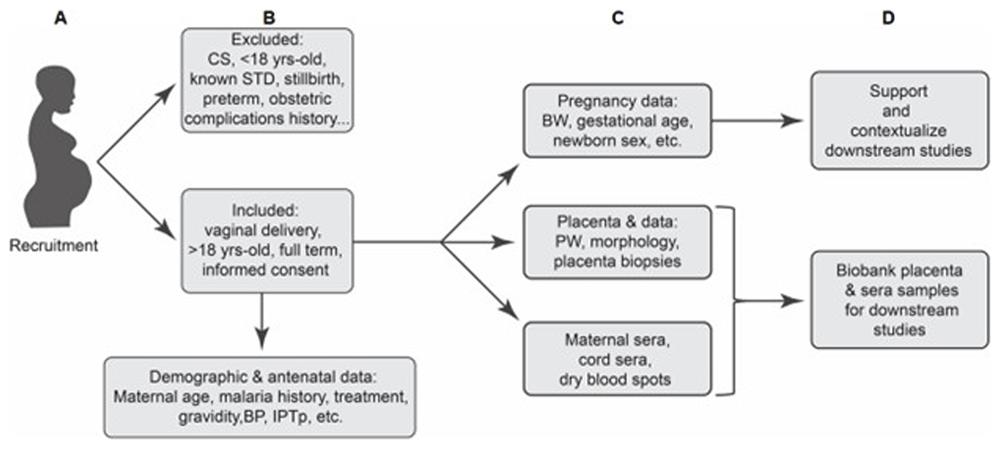
Figure 1. A schematic of sample donor recruitment and underlying biobank data and sample collection.
Data and samples were obtained from participants who met the set inclusion criteria and gave written informed consent to participate. CS: cesarean section, STD: sexually transmitted disease, BP: blood pressure, IPTp: intermittent presumptive treatment, PW: placenta weight.
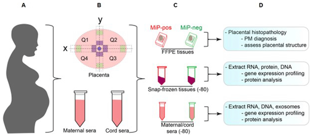
Figure 2. Schematic representation of sample collection for the biobank.
A) Samples were donated by residents of a malaria and a non-malaria endemic region of Kenya. B) Placentas (upper panel) were biopsied in the four quadrants at the sites coded in green (peripheral) and purple (central). Maternal and cord sera were also obtained (lower panel). The blue spot indicates cord insertion. C) Some biopsies were formalin-fixed and embedded, while other biopsies and sera were snap-frozen and stored at -80 °C. D) Biobank can support various types of analyses.
2.2 Histology
Fixed placental biopsies were embedded in paraffin as described previously19, using a MediMeas embedding system. The presence of infected erythrocytes in placental biopsies was examined using hematoxylin and eosin staining. Briefly, 5-µm-thick placental sections were placed on charged slides (Dako, Cat No. K8020) and then dried overnight on a slide warmer at 37 °C. They were then dewaxed using xylene (Finar Chemicals, Cat No. 21940LC250) and rehydrated through sequential dipping in absolute, 95%, 70%, and 50% ethanol (Scharlau, Cat No. ET00052500), and distilled water. Next, they were dipped in hematoxylin (Loba Chemie, Cat No. 04023) for seven min and rinsed under running water, followed by destaining in acid alcohol (1% hydrochloric acid in 70% ethanol, 10 dips). The slides were then dipped in eosin (Griffchem, Cat No. 45380) for 45 s, washed in distilled water (one dip), and dehydrated in ethanol (95% and absolute, 10 dips each) before clearance in two xylene washes, five min each. The samples were then cover-slipped using dibutylphthalate polystyrene xylene mountant (Finar Chemicals, Cat No. 10525LM250), followed by examination under a microscope (Richter Optica UX1, M2 Scientific) and imaging (40X magnification) in at least 10 fields of view per slide using a Moticam microscope camera (Motic Scientific). Placental malaria status was determined based on the presence of malaria parasite-infected erythrocytes, as described previously20. Past placental malaria was indicated by the absence of infected erythrocytes and the presence of fibrin-enmeshed hemozoin and pigmented macrophages21, while chronic placental malaria was indicated by signs of past placental malaria in the presence of infected erythrocytes22.
2.3 DNA extraction
Parasite DNA was extracted from dry blood spots (DBS) using the Chelex method, as described previously23. Briefly, a 3-mm diameter punch of each DBS was placed into a 1.5 mL microcentrifuge tube, incubated in 1 ml of PBS 1X for 40 min at room temperature, centrifuged at 14,000 rpm for two minutes at 4 °C, and the supernatant discarded. These steps were repeated for a second wash, after which the pellet was resuspended in 50 µL of nuclease-free water. Next, 20 µL of 20% Chelex ®100–sodium (Sigma–Aldrich, Cat. No. 95577) was added to the samples, followed by incubation at 100 °C for 10 min, and centrifugation at 14,000 rpm for one min, at room temperature. The supernatants (containing DNA) were then transferred into new 1.5 mL microcentrifuge tubes and stored at -20 °C.
To extract DNA from placental samples, approximately 10 mg of a cross-sectional placental tissue sample (minced with a sterile scalpel) was placed in a 1.5 mL microcentrifuge tube containing 195 µL of 10% Chelex ®100–sodium and 5 µL of proteinase K (NEB, Cat No. P8107S) and incubated at 60 °C for three h. They were then incubated at 95 °C for 15 min, vortexed briefly, and centrifuged at 14,000 rpm for two min. The supernatants (containing DNA) were then carefully transferred into new microcentrifuge tubes and stored at -20 °C. The extracted DNA was cleaned by precipitation through overnight incubation in a solution of 3 M sodium acetate (ThermoFisher, Cat No. AM9740) and ice-cold absolute ethanol (Scharlau, Cat No. ET00052500) at 0.1 and 2.5 times the sample volumes, respectively. The samples were then centrifuged at 14,000 rpm (4 °C) for 30 min, and the supernatants were discarded. The DNA pellets were then washed twice with 500 µL of ice-cold 70% ethanol (Scharlau, Cat No. ET00052500) and centrifuged at 14,000 rpm for 10 min at 4 °C. After air-drying for 30 min, the DNA pellet was resuspended in 50 µL of nuclease-free water, quantified using a nano-spectrophotometer (Labtron, LNS-C10), and stored at -20 °C.
2.4 PCR analysis
To detect P. falciparum, PCR was conducted using the following P. falciparum-specific primers (expected band size: 205 bp)24: rFAL1 5’-TTAAACTGGTTTGGGAAACCAAATATATT-3’ (forward) and rFAL2 5’-ACACAATGAACTCAATCATGACTACCCGTC-3′ (reverse). The following PCR program was used: initial denaturation at 94 °C for 30 s, final denaturation at 94 °C for 30 s, annealing at 51 °C for 45 s, extension at 68 °C for one min, and a final extension at 68 °C for five min. To confirm the PCR results, samples that were negative using the initial PCR were subjected to nested PCR analysis25 to enhance sensitivity. To this end, a primary PCR was first performed using the primers rPLU6 5’-CTTGTTGTTGCCTTAAACTTC-3’ (forward) and rPLU6 5’-TTAAAATTGTTGCAGTTAAAACG-3’ (reverse), which produced a 1.2 kb product26 that was then subjected to the P. falciparum-detecting PCR described above (expected band size: 205 bp). The following primary nested PCR program was used: initial denaturation at 94 °C for five min, final denaturation at 94 °C for 30 s, annealing at 55 °C for one min and 15 s, extension at 68 °C for one minute, and a final extension at 68 °C for five min. PCR was performed using One Taq® Quick-Load® 2X Master Mix with standard buffer (NEB, Cat No. M0486S) on a MiniAmp Plus thermal cycler (Applied Biosystems). PCR products were examined using 2% agarose (Cleaver Scientific, Cat No. CSL-AG 100) after staining with SmartGlowTM (Accuris, Cat No. E4500-PS) Fluorescent DNA Dye.
2.5 Identical MultiRepeat Sequence (IMRS) analysis
IMRS is a highly sensitive tool that can detect very low P. falciparum densities (asymptomatic) by simultaneously amplifying identical multi-repeat genomic regions16. Our group recently coupled this powerful technique with isothermal DNA amplification, which allows the use of a constant IMRS DNA amplification temperature27 (termed iso-IMRS) for P. falciparum DNA detection15 using the following reagents: Bst 2.0 WarmStart® DNA polymerase (NEB, Cat No. M0538L), isothermal amplification buffer (NEB, Cat No. B0537S), ficoll (Sigma–Aldrich, Cat No. F5415), betaine (Sigma–Aldrich, Cat No. B0300), and dNTPs (NEB, Cat No. N0447S), nuclease-free water (Promega, Cat No. P119E), and forward (5’-CTGGAGGTCAGTTCACAGTACCC-3’) and reverse (5’-CTCTACAATCCGTAGAGTTACTGG-3’) primers, as described previously15. The iso-IMRS amplification products were examined using 2% agarose gel (Cleaver Scientific, Cat No. CSL-AG 100) at 100V for 40 min after staining with SmartGlowTM (Accuris, Cat No. E4500-PS) Fluorescent DNA Dye. We used this method to test for P. falciparum in the placental tissue, placenta dry blood spots, and maternal dry blood spots.
2.6 Data analyses
GraphPad Prism version 9 was used for statistical analyses and data visualization. The sample size for the analysis comparing the effectiveness of conventional malaria diagnostic tools with that of iso-IMRS in placental malaria detection (Section 3.4) was calculated assuming a confidence level of 95%, a placental malaria population proportion of 54% (based on our placental malaria rate determined using histological analysis of samples from the malaria endemic region), and a population size of 159 (histology-examined placenta samples from the malaria-endemic region).
3. Results
3.1 Placental malaria diagnosis
Hematoxylin and eosin staining was used for placental malaria diagnosis in placental biopsies from 174 donors: 77 with a history of malaria during the current pregnancy and 97 without. Of the donors, 159 and 15 were residents of malaria endemic and non-malaria endemic regions, respectively. Based on histological examination, the biobank placenta samples were classified into placental malaria-negative (no infected erythrocytes observed, n = 54, including 15 from the non-malaria-endemic region), placental malaria-positive (active [at least one infected erythrocyte observed] and chronic-active [at least one infected erythrocyte observed along with fibrin-enmeshed hemozoin and macrophages], n = 92), and past placental malaria (only hemozoin present, n = 28) groups, as described previously21,22 (Figure 3A–D, Raw data File 1).
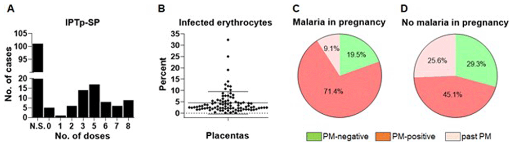
Figure 3. Most residents of the malaria endemic region had placental malaria (PM).
A) Characterization of the data underlying the biobank revealed that frequency of anti-malaria chemoprophylaxis varied widely among the sample donors. N.S.: number of IPTp-SP (intermittent presumptive treatment with sulfadoxine-pyrimethamine) doses not specified. B) Histological analysis indicates that the number of infected erythrocytes varies widely across asymptomatic placenta malaria cases. C–D) Histological analysis indicated that the majority of cases with (C) or without (D) a history of malaria in pregnancy had ongoing or past placental malaria.
3.2 Placental malaria is frequently asymptomatic
At the time of sample donation, participants did not exhibit symptoms associated with malaria and they did not test positive for malaria using rapid diagnostic tests. Almost all placental donors from the malaria-endemic region (97%, Raw data File 1) had received IPTp-SP during pregnancy, although the number of administered doses varied widely (Figure 3A). Moreover, based on histological analysis of the samples from the malaria endemic region, the number of infected erythrocytes in the placentas varied widely (Figure 3B). Analysis of the data stratified by the history of malaria infection during the current pregnancy revealed that 71.4% of the participants who reported a history of malaria during the current pregnancy also had placental malaria, as revealed by histological examination, while 9.1% of the cases exhibited signs of past placental malaria infection (Figure 3C), which was indicated by the presence of hemozoin (malaria pigment) but not infected erythrocytes (Figure 4). Importantly, this analysis showed that among those who reported having no history of malaria during the current pregnancy, 45.1% had placental malaria, while 25.6% exhibited past placental malaria (Figure 3D). Taken together, these data indicate that placental malaria is frequent even in the absence of overt malaria symptoms during pregnancy.
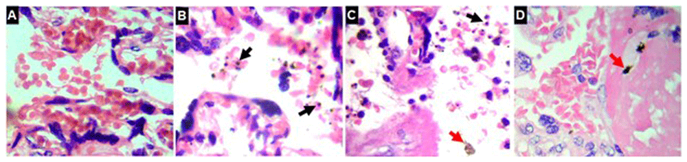
Figure 4. Histological analysis was used to diagnose and classify placental malaria.
A) Hematoxylin and eosin (H&E) staining of a placental malaria-free sample. B) H&E shows an active case of placental malaria (black arrow: infected erythrocytes). C) H&E shows a case of chronic-active placental malaria (black arrow: infected erythrocytes, red arrow: hemozoin). D) H&E shows a case of past placental malaria (red arrows: fibrin-entrapped hemozoin).
3.3 Conventional techniques fail to detect some placental malaria cases
Participants were grouped into malaria in pregnancy and non-malaria in pregnancy groups based on their response to the question of whether they had at least one microscopy-confirmed malaria episode during the current pregnancy (before recruitment into this study). Moreover, during sample donation, rapid malaria diagnostic tests, which are widely used in endemic regions28, did not detect malaria infection in any of the participants. Because of the constraints in the low-resource settings where the samples were collected, it was not feasible to use microscopy, the gold standard for malaria diagnosis29, to examine maternal blood films at the time of recruitment and sample collection. However, because rapid diagnostic tests and microscopy frequently fail to detect placental malaria29, we collected maternal dry blood spots for downstream PCR-based malaria diagnosis, which is highly sensitive30.
3.4. A comparison of conventional methods identifies iso-IMRS as a highly sensitive placental malaria diagnostic tool
Since rapid diagnostic testing at the time of sample donation did not detect maternal malaria in the sample donors, and yet, histology, the gold standard for placental malaria diagnosis4, revealed infected erythrocytes in most placentas, we wondered how the efficiency of histology-based placental malaria diagnosis compared with that of rapid diagnostic tests and PCR. The comparison was performed using 59 samples (the sample size was determined as described in Section 2.6), of which, based on histology, 22 (37.3%) and 37 (62.7%) were placental malaria positive and negative, respectively. A higher number of placental malaria negative samples (based on histology) was included to better assess if and at which rate, histology might miss placental malaria positive cases. Compared with rapid diagnostic tests, which did not detect any malaria in pregnancy cases (Figure 5A), PCR analysis of the maternal dry blood spots detected eight (13.6%) P. falciparum-positive cases (Figure 5B; a representative PCR image is shown in Figure 5K). Of these, two cases were positive for placental malaria based on histology (Raw data File 2). This indicates that rapid tests miss asymptomatic malaria in pregnancy and that during pregnancy, they are not a reliable indicator of ongoing placental malaria. Next, we examined how placental histology compared with PCR analyses of placental blood and tissue. This analysis revealed that PCR analysis of placental dry blood spots detected P. falciparum DNA in 13 of the 59 samples (22%), and of the 13, seven also had histology-confirmed placental malaria (Figure 5D) while six were malaria in pregnancy positive based on maternal dry blood spot PCR (Raw data File 2). PCR analysis of DNA extracted from placental tissues detected P. falciparum DNA in 10 of the 59 samples (17%), of which, five were placental malaria positive based on histology (Figure 5E), four were positive for malaria in pregnancy based on maternal dry blood spot PCR, and six were P. falciparum positive based on placental dry blood spot PCR (Raw data File 2).
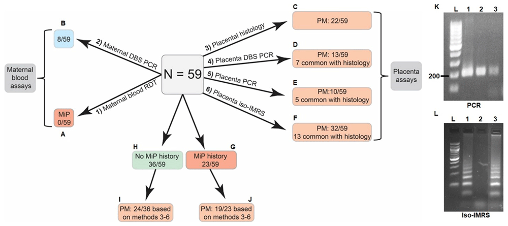
Figure 5. A comparison of the efficiencies of various tools in malaria in pregnancy (MiP) detection.
A–B) Rapid diagnostic tests (RDT) vs PCR analysis of maternal blood (DBS: dry blood spot). C) Placenta tissue histology. D–E) Placenta blood and tissue PCR. F) Placenta tissue iso-IMRS (isothermal identical multirepeat sequence analysis). G–J) Combined, these approaches (3–6) detected placental malaria in 43 (72.9%) of the analyzed samples. K–L) representative images of P. falciparum detection in placenta tissue biopsies using PCR (K; PCR product: 205 base pair; L: ladder, 1: positive control DNA sample, 2: dry blood spot DNA sample, 3: placenta tissue DNA sample), and iso-IMRS (L, L: 100 base pair ladder, 1: positive control DNA sample, 2: negative control sample, 3: placenta DNA sample), which generates amplicons of various sizes because multiple sequences are targeted.
PCR analysis of P. falciparum using IMRS has been proposed as a highly sensitive means of detecting asymptomatic malaria16. Recently, our group coupled isothermal amplification and IMRS to develop iso-IMRS, an ultrasensitive P. falciparum detection tool that is highly suitable for low-resource settings31. Therefore, we investigated whether iso-IMRS could detect placental malaria more sensitively than conventional approaches. Remarkably, iso-IMRS detected P. falciparum DNA in 32 of the 59 placentas examined (54.2%; Figure 5F; Figure 5K shows a representative image of iso-IMRS results), including 13 that were placental malaria positive based on histology and 19 that were placental malaria negative based on histology (Raw data File 2). These data indicate that iso-IMRS is more sensitive than conventional tools for detecting placental malaria. Combined, histology, placental dry blood spot PCR, placental tissue PCR, and iso-IMRS indicated that 43 of the 59 placentas (72.9%) were affected by placental malaria (Figure 5G–J, Raw data File 2).
4. Discussion
Pregnancy is associated with an increased risk of infection by various pathogens, which may result in severely adverse pregnancy outcomes32. However, the mechanisms through which such infections disrupt placental health and function are unclear, limiting the ability to diagnose and intervene against placental infections during pregnancy9. This is partly because placental infections occur disproportionately in resource-limited settings, which have significant research capacity constraints, and because the placenta is inaccessible for research during pregnancy. Nonetheless, biobanks of naturally infected samples from endemic regions may offer vital resources for bridging crucial research gaps.
In malaria-endemic regions, placental malaria, which is characterized by the sequestration of malaria parasite-infected erythrocytes (mainly P. falciparum) in placental intervillous spaces, is associated with poor pregnancy outcomes, including fetal growth restriction, preterm birth, and stillbirth4. However, the mechanisms by which placental malaria affects placental health and function are unclear, and currently, no tools are available for placental malaria detection and treatment during pregnancy. Here, focusing on placental malaria, we describe the establishment of a biobank to support placental health studies in resource-limited contexts.
As adult residents of malaria-endemic regions possess partial immunity33, many cases are asymptomatic14, making it difficult to accurately identify patients with malaria. This challenge is exacerbated by the fact that microscopy, the gold standard for malaria diagnosis, is subjective, labor- and skill-intensive34, and relatively cost-intensive in resource-limited settings. Consequently, here, rapid diagnostic tests, which are the main malaria testing tools in Africa28, were used to screen for malaria in pregnancy cases during sample donor recruitment. However, these tests did not detect any malaria-positive cases in those with or without a known history of malaria infection during pregnancy. This was probably because rapid tests are prone to false-negative readings in cases of low parasite-density or asymptomatic malaria35,36, which is common in malaria-endemic regions and is estimated to be present in over 80% of residents of highly endemic regions36, or deletions of the target proteins, HRP2/337. Moreover, in such cases, parasite levels are often below the limit of microscopic detection36, while driving significant chronic pathogenesis, including in pregnant women38. Indeed, PCR analysis of maternal dry blood spots identified malaria in pregnancy cases that were missed by rapid diagnostic tests (Figure 5A–B), indicating that the use of more sensitive techniques, such as nucleic acid-based assays, to detect malaria in pregnancy is critical for placental malaria research and clinical purposes.
Importantly, histological examination revealed that 120 (75.5%) of the 159 placentas from the malaria-endemic region, including those without a known history of malaria during pregnancy, were affected by placental malaria (92 active and 28 past cases, Raw data File 1). Crucially, almost all sample donors had received malaria chemoprophylaxis during pregnancy, although the number of doses used varied widely, and the burden of infected erythrocytes was highly variable across placentas (Figure 4A–B). Therefore, such variables should be considered in studies on placental malaria. Moreover, for research and clinical purposes, it may be important to accurately identify past placental malaria cases because hemozoin, a hallmark of chronic-active and past placental malaria22, is reported to be immunomodulatory39. Together, these observations indicate that in malaria endemic areas, even with chemoprophylaxis, the burden of placental malaria is remarkably high in asymptomatic malaria in pregnancy cases, including those found to be malaria negative using rapid diagnostic tests, which in Africa are used to test up to 87% of suspected malaria cases28.
Although histology is the gold standard for placental malaria diagnosis4, the biopsies examined represent a very small proportion of the whole placenta, which, especially in cases with low parasite densities, may lead to false-negative findings because infected erythrocytes may be absent in the randomly sampled area but present in other parts of the placenta. Because nucleic acid amplification is highly sensitive, we compared its P. falciparum detection efficiency in 59 placental biopsies and placental blood samples with that of histological analysis (Figure 5C–E). Placental dry blood spot PCR detected P. falciparum in 13 samples, compared with 22 based on histology, indicating that histology is superior. However, only seven cases were common between the two methods. Considering that placental biopsies are quite small relative to placenta size, it is likely that placental DBS preparations sampled unique areas that contained parasites. Similarly, PCR analysis of placental tissue biopsies detected P. falciparum DNA in 10 of the 59 samples examined, including five that were not detected by histology. These additional cases may have resulted from PCR detection of P. falciparum DNA on hemozoin40 in samples with hemozoin only, which were classified as past placental malaria based on histology.
As it simultaneously amplifies numerous repeats of a genomic sequence, P. falciparum IMRS PCR is an ultrasensitive tool for malaria diagnosis16, and when coupled with isothermal amplification (iso-IMRS), it may be deployed in resource-limited point-of-care settings. The detection of P. falciparum DNA using iso-IMRS PCR in tissue biopsies from 32 of 59 placentas (Figure 5F) highlights iso-IMRS as the most sensitive tool for placental malaria detection. Of the 32 P. falciparum-positive cases, 13 were also identified by histology, indicating that relative to histology, iso-IMRS detected markedly more unique cases than placenta dry blood spots and placenta tissue PCR. This is attributable to the higher sensitivity of iso-IMRS, which may have facilitated P. falciparum DNA detection in tissues with very low parasite densities or in cases of past placental malaria, with very low levels of parasite DNA in hemozoin deposits. Taken together, our observations indicate that iso-IMRS is the most sensitive tool for placental malaria detection, followed by histology and PCR. Importantly, considering that the results obtained using different techniques did not overlap fully, when using a single technique, it is necessary to extensively sample the placenta to reduce the likelihood of false negative findings. Alternatively, combining histology and iso-IMRS may achieve high placental malaria detection rates, which would be highly valuable for the accurate characterization of placental samples in placental malaria research. Finally, considering that most placental samples from our malaria endemic region had ongoing or past placental malaria, when setting up biobanks, it may be highly beneficial to include samples from a non-malaria-endemic region for use as placental malaria-naïve controls. However, such control samples should undergo similar rigorous characterization.
Ethics statement
This study was approved by Mount Kenya University’s Ethics Review Committee, approval number 1314 (date: 17.06.2022) and was conducted in adherence to the Declaration of Helsinki guidelines.
Consent to participate
All study participants gave written informed consent.